. 2002 Jul 23;99(15):9819-24.
doi: 10.1073/pnas.152205299. Epub 2002 Jul 15.

Multiple centrosomes arise from tetraploidy checkpoint failure and mitotic centrosome clusters in p53 and RB pocket protein-compromised cells

Affiliations

Multiple centrosomes arise from tetraploidy checkpoint failure and mitotic centrosome clusters in p53 and RB pocket protein-compromised cells

Franck Borel et al. Proc Natl Acad Sci U S A. .

Abstract

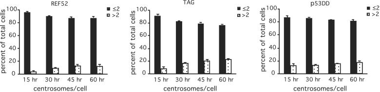
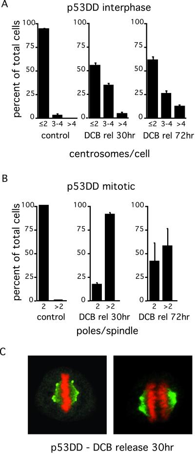
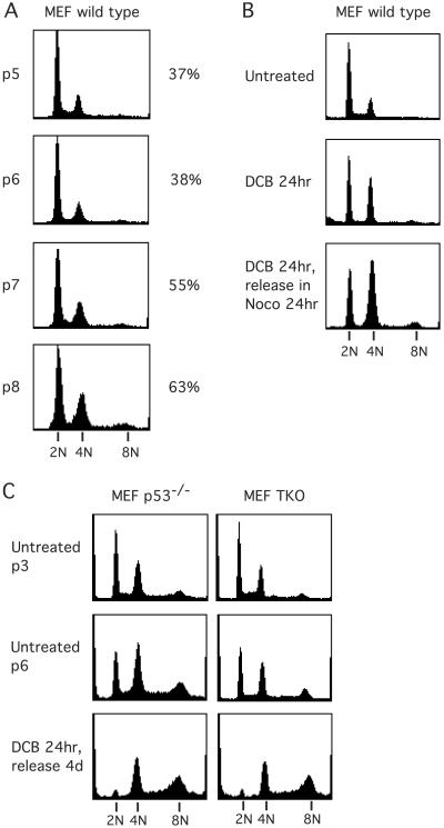
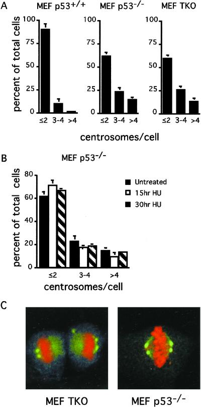
A high degree of aneuploidy characterizes the majority of human tumors. Aneuploid status can arise through mitotic or cleavage failure coupled with failure of tetraploid G(1) checkpoint control, or through deregulation of centrosome number, thus altering the number of mitotic spindle poles. p53 and the RB pocket proteins are important to the control of G(1) progression, and p53 has previously been suggested as important to the control of centrosome duplication. We demonstrate here that neither suppression of p53 nor of the RB pocket protein family directly generates altered centrosome numbers in any of several mammalian primary cell lines. Instead, amplification of centrosome number occurs in two steps. The first step is failure to arrest at a G(1) tetraploidy checkpoint after failure to segregate the genome in mitosis, and the second step is clustering of centrosomes at a single spindle pole in subsequent tetraploid or aneuploid mitosis. The trigger for these events is mitotic or cleavage failure that is independent of p53 or RB status. Finally, we find that mouse embryo fibroblasts spontaneously enter tetraploid G(1), explaining the previous demonstration of centrosome amplification by p53 abrogation alone in these cells.


Figures

Figure 1
Figure 1
Absence of p53 and RB pocket protein family functions does not modify ploidy nor centrosome number in fibroblast cells. (A) DNA content of random cycling REF-52, p53DD REF-52, and TAG cells, assayed by flow cytometry. Centrosome (B) and spindle pole (C) numbers in the indicated cell lines, determined by microscopic analysis of random cycling populations stained with γ-tubulin and PI. (B) Interphase cells were scored as having a normal number of centrosomes (1 or 2 γ-tubulin spots; black bars) or numbers in excess of two (white bars). (C) Mitotic cells were counted as having either two (black bars) or more than two spindle poles (white bars). In both B and C, values are means of three counts of 100–200 cells each, ± SD.
Figure 2
Figure 2
Absence of p53 and RB pocket protein family functions does not lead to centrosome amplification in S phase arrested cells. REF-52, p53DD REF-52, and TAG cells were exposed to 2 mM hydroxyurea for varying times and analyzed for centrosome numbers by using anti-γ tubulin antibodies. Cells were counted as having normal number of centrosomes (1 or 2 γ-tubulin spots) or excessive numbers of centrosomes (>2 spots) at each time. Values are means of three counts of at least 200 cells each, ±SD.
Figure 3
Figure 3
Centrosome amplification correlates with induction of cleavage failure by DCB in p53-incompetent cells (p53DD REF-52). At the indicated times after release from DCB and after induction of cleavage failure, centrosomes were labeled with anti-γ-tubulin antibodies, and cells were counterstained for DNA with PI. Interphase cells (A) were counted as having 2 or less, 3 or 4, or >4 centrosomes (γ-tubulin spots), and mitotic cells (B) were counted as having 2 or >2 spindle poles. Control refers to random cycling cells. Histograms in A and B represent three counts of 100–200 cells each; bars represent SD. (C) Representative metaphase (Left) and mid-anaphase (Right) cells 30 h after release from DCB, demonstrating bipolar spindles with centrosome clustering at the spindle poles.
Figure 4
Figure 4
Spontaneous induction of tetraploidy in MEF cells. (A) DNA content of randomly cycling wild-type MEFs at different passages in culture (as determined by flow cytometry) show that those cells become increasingly tetraploid between passages 5 and 8. Passage number is indicated by “p5-p8” (at left), and the percent indicates the ratio of 4N to 2N cells at that passage (at right). (B) Exposure of wild-type MEFs to DCB at passage 3 leads to accumulation of a subpopulation with 4N DNA. Release from DCB into nocodazole shows no further progression to >4N DNA content, indicating that wild-type MEF cells have a functioning tetraploidy checkpoint. Even at passage 3, a substantial subpopulation remains 2N after DCB and nocodazole exposure and, thus, is not cycling. (C) p53−/− and TKO MEFs also become increasingly tetraploid in culture. These passage 6 cells do not arrest when made tetraploid after induction of cleavage failure by DCB. They continue dividing (data not shown) and progress to hyperdiploid status.
Figure 5
Figure 5
Neither p53 nor RB pocket protein status per se generates centrosome amplification in MEF cells. (A) Random cycling wild-type, p53−/−, and TKO MEFs were analyzed by microscopy for centrosome number at passage 6. (B) Alternatively, p53−/− MEFs were exposed to 2 mM hydroxyurea (HU) for the indicated times, followed by centrosome number counts. Cells were counted as having a normal number of centrosomes: 1 or 2, 3 or 4, or >4 centrosomes (γ-tubulin spots). Values represent the average of three counts of 200 cells each ± SD. (C) Representative random cycling TKO and p53−/− MEFs that have formed bipolar spindles with centrosome clustering at spindle poles.

References

    1. Cahill D P, Lengauer C, Yu J, Riggins G J, Willson J K, Markowitz S D, Kinzler K W, Vogelstein B. Nature (London) 1998;392:300–303. - PubMed
    1. Sandberg A A. Cancer Res. 1977;37:2950–2956. - PubMed
    1. Rabinovitch P S, Reid B J, Haggitt R C, Norwood T H, Rubin C E. Lab Invest. 1989;60:65–71. - PubMed
    1. Giaretti W. Lab Invest. 1994;71:904–910. - PubMed
    1. Schutte B, Reynders M M, Wiggers T, Arends J W, Volovics L, Bosman F T, Blijham G H. Cancer Res. 1987;47:5494–5496. - PubMed

Publication types

Substances