. 2002 Jul 23;99(15):10156-61.
doi: 10.1073/pnas.152259999. Epub 2002 Jul 15.

Energetic communication between mitochondria and nucleus directed by catalyzed phosphotransfer

Affiliations

Energetic communication between mitochondria and nucleus directed by catalyzed phosphotransfer

Petras P Dzeja et al. Proc Natl Acad Sci U S A. .

Abstract

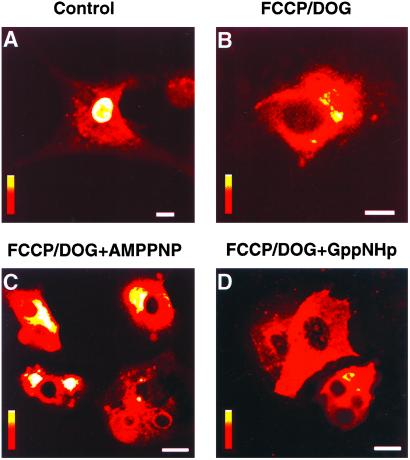
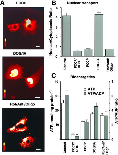
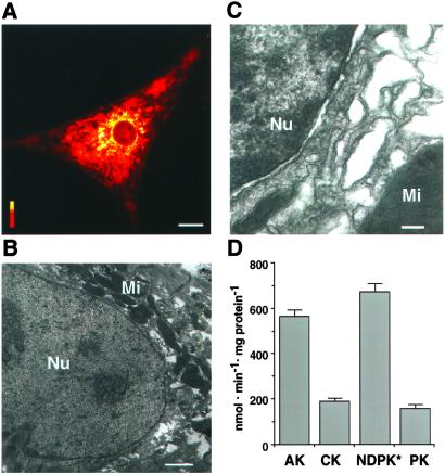
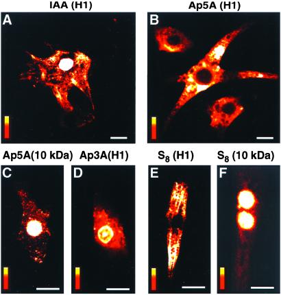
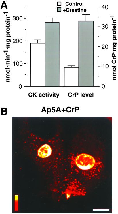
Exchange of information between the nucleus and cytosol depends on the metabolic state of the cell, yet the energy-supply pathways to the nuclear compartment are unknown. Here, the energetics of nucleocytoplasmic communication was determined by imaging import of a constitutive nuclear protein histone H1. Translocation of H1 through nuclear pores in cardiac cells relied on ATP supplied by mitochondrial oxidative phosphorylation, but not by glycolysis. Although mitochondria clustered around the nucleus, reducing the distance for energy transfer, simple nucleotide diffusion was insufficient to meet the energetic demands of nuclear transport. Rather, the integrated phosphotransfer network was required for delivery of high-energy phosphoryls from mitochondria to the nucleus. In neonatal cardiomyocytes with low creatine kinase activity, inhibition of adenylate kinase-catalyzed phosphotransfer abolished nuclear import. With deficient adenylate kinase, nucleoside diphosphate kinase, which secures phosphoryl exchange between ATP and GTP, was unable to sustain nuclear import. Up-regulation of creatine kinase phosphotransfer, to mimic metabolic conditions of adult cardiac cells, rescued H1 import, suggesting a developmental plasticity of the cellular energetic system. Thus, mitochondrial oxidative phosphorylation coupled with phosphotransfer relays provides an efficient energetic unit in support of nuclear transport.


Figures

Figure 1
Figure 1
Nuclear import of histone H1 requires energy of ATP and is not supported by nonhydrolyzable ATP or GTP analogs. Nuclear transport of fluorescein-tagged histone H1 was monitored by laser confocal microscopy after microinjection of H1 into the cytosol of neonatal cardiomyocytes. Cellular ATP/GTP levels were depleted by a 30-min treatment (37°C) with the mitochondrial uncoupler, FCCP (1 μM), and the inhibitor of glycolysis, DOG (6 mM). H1 is transported in the nucleus of control (A), but not ATP/GTP-depleted (B) cells. Microinjection into ATP/GTP-depleted cells of nonhydrolyzable ATP and GTP analogs, AMPPNP (C) or GppNHp (D), failed to rescue nuclear import of H1. Vertical bars = fluorescence scale of 0–255 arbitrary units, with red indicating lowest and white indicating highest intensity. (AD) Magnification = 10 μm.
Figure 2
Figure 2
Mitochondrial ATP production but not glycolysis is sufficient to support nuclear transport. (A) Nuclear import of H1 inhibited by an uncoupler of mitochondrial oxidative phosphorylation (Top, 1 μM FCCP) or by a mixture of mitochondrial respiratory chain and F0F1-ATPase inhibitors (Bottom, 1 μg/ml each of rotenone, antimycin, oligomycin; Rot/Anti/Oligo), but not by inhibitors of glycolysis (Middle, 6 mM 2-deoxyglucose plus 0.5 mM iodacetate, DOG/IA). Exposure time to inhibitors was 30 min. Horizontal bars = 10 μm; vertical bars = fluorescence scale of 0–255 arbitrary units as in Fig. 1. (B) Average nuclear/cytoplasmic fluorescence ratio for fluorescein-isothiocyanate-labeled H1 injected into the cytosol of controls and cells treated with FCCP/DOG, FCCP, DOG/IA, and Rot/Anti/Oligo. (C) Cellular ATP levels and ATP/ADP ratios in cardiomyocytes after treatments with mitochondrial or glycolytic inhibitors.
Figure 3
Figure 3
Mitochondrial distribution and phosphotransfer enzyme activity in cardiac cells. (A) Mitochondria cluster around the nucleus as detected by the mitochondrial marker MitoTracker (0.5 μM, 20-min incubation) using confocal microscopy. Horizontal bar = 10 μm; vertical bar = fluorescence scale of 0–255 arbitrary units. (B and C) Electron micrographs of cardiac cells show mitochondria (Mi) around the nucleus (B, Nu) and a crowded perinuclear space filled with membranes (C). Bars = 1 μm (B) and 200 nm (C). (D) Average catalytic activity of phosphotransfer enzymes expressed in nmol/min/mg protein. AK, adenylate kinase; CK, creatine kinase; NDPK, nucleoside diphosphate kinase; PK, pyruvate kinase. * indicates that the value of NDPK activity should be multiplied by three.
Figure 4
Figure 4
Nuclear import of H1 abolished by inhibitors of adenylate kinase. (A) Treatment with 0.5 mM IAA, which inhibits creatine kinase, did not affect nuclear import of H1. (B) Cytosolic microinjection of diadenosine pentaphosphate (Ap5A, 0.5 mM in pipette solution), which inhibits adenylate kinase, abolished nuclear import of H1. (C) Ap5A did not affect transport of 10-kDa dextrans, which occurs by passive diffusion. (D) Microinjection of the Ap5A analog Ap3A (0.5 mM in pipette solution), which does not inhibit adenylate kinase, did not prevent import of H1. (E and F) Treatment with elemental sulfur (0.01 mM S8), which, like Ap5A, inhibits adenylate kinase, inhibited nuclear import of H1 (E) but not passive diffusion of 10-kDa dextrans (F). Bar = 10 μm (A, B) or 20 μm (CF); vertical bars = fluorescence scale of 0–255 arbitrary units.
Figure 5
Figure 5
Suppressed nuclear import under adenylate kinase deficit, rescued by activation of creatine kinase phosphotransfer. (A) Creatine kinase (CK) activity and creatine phosphate (CrP) levels in neonatal cardiomyocytes grown without (Control) and with 20 mM creatine (+Creatine). (B) Nuclear import of H1 is maintained in cells grown with creatine after microinjection of the adenylate kinase inhibitor, Ap5A (0.5 mM in pipette solution), in the presence of the creatine kinase substrate CrP (67 mM in pipette solution). Horizontal bar = 10 μm; vertical bar = fluorescence scale of 0–255 arbitrary units.

References

    1. Mattaj I W, Englmeier L. Annu Rev Biochem. 1998;67:265–306. - PubMed
    1. Gorlich D, Kutay U. Annu Rev Cell Dev Biol. 1999;15:607–660. - PubMed
    1. Perez-Terzic C, Pyle J, Jaconi M, Stehno-Bittel L, Clapham D E. Science. 1996;273:1875–1877. - PubMed
    1. Perez-Terzic C, Gacy A M, Bortolon R, Dzeja P P, Puceat M, Jaconi M, Prendergast F G, Terzic A. Circ Res. 1999;84:1292–12301. - PubMed
    1. Talcott B, Moore M S. Trends Cell Biol. 1999;9:312–318. - PubMed

Publication types

MeSH terms

Substances

LinkOut - more resources