. 2002 Jul 23;99(15):9852-7.
doi: 10.1073/pnas.152621799. Epub 2002 Jul 15.

Association of calnexin with mutant peripheral myelin protein-22 ex vivo: a basis for "gain-of-function" ER diseases

Affiliations

Association of calnexin with mutant peripheral myelin protein-22 ex vivo: a basis for "gain-of-function" ER diseases

K M Dickson et al. Proc Natl Acad Sci U S A. .

Abstract

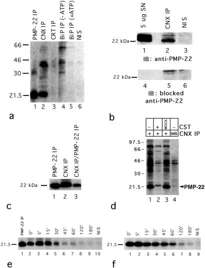
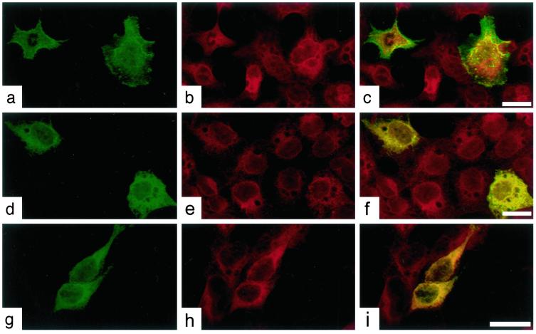
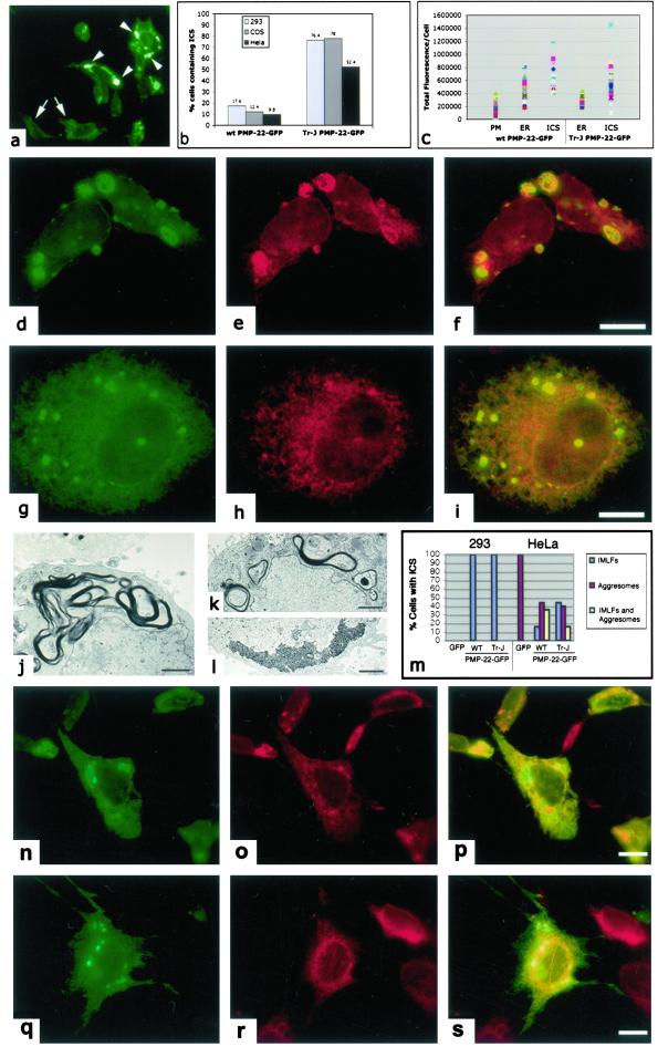
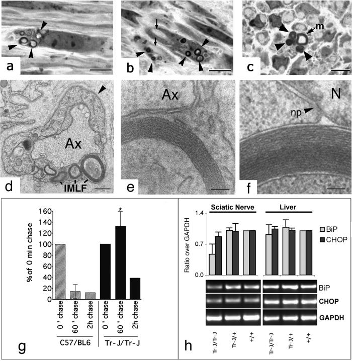
Schwann cell-derived peripheral myelin protein-22 (PMP-22) when mutated or overexpressed causes heritable neuropathies with a previously unexplained "gain-of-function" endoplasmic reticulum (ER) retention phenotype. In wild-type sciatic nerves, PMP-22 associates in a specific, transient (t(1/2 ) approximately equal to 11 min), and oligosaccharide processing-dependent manner with the lectin chaperone calnexin (CNX), but not calreticulin nor BiP. In Trembler-J (Tr-J) sciatic nerves, prolonged association of mutant PMP-22 with CNX is found (t(1/2) > 60 min). In 293A cells overexpressing PMP-22(Tr-J), CNX and PMP-22 colocalize in large intracellular structures identified at the electron microscopy level as myelin-like figures with CNX localization in the structures dependent on PMP-22 glucosylation. Similar intracellular myelin-like figures were also present in Schwann cells of sciatic nerves from homozygous Trembler-J mice with no detectable activation of the stress response pathway as deduced from BiP and CHOP expression. Sequestration of CNX in intracellular myelin-like figures may be relevant to the autosomal dominant Charcot-Marie-Tooth-related neuropathies.


Figures

Figure 1
Figure 1
CNX coimmunoprecipitation with PMP-22 in rat sciatic nerves. (a) Lysates (200 μg/immunoprecipitation) from metabolically labeled (0.4 mCi/ml; specific activity = 1,175 Ci/mmol) sciatic nerves were immunoprecipitated with either anti-PMP-22 (lane 1), anti-CNX (lane 2), anticalreticulin (lane 3), anti-BiP (lanes 4 and 5) Abs, or nonimmune serum (NIS; lane 6). As a specificity control, anti-BiP immunoprecipitates were incubated with Mg2+ ATP to release substrates because of ATPase activity (lane 5). (b) Unlabeled sciatic nerve lysates (400 μg/immunoprecipitation) were immunoprecipitated with either anti-CNX Ab (lanes 2 and 5) or nonimmune serum (lanes 3 and 6) and Western blotted. Total protein from rat sciatic nerves were included as positive controls (lanes 1 and 4). Blots were probed with either anti-PMP-22 (lanes 1–3) or anti-PMP-22 (lanes 4–6) preblocked with the immunizing peptide. (c) Metabolically labeled sciatic nerve lysates (30 min) were either immunoprecipitated with anti-PMP-22 (lane 1) or anti-CNX (lane 2). CNX immunoprecipitates were resolubilized and reimmunoprecipitated with anti-PMP-22, confirming the identity of the 22-kDa band as PMP-22 (lane 3). (d) Treatment of sciatic nerves with 1 mM CST 60 min before and during metabolic labeling reduced the association of PMP-22 and CNX (compare lanes 1 and 2) by 90% as determined by densitometry (data not shown). In mock CST controls (lane 3), sciatic nerves were simultaneously incubated in DMEM in the absence of CST. After treatment with/without CST, lysates were immunoprecipitated with either anti-CNX (lane 1–3) or nonimmune serum (lane 4). (e and f) Sciatic nerves were metabolically labeled and then “chased” with 2 mM l-methionine in DMEM for the indicated time intervals. Lysates were subsequently immunoprecipitated with anti-CNX (e, lanes 2–9), anti-PMP-22 (e, lane 1; f, lanes 1–8), or nonimmune serum (e, lane 10; f, lane 9). Exposure = 21 d for a and d; 10 d for c, e, and f.
Figure 2
Figure 2
CNX associates with PMP-22wt, PMP-22Tr, and PMP-22Tr-J in transiently transfected 293A cells. Cells were transfected with either PMP-22wt-GFP (ac), PMP-22Tr-GFP (df), and PMP-22Tr-J-GFP (gi) and processed for anti-CNX immunofluorescence (b, e, and h). Confocal overlay images for PMP-22wt-GFP and CNX show a distinct plasmalemmal colocalization (c). By contrast, PMP-22Tr-GFP (f) and PMP-22Tr-J-GFP (i) show a colocalization with CNX. (Bar = 25 μm.)
Figure 3
Figure 3
CNX colocalizes with IMLFs in cells overexpressing PMP-22wt and PMP-22Tr-J. (a) The presence of large ICS (arrowheads) within 293 cells transiently transfected with PMP-22Tr-J -GFP in comparison to cells having an ER-like localization of the PMP-22-GFP protein (arrows). Quantitation (total of 1,000 cells counted/cell-type) of the presence of ICS/cell-type confirmed the greater incidence of these structures residing in cells transfected with mutant PMP-22Tr-J-GFP as compared to cells transfected with PMP-22wt-GFP (b). The total fluorescence/cell (c; described in Materials and Methods) assessed the level of PMP-22-GFP protein expression in transfected 293 cells with either plasma membrane (PM), ER, or ICS scored. Two-way ANOVA determined the amounts of PMP-22wt-GFP protein expressed with PM, ER, and ICS localizations as significantly different (P < 0.001). Likewise, the amounts of PMP-22Tr-J-GFP protein with ER and ICS localizations were also significantly different (P < 0.001). Immunofluorescence microscopy of transfected 293A (df) and HeLa (gi) cells revealed a colocalization of CNX with ICS in cells overexpressing PMP-22Tr-J-GFP (f and i). EM of 293A cells overexpressing PMP-22Tr-J-GFP, revealed the ultrastructure of these IMLFs in the cytoplasm of cells (j). Conversely, HeLa cells transfected with PMP-22Tr-J-GFP led to the formation of either IMLFs (k), aggresomes (l), or a combination of both structures within one cell (see m). Although aggresomes could be found in all transfected HeLa cells, no aggresomes were seen in 293 cells (m). Immunofluorescence of the ER protein disulfide isomerase (o) did not reveal a colocalization with IMLFs (n and p). Removal of the N-linked glucosylation site (N41A) of PMP-22wt-GFP revealed ICS (q) but these were unreactive for CNX (r and s) revealing that formation of IMLFs was not dependent on its association. (Bar = df and gi,15 μm; j, k, and l, 2 μm; and ns, 15 μm.)
Figure 4
Figure 4
Presence of IMLFs in Tr-J/Tr-J nerves and prolonged association of CNX with PMP-22Tr-J in vivo. IMLFs were visualized in Schwann cells of Tr-J homozygous mice at the light microscope (ac) and EM levels (df). (a) IMLFs (arrowheads) present in a perinuclear position of a Schwann cell. (b) IMLFs (arrowheads) in Schwann cells and the consequent lack of myelination in nearby axons (arrows). (c) Cross section of a partially myelinated fiber (m) and a closely associated Schwann cell with IMLFs (arrowheads). (d) IMLFs in a Schwann cell identified by its basal lamina (arrowhead). An unmyelinated axon (Ax) is indicated. At higher magnification (e), parallel arrays of membranes in an IMLF of the Schwann cell can be visualized. The ultrastructure of the IMLF seen in Tr-J sciatic nerves (e) is similar to that seen in 293 cells overexpressing PMP-22Tr-J-GFP (f). (g) Sciatic nerves from wt and Tr-J/Tr-J mice were pulse-labeled for 30 min with Tran35S-label (0.4 mCi/ml; specific activity = 1,175 Ci/mmol) chased with 2 mM l-methionine supplemented DMEM for either 60 or 120 min, followed by immunoprecipitation of lysates with anti-CNX. In wt (C57BL/6) sciatic nerves, the association of PMP-22wt and CNX is greatly diminished after a 60-min chase period, as evaluated by densitometry. In Tr-J/Tr-J sciatic nerves, PMP-22Tr-J/CNX associations are not significantly decreased until 120 min after chase. The SD for the 60-min time point is shown. Data points (taken from three separate experiments; three animals/experiment) at the 60-min time point in Tr-J/Tr-J animals were significantly different from wt control (*, P < 0.001). (h) Relative expression levels of BiP and CHOP (normalized to glyceraldehyde-3-phosphate dehydrogenase) in wt, heterozygous, and homozygous Tr-J mice showed no increase in CHOP mRNA levels, indicating a lack of an unfolded protein response in Tr-J/Tr-J sciatic nerves (h). Values ± half variation are the mean of two PCRs performed for sciatic nerve and liver, respectively. m, myelinated fiber; Ax, axon; np, nuclear pore; N, nucleus. (Bar = ac, 5 μm; d, 2 μm; and e and f, 100 nm.)

References

    1. Naef R, Suter U. Microsc Res Tech. 1998;41:359–371. - PubMed
    1. Lupski J R, de Oca-Luna R M, Slaugenhaupt S, Pentao L, Guzzetta V, Trask B J, Saucedo-Cardenas O, Barker D F, Killian J M, Garcia C A, et al. Cell. 1991;66:219–232. - PubMed
    1. Chance P F, Alderson M K, Leppig K A, Lensch M W, Matsunami N, Smith B, Swanson P D, Odelberg S J, Disteche C M, Bird T D. Cell. 1993;72:143–151. - PubMed
    1. Warner L E, Garcia C A, Lupski J R. Annu Rev Med. 1999;50:263–275. - PubMed
    1. Suter U, Welcher A A, Ozcelik T, Snipes G J, Kosaras B, Francke U, Billings-Gagliardi S, Sidman R L, Shooter E M. Nature (London) 1992;356:241–244. - PubMed

Publication types

MeSH terms