. 2002 Jul 23;99(15):9662-7.
doi: 10.1073/pnas.152667399. Epub 2002 Jul 15.

Self-assembling biomaterials: liquid crystal phases of cholesteryl oligo(L-lactic acid) and their interactions with cells

Affiliations

Self-assembling biomaterials: liquid crystal phases of cholesteryl oligo(L-lactic acid) and their interactions with cells

Julia J Hwang et al. Proc Natl Acad Sci U S A. .

Abstract

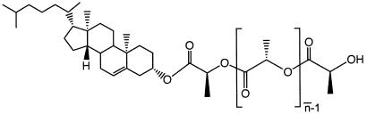
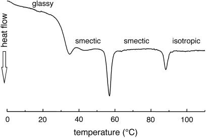
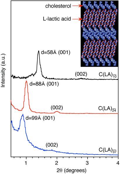
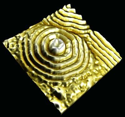
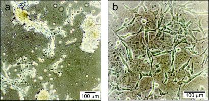
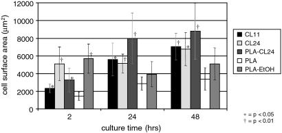
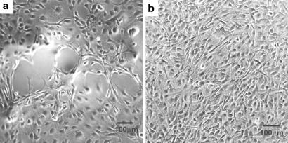
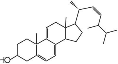
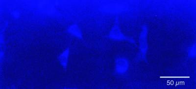
We report here on the synthesis and characterization of a series of self-assembling biomaterials with molecular features designed to interact with cells and scaffolds for tissue regeneration. The molecules of these materials contain cholesteryl moieties, which have universal affinity for cell membranes, and short chains of lactic acid, a common component of biodegradable tissue engineering matrices. The materials were synthesized in good yields with low polydispersities in the range of 1.05-1.15, and their characterization was carried out by small-angle x-ray diffraction, transmission electron microscopy, electron diffraction, differential scanning calorimetry, and atomic force microscopy. These molecular materials form layered structures that can be described as smectic phases and can also order into single-crystal stacks with an orthorhombic unit cell. Their layer spacings range from 58 to 99 A, corresponding to bilayers of oligomers with an average of 10 and 37 lactic acid residues, respectively. The self-organized layered structures were found to promote improved fibroblast adhesion and spreading, although the specific mechanism for this observed response remains unknown. The ability of self-assembling materials to present ordered and periodic bulk structures to cells could be a useful strategy in tissue engineering.


Figures

Figure 1
Figure 1
Chemical structure of C-LA.
Figure 2
Figure 2
Characteristic birefringent texture under crossed polars for C-LAformula image at 25°C.
Figure 3
Figure 3
DSC scan of C-LAformula image revealing a glass transition at 32°C, a thermotropic liquid crystalline transition (smectic to smectic phase) at 57°C, and isotropization at 88°C.
Figure 4
Figure 4
SAXS scans of C-LA (n̄ = 10, 24, 37), reveal (001) and (002) reflections indicating lamellar organization. (Inset) A molecular graphics model showing the proposed packing of C-LAformula image molecules into a highly interdigitated bilayer with spacing of 58 Å. [Reproduced with permission from ref. (Copyright 2002, Am. Chem. Soc.).]
Figure 5
Figure 5
(a) TEM image and electron diffraction pattern of single crystal C-LAformula image. (b) TEM image of a film of C-LA revealing multilayer domains formed by spiral dislocations. (Inset) Electron diffraction pattern (rings correspond to evaporated Au), indicating that the material is not a single crystal but a smectic Eh liquid crystalline phase. (c) TEM image of a precipitate C-LA showing a multilayered lozenge-shaped single crystal formed by spiral dislocations. (Inset) Electron diffraction pattern revealing an orthorhombic unit cell.
Figure 6
Figure 6
AFM image showing the surface topography of C-LAformula image films. The image is 106 × 106 microns.
Figure 7
Figure 7
(a) 3T6 fibroblasts on a PLA surface 72 hr after cell seeding. The fibroblasts aggregate and do not spread on these surfaces. (b) 3T6 fibroblasts on C-LAformula image 72 hr after seeding. The cells spread preferentially along the boundaries of focal conic domains.
Figure 8
Figure 8
Surface area of adhered cells on various biomaterials. Error bars are ± a 95% confidence level. All sample sets at each timepoint were compared by using ANOVA to the PLA control. *, P < 0.05; †, P < 0.01.
Figure 9
Figure 9
3T3 fibroblasts after 3 days of exposure to (a) a PLA surface and (b) a PLA surface modified with C-LAformula image. A higher population of cells is found on the surface in (b) modified with self-assembling multilayers.
Figure 10
Figure 10
Chemical structure of dehydroergosterol.
Figure 11
Figure 11
3T3 fibroblasts after 6 hr of exposure to a surface of DHE-(l-lactic acid)n. Image was taken under epifluorescence illumination by using a 4′,6-diamidino-2-phenylindole-type filter set. The fluorescent material is visible throughout the cell cytoplasm and membrane.

References

    1. Whitesides G M, Mathias J P, Seto C T. Science. 1991;254:1312–1319. - PubMed
    1. Lehn J M. Supramolecular Chemistry. New York: VCH; 1995.
    1. Stupp S I, LeBonheur V, Walker K, Li L S, Huggins K, Keser M, Amstutz A. Science. 1997;276:384–389. - PubMed
    1. Kricheldorf H R, Kreiser-Saunders I. Polymer. 1994;35:4175–4180.
    1. Dahl J S, Dahl C E, Bloch K. J Biol Chem. 1981;256:87–91. - PubMed

Publication types