

doi: 10.1523/JNEUROSCI.22-14-05797.2002.
Role for reelin in the development of granule cell dispersion in temporal lobe epilepsy
Affiliations
- PMID: 12122039
- PMCID: PMC6757930
- DOI: 10.1523/JNEUROSCI.22-14-05797.2002
Item in Clipboard
Role for reelin in the development of granule cell dispersion in temporal lobe epilepsy
J Neurosci.
.
Abstract
The reelin signaling pathway plays a crucial role during the development of laminated structures in the mammalian brain. Reelin, which is synthesized and secreted by Cajal-Retzius cells in the marginal zone of the neocortex and hippocampus, is proposed to act as a stop signal for migrating neurons. Here we show that a decreased expression of reelin mRNA by hippocampal Cajal-Retzius cells correlates with the extent of migration defects in the dentate gyrus of patients with temporal lobe epilepsy. These results suggest that reelin is required for normal neuronal lamination in humans, and that deficient reelin expression may be involved in migration defects associated with temporal lobe epilepsy.
Figures
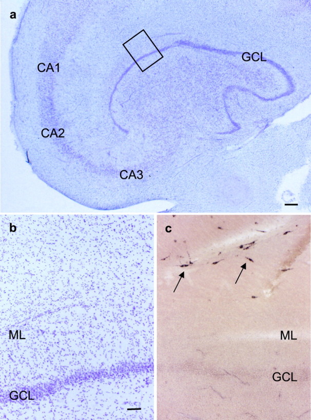
Reelin mRNA expression in a representative sample of a human control hippocampus. a, Hippocampal areas in cresyl violet stain. The inset is shown inb. Scale bar, 400 μm. b, Portion of the dentate gyrus framed in a. The GCL is densely packed. Scale bar, 80 μm. c, ISH for reelin mRNA. Many reelin mRNA-positive cells with bipolar morphology resembling CR cells are observed at the hippocampal fissure (arrows). Same magnification as in b. CA1, CA2, CA3, Hippocampal subfields; ML, Dentate molecular layer.
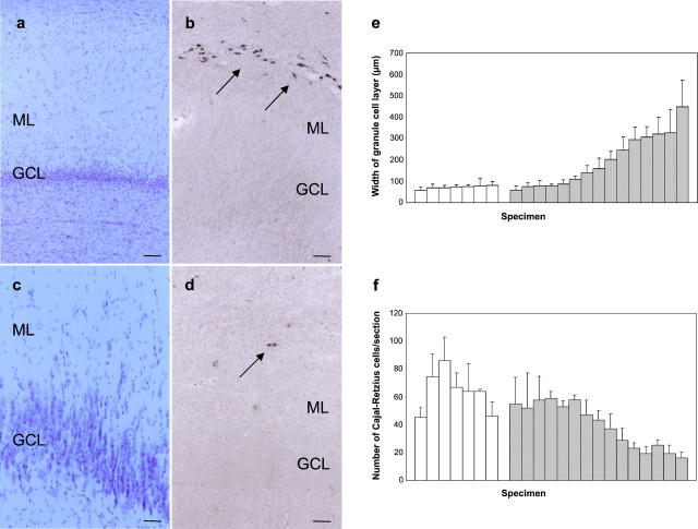
Reelin mRNA expression in the dentate gyrus of control and epileptic cases. a–d, Reelin mRNA expression in representative samples of epileptic hippocampi with mild (a, b) and severe (c, d) GCD. Consecutive sections are shown in cresyl violet stain and after ISH for reelin mRNA. Arrows point to reelin mRNA-positive cells at the hippocampal fissure. Scale bars: a, b, 100 μm;c, d, 50 μm. e, f, Correlation of the GCD (width of the GCL) and number of reelin mRNA-expressing CR cells in sections of seven control hippocampi (white bars) and 15 epileptic hippocampi with AHS (gray bars). Data are represented as mean ± SD. ML, Dentate molecular layer.
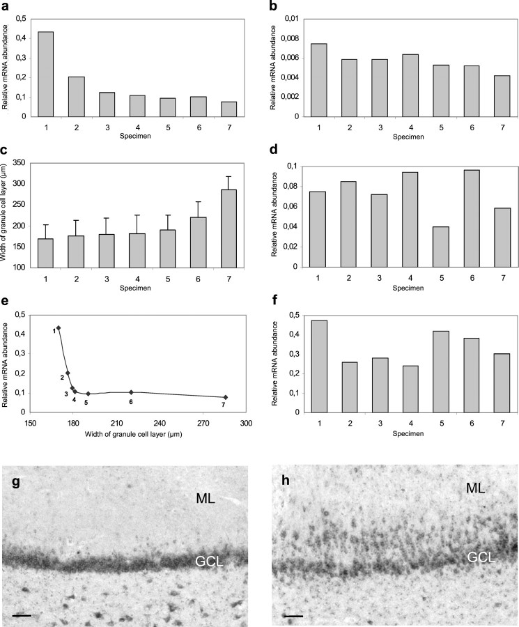
Expression profiles of reelin signal-transduction components in epileptic human hippocampi.a, Histogram showing the amounts of reelin mRNA in the dentate gyrus of individual epileptic human hippocampi (n = 7). Reelin mRNA levels were determined by quantitative RT-PCR in extracts from microdissected dentate gyri. Two measurements for each individual case were performed. Quantification included the addition of an external standard and volume measurement of the microdissected region. b, d, f, Histograms showing the expression levels of VLDLR (b), ApoER2 (d), and dab1 (f) mRNA in the dentate gyrus of individual epileptic human hippocampi. The epileptic cases (n = 7) were identical to the ones shown in a. c, Histogram showing the width of the GCL in the dentate gyrus of individual epileptic hippocampi. The width of the GCL was measured in cresyl violet-stained sections of the same cases (n = 7) as shown ina. e, Correlation of reelin mRNA levels and the width of the GCL. The reelin mRNA levels shown ina were plotted against the width of the GCL (c). g, Expression of dab1 mRNA in granule cells of a normal human dentate gyrus visualized by ISH.h, Expression of dab1 mRNA in the dentate gyrus of a TLE patient. Note dab1 mRNA in dispersed granule cells. Scale bars, 80 μm (g, h). ML, Dentate molecular layer.
References
-
- Altman J, Das GD. Autoradiographic and histological evidence of postnatal hippocampal neurogenesis in rats. J Comp Neurol. 1965;124:319–335. - PubMed
-
- Armstrong DD. The neuropathology of temporal lobe epilepsy. J Neuropath Exp Neurol. 1993;52:433–443. - PubMed
-
- Blümcke I, Beck H, Suter B, Hoffmann D, Fodisch HJ, Wolf HK, Schramm J, Elger CE, Wiestler OD. An increase of hippocampal calretinin-immunoreactive neurons correlates with early febrile seizures in temporal lobe epilepsy. Acta Neuropathol. 1999;97:31–39. - PubMed
-
- Bouillert V, Loup F, Kiener T, Marescaux C, Fritschy JM. Early loss of interneurons and delayed subunit-specific changes in GABA(A)-receptor expression in a mouse model of mesial temporal lobe epilepsy. Hippocampus. 2000;10:305–324. - PubMed
Publication types
MeSH terms
Substances
LinkOut - more resources
Full Text Sources