. 2002 Jul 15;22(14):5946-54.
doi: 10.1523/JNEUROSCI.22-14-05946.2002.

Sequential nuclear accumulation of the clock proteins period and timeless in the pacemaker neurons of Drosophila melanogaster

Affiliations

Sequential nuclear accumulation of the clock proteins period and timeless in the pacemaker neurons of Drosophila melanogaster

Orie T Shafer et al. J Neurosci. .

Abstract

Antisera against the circadian clock proteins Period (PER) and Timeless (TIM) were used to construct a detailed time course of PER and TIM expression and subcellular localization in a subset of the ventrolateral neurons (vLNs) in the Drosophila accessory medulla (AMe). These neurons, which express pigment-dispersing factor, play a central role in the control of behavioral rhythms. The data revealed several unexpected features of the circadian clock in Drosophila. First, TIM but not PER was restricted to the cytoplasm of vLNs throughout most of the early night. Second, the timing of TIM and PER nuclear accumulation was substantially different. Third, the two subsets of vLNs, the large and small vLNs, had a similar timing of PER nuclear accumulation but differed by 3-4 hr in the phase of TIM nuclear accumulation. These aspects of PER and TIM expression were not predicted by the current mechanistic model of the circadian clock in Drosophila and are inconsistent with the hypothesis that PER and TIM function as obligate heterodimers. The differing profiles of TIM and PER nuclear accumulation suggest that PER and TIM have distinct functions in the nuclei of vLNs.


Figures

Fig. 1.
Fig. 1.
Expression of PER and TIM in the vLNs of wild-type (WT) and mutant flies. Typical vLNs of wild-type (Canton S), per01, andtim01 null mutant flies dissected at ZT 00/24 are shown. Scale bar, 5 μm.
Fig. 2.
Fig. 2.
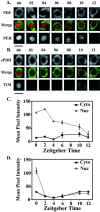
Daytime expression of PER and TIM in the large vLNs. A, Typical optical sections of large vLNs stained for PDF and PER. ZTs are indicated above each optical section. B, Typical optical sections of large vLNs stained for cPDH and TIM. Scale bars, 5 μm. C, Quantification of cytoplasmic (Cyto) and nuclear (Nuc) PER staining in the large vLNs as a function of ZT. D, Quantification of cytoplasmic and nuclear TIM staining in the large vLNs as a function of ZT. For each ZT, five brains were processed for each protein, and four large vLNs were imaged from one hemisphere of each brain.
Fig. 3.
Fig. 3.
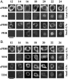
Night-time expression of PER and TIM in the large vLNs. A, Typical optical sections of large vLNs stained for PDF and PER. ZTs are indicated above each set of optical sections. B, Typical optical sections of large vLNs stained for cPDH and TIM. Scale bars, 5 μm. C, Quantification of cytoplasmic (Cyto) and nuclear (Nuc) PER staining in the large vLNs as a function of ZT. D, Quantification of cytoplasmic and nuclear TIM staining in the large vLNs as a function of ZT. For each ZT, five brains were processed for each protein and four large vLNs were imaged from one hemisphere of each brain. E, Inverted and quantified images of a large vLN double-labeled for cPDH and TIM at ZT 17. Staining values for the edge (blue) and center (red) of the nucleus are indicated. F, vLNs double-labeled for TIM and PER at ZT 17. Scale bar, 5 μm.
Fig. 4.
Fig. 4.
Night-time expression of PER and TIM in the small vLNs at ZTs 16, 18, and 20. A, Typical optical sections of small vLNs stained for cPDH and PER. ZTs are indicatedabove each set of optical sections. B, Typical optical sections of small vLNs stained for cPDH and TIM. Scale bars, 5 μm. C, Quantification of cytoplasmic (Cyto) and nuclear (Nuc) PER staining in the small vLNs. D, Quantification of cytoplasmic and nuclear TIM staining in the small vLNs. The quantification is based on images of four brains per time point per protein and three to four small vLNs per brain.
Fig. 5.
Fig. 5.
Comparison of nuclear accumulation in the large and small vLNs throughout the night. A, Typical optical sections of large and small vLNs stained for cPDH and PER. ZTs are indicated above each set of optical sections.B, Typical optical sections for large and small vLNs stained for cPDH and TIM. Scale bars, 5 μm.
Fig. 6.
Fig. 6.
TIM expression in the large and small vLNs under constant darkness and temperature. A, Typical optical sections of large vLNs stained for cPDH and TIM. CTs are indicatedabove each set of optical sections. B, Typical optical sections of small vLNs stained for cPDH and TIM. Scale bars, 5 μm. C, Quantification of cytoplasmic (Cyto) and nuclear (Nuc) TIM staining in the large vLNs. D, Quantification of cytoplasmic and nuclear TIM staining in the small vLNs. The quantifications are based on images of four brains per time point per protein and three to four large or small vLNs per brain.
Fig. 7.
Fig. 7.
PER expression in the large and small vLNs under constant darkness and temperature. A, Typical optical sections of large vLNs stained for cPDH and PER. CTs are indicatedabove each set of optical sections. B, Typical optical sections of small vLNs stained for cPDH and PER. Scale bars, 5 μm. C, Quantification of cytoplasmic (Cyto) and nuclear (Nuc) PER staining in the large vLNs. D, Quantification of cytoplasmic and nuclear PER signal in the small vLNs. The quantifications are based on images of four brains per time point per protein and three to four large or small vLNs per brain.

References

    1. Allada R, White NE, So WV, Hall JC, Rosbash M. A mutant Drosophila homolog of mammalian Clock disrupts circadian rhythms and transcription of period and timeless. Cell. 1998;93:791–804. - PubMed
    1. Curtin KD, Huang ZJ, Rosbash M. Temporally regulated nuclear entry of the Drosophila Period protein contributes to the circadian clock. Neuron. 1995;14:365–372. - PubMed
    1. Darlington TK, Wager-Smith K, Ceriani MF, Staknis D, Gekakis N, Steeves TDL, Weitz CJ, Takahashi JS, Kay SA. Closing the circadian loop: CLOCK-induced transcription of its own inhibitors per and tim. Science. 1998;280:1599–1603. - PubMed
    1. Dircksen H, Zahnow CA, Gaus G, Keller R, Rao KR, Riehm JP. The ultrastructure of nerve endings containing the pigment-dispersing hormone (PDH) in crustacean sinus glands: identification by an antiserum against synthetic PDH. Cell Tissue Res. 1987;250:377–387.
    1. Dunlap JC. Molecular bases for circadian clocks. Cell. 1999;96:271–290. - PubMed

Publication types

LinkOut - more resources