. 2002 Jul 15;22(14):5955-65.
doi: 10.1523/JNEUROSCI.22-14-05955.2002.

Mapping the binding site of the neuroprotectant ifenprodil on NMDA receptors

Affiliations

Mapping the binding site of the neuroprotectant ifenprodil on NMDA receptors

Florent Perin-Dureau et al. J Neurosci. .

Abstract

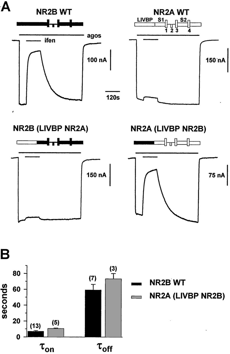
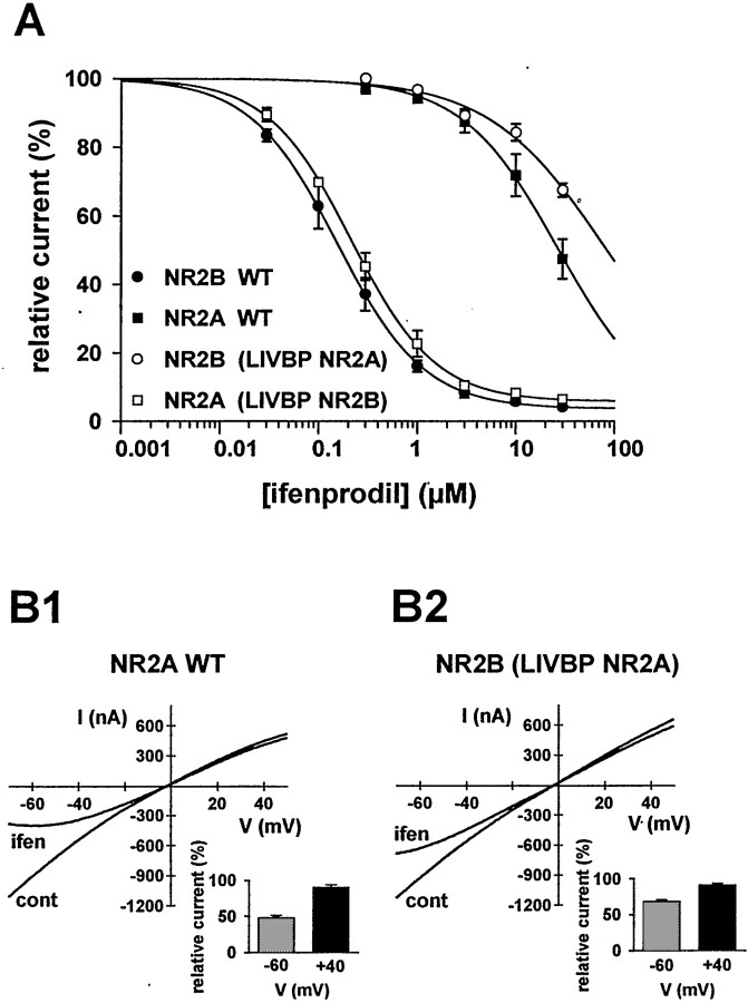
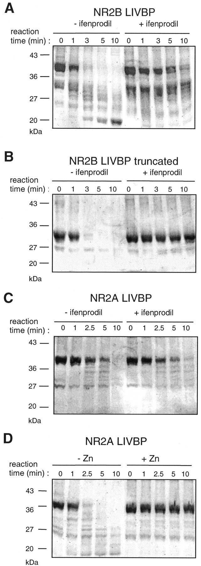
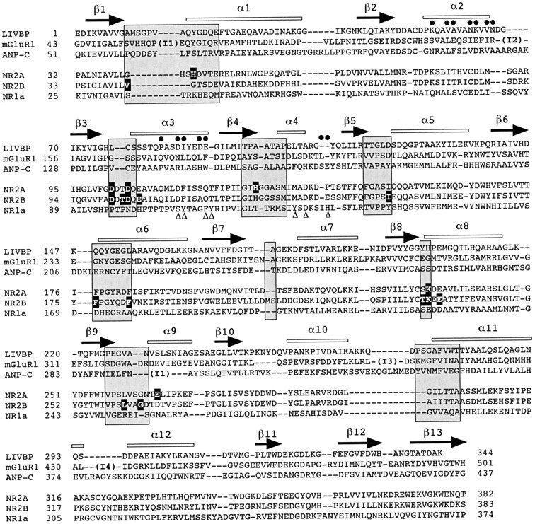
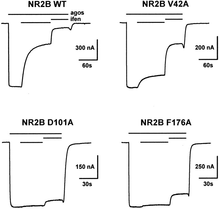
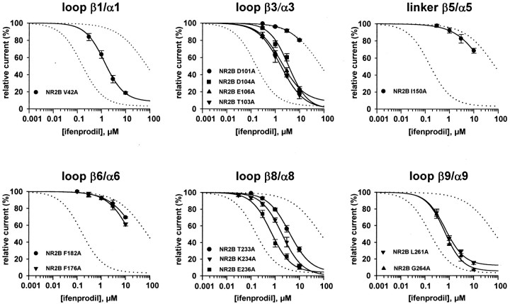
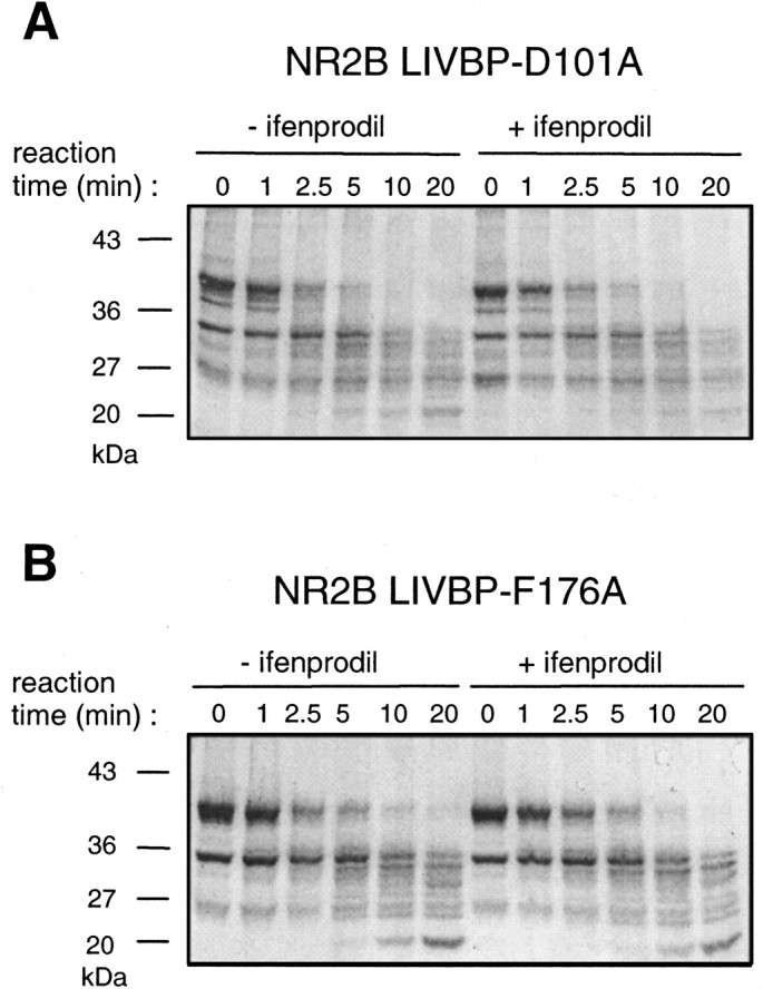
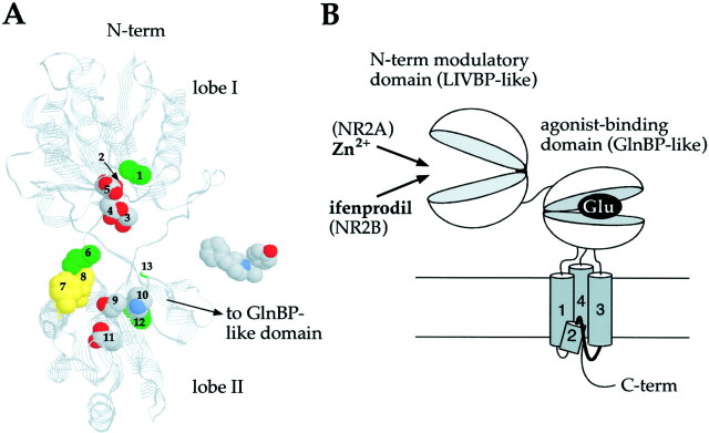
Ifenprodil is a noncompetitive antagonist of NMDA receptors highly selective for the NMDA receptor 2B (NR2B) subunit. It is widely used as a pharmacological tool to discriminate subpopulations of NMDA receptors, and derivatives are currently being developed as candidate neuroprotectants. Despite numerous studies on the mechanism of action of ifenprodil on NMDA receptors, the structural determinants responsible for the subunit selectivity have not been identified. By combining functional studies on recombinant NMDA receptors and biochemical studies on isolated domains, we now show that ifenprodil binds to the N-terminal leucine/isoleucine/valine-binding protein (LIVBP)-like domain of NR2B. In this domain, several residues, both hydrophilic and hydrophobic, were found to control ifenprodil inhibition. Their location in a modeled three-dimensional structure suggests that ifenprodil binds in the cleft of the LIVBP-like domain of NR2B by a mechanism (Venus-flytrap) resembling that of the binding of Zn on the LIVBP-like domain of NR2A. These results reinforce the proposal that the LIVBP-like domains of NMDA receptors, and possibly of other ionotropic glutamate receptors, bind modulatory ligands. Moreover, they identify the LIVBP-like domain of the NR2B subunit as a promising therapeutic target and provide a framework for designing structurally novel NR2B-selective antagonists.


Figures

Fig. 1.
Fig. 1.
The LIVBP-like domain of NR2B controls the high-affinity ifenprodil inhibition of NMDA receptors.A, Swapping the LIVBP-like domain between NR2B and NR2A subunits transfers the NR2B-specific high-affinity ifenprodil (ifen) inhibition from NR2B to NR2A. Eachtrace shows the inhibition of the current response to agonists (agos; glutamate and glycine, 100 μm each) by 1 μm ifenprodil inXenopus oocytes coexpressing either NR2B wt, NR2A wt, or chimeric NR2B/NR2A subunits with the NR1 wt subunit. Note that the slow kinetics of ifenprodil inhibition of NR1/NR2B wt (most particularly the recovery rate) are also observed in NR1/NR2A-(LIVBP NR2B) receptors. The recordings were made at −60 mV. The bars above the current traces indicate the duration of agonists and ifenprodil applications. A schematic diagram of the NR2 construct is shown on top of each trace:LIVBP, LIVBP-like domain; S1 S2, agonist-binding GlnBP-like domain; 1, 3, 4, transmembrane segments; 2, reentrant pore loop.B, NR1/NR2B wt and NR1/NR2A-(LIVBP NR2B) receptors display similar kinetic parameters of ifenprodil inhibition. The time constants of onset (τon) and offset (τoff) of the inhibition of agonist-induced currents by 1 μm ifenprodil were estimated by single-exponential fits to traces such as those shown inA (see Materials and Methods for the fitting procedures). The mean τon and τoff values are as follows: NR2B wt, 7.1 ± 1 sec (n = 13) and 59 ± 7 (n = 7), respectively; NR2A-(LIVBP NR2B), 10.6 ± 0.6 sec (n = 5) and 73 ± 7 sec (n = 3), respectively.
Fig. 2.
Fig. 2.
Parameters of ifenprodil inhibition of the chimeric and wild-type NMDA receptors. A, Concentration–response curves at equilibrium and at −60 mV. Datapoints were fitted with Hill equations (see Materials and Methods). Each point is the mean value of 3–15 oocytes. The estimated values of IC50,nH, and maximal inhibition are, respectively, 155 nm, 0.98, and 96% for NR2B wt and 215 nm, 1.00, and 94% for NR2A-(LIVBP NR2B). For NR2A wt and NR2B-(LIVBP NR2A), the estimated IC50 values are 28 and 75 μm, respectively. B, The low-affinity ifenprodil inhibition of NR2A- and NR2B-(LIVBP NR2A)-containing receptors is voltage-dependent. Leak-subtracted agonist-induced NMDA currents were recorded in the absence (cont) and presence of 30 μm ifenprodil (ifen) during 2 sec voltage ramps from −70 to +50 mV. Insets, Mean relative currents (percentage) measured at −60 and +40 mV.
Fig. 3.
Fig. 3.
Ifenprodil protects the isolated LIVBP-like domain of NR2B but not that of NR2A against hydrolysis by trypsin. Isolated LIVBP-like domains of NR2A and NR2B were produced in E. coli and subjected to trypsinization with or without ifenprodil or Zn for various amounts of time (up to 10 min). Fifty micrograms of the purified LIVBP-like domains were mixed with 0.1 μg of trypsin. The samples were analyzed on 12% SDS-PAGE gels. Lane 0corresponds to the protein solution just before trypsin addition.A, Ifenprodil (10 μm) protects the NR2B LIVBP-like domain (main band at ∼40 kDa) against trypsin digestion.B, Ifenprodil (10 μm) also protects a C-terminal truncated NR2B LIVBP-like domain (main band at ∼30 kDa) against trypsin digestion. C, Ifenprodil (100 μm) does not protect the NR2A LIVBP-like domain (main band at ∼40 kDa) against trypsin digestion. D, Zn (10 μm) protects the NR2A LIVBP-like domain against trypsin digestion.
Fig. 4.
Fig. 4.
Ligand-contacting regions in proteins containing LIVBP-like domains. Amino acid sequence alignment of the LIVBP-like domains of the NMDA receptor subunits NR2A, NR2B, and NR1 and the LIVBP-like domains of known structures from LIVBP (Sack et al., 1989), mGluR1 (Kunishima et al., 2000), and ANP-C (He et al., 2001). For LIVBP (E. coli), mGluR1, and ANP-C, the alignments were obtained from structure superimposition; for NR2A and NR2B, the alignments were adapted from those of Paoletti et al. (2000); for NR1, the alignment was based on the conserved pattern of a cluster of hydrophobic residues within LIVBP-like domains (see Materials and Methods). The β strands (arrows) and α helices (open bars) identified in LIVBP, mGluR1, and ANP-C crystal structures are indicated on top of the alignment. The insertions found in mGluR1 with respect to LIVBP are indicated by I1 (14 residues), I2 (31 residues), I3 (47 residues), and I4 (13 residues) and in ANP-C by I1 (21 residues).Shaded boxes indicate regions (mostly loops) known to contact the ligand molecules in LIVBP-like domains (adapted fromPaoletti et al., 2000). Residues of NR2A controlling high-affinity Zn inhibition (see Paoletti et al., 2000) and residues of NR2B identified as controlling ifenprodil inhibition (present study) are highlighted. Residues of NR1 mutated by Masuko et al. (1999) and affecting ifenprodil inhibition are indicated by triangles. Residues of mGluR1 and ANP-C participating in dimerization of the LIVBP-like domains (Kunishima et al., 2000; He et al., 2001) are indicated by closed circles.
Fig. 5.
Fig. 5.
Identification in the LIVBP-like domain of NR2B critical residues controlling high-affinity ifenprodil inhibition. A comparison of the current traces obtained from oocytes coexpressing NR1 with either wt or mutated NR2B subunits is shown. Ifenprodil was applied at two increasing concentrations (300 nm and 3 μm) during an application of agonists. Thebars above the current traces indicate the duration of agonists (agos) and ifenprodil (ifen) applications. The holding potential was −60 mV. These current traces are typical of NR2B mutants having an intermediate (V42A) or strong (D101A, F176A) effect on ifenprodil sensitivity.
Fig. 6.
Fig. 6.
Ifenprodil concentration–response curves of receptors mutated at critical residues. Each graphcorresponds to a region in the LIVBP-like domain of NR2B in which one or more critical residues controlling ifenprodil inhibition were identified. The dotted curves are the fits of the ifenprodil concentration–response curves of the NR1/NR2B wt receptors (left dotted curve) and the chimeric NR1/NR2B-(LIVBP NR2A) receptors (right dotted curve) obtained in Figure2B. The estimated values of IC50 of the different mutated receptors are listed in Table 2. Estimated values of Hill coefficients (0.92–1.10) and maximal inhibitions (88–100%) are in the same range as those obtained with NR2B wt receptors (Fig.2A). Each data point is the mean value obtained from 3 to 22 oocytes. The recordings were made at −60 mV.
Fig. 7.
Fig. 7.
Lack of ifenprodil-induced protection against trypsin digestion of NR2B LIVBP-like domains mutated at critical positions. Point mutations (D101A and F176A) were introduced in the plasmid pGEX-2T-LIVBP NR2B and the mutated domains produced inE. coli. Protection by ifenprodil (10 μm) against trypsin digestion was tested using the protocol described in Figure 3. A, Mutation D101A. B, Mutation F176A.
Fig. 8.
Fig. 8.
LIVBP-like domain of NR2 subunits as binding domains for extracellular modulators of NMDA receptor activity.A, 3D model of the LIVBP-like domain of NR2B and the putative ifenprodil binding site. This model was produced by homology modeling using the sequence alignment shown in Figure 4 and the Protein Data Bank coordinates of LIVBP (2LIV), the unliganded form of LIVBP (Sack et al., 1989). The residues identified as critical for high-affinity ifenprodil inhibition (numbered 1–13 for clarity) are displayed in the space fill representation and according to the following color code: Corey–Pauling–Koltun (CPK) for polar and charged residues (lobe I: Asp101(2), Thr103(3), Asp104(4), Glu106(5); lobe II: Thr233(9), Lys234(10), Glu236(11)), greenfor aliphatic residues (lobe I: Val42(1); lobe II: Leu261(12), Gly264(13); hinge: Ile150(6)), andyellow for aromatic residues (lobe II: Phe176(7), Phe182(8)). On theright of the model is a space fill representation of the ifenprodil molecule (CPK color code). B, Functional organization of the extracellular regions of the NR2 subunits. See Discussion for more details.

References

    1. Armstrong N, Sun Y, Chen GQ, Gouaux E. Structure of a glutamate-receptor ligand-binding core in complex with kainate. Nature. 1998;395:913–917. - PubMed
    1. Armstrong N, Gouaux E. Mechanisms for activation and antagonism of an AMPA-sensitive glutamate receptor: crystal structures of the GluR2 ligand binding core. Neuron. 2000;28:165–181. - PubMed
    1. Ayalon G, Stern-Bach Y. Functional assembly of AMPA and kainate receptors is mediated by several discrete protein-protein interactions. Neuron. 2001;31:103–113. - PubMed
    1. Callebaut I, Labesse G, Durand P, Poupon A, Canard L, Chomilier J, Henrissat B, Mornon JP. Deciphering protein sequence information through hydrophobic cluster analysis (HCA): current status and perspectives. Cell Mol Life Sci. 1997;53:621–645. - PMC - PubMed
    1. Carter C, Benavides J, Legendre P, Vincent JD, Noel F, Thuret F, Lloyd KG, Arbilla S, Zivkovic B, MacKenzie ET, Scatton B, Langer SZ. Ifenprodil and SL 82.0715 as cerebral anti-ischemic agents: II. Evidence for N-methyl-d-aspartate receptor antagonist properties. J Pharmacol Exp Ther. 1988;247:1222–1232. - PubMed

Publication types

MeSH terms

LinkOut - more resources