. 2002 Jul 15;22(14):6071-82.
doi: 10.1523/JNEUROSCI.22-14-06071.2002.

The type 1 interleukin-1 receptor is essential for the efficient activation of microglia and the induction of multiple proinflammatory mediators in response to brain injury

Affiliations

The type 1 interleukin-1 receptor is essential for the efficient activation of microglia and the induction of multiple proinflammatory mediators in response to brain injury

Anirban Basu et al. J Neurosci. .

Abstract

Interleukin-1 (IL-1) is induced immediately after insults to the brain, and elevated levels of IL-1 have been strongly implicated in the neurodegeneration that accompanies stroke, Alzheimer's disease, and multiple sclerosis. In animal models, antagonizing IL-1 has been shown to reduce cell death; however, the basis for this protection has not been elucidated. Here we analyzed the response to penetrating brain injury in mice lacking the type 1 IL-1 receptor (IL-1R1) to determine which cellular and molecular mediators of tissue damage require IL-1 signaling. At the cellular level, fewer amoeboid microglia/macrophages appeared adjacent to the injured brain tissue in IL-1R1 null mice, and those microglia present at early postinjury intervals retained their resting morphology. Astrogliosis also was mildly abrogated. At the molecular level, cyclooxygenase-2 (Cox-2) and IL-6 expression were depressed and delayed. Interestingly, basal levels of Cox-2, IL-1, and IL-6 were significantly lower in the IL-1R1 null mice. In addition, stimulation of vascular cell adhesion molecule-1 mRNA was depressed in the IL-1R1 null mice, and correspondingly, there was reduced diapedesis of peripheral macrophages in the IL-1R1 null brain after injury. This observation correlated with a reduced number of Cox-2+ amoeboid phagocytes adjacent to the injury. In contrast, several molecular aspects of the injury response were normal, including expression of tumor necrosis factor-alpha and the production of nerve growth factor. Because antagonizing IL-1 protects neural cells in experimental models of stroke and multiple sclerosis, our data suggest that cell preservation is achieved by abrogating microglial/macrophage activation and the subsequent self-propagating cycle of inflammation.


Figures

Fig. 1.
Fig. 1.
Microglia and macrophages are less responsive to stab wound in the IL-1R1 null mice. Adult IL-1R1 mice (A) or age-matched wild-type mice (B) received a penetrating brain injury. After 24 hr, the animals were killed by perfusion and processed for tomato lectin histofluorescence. In these panels the lesion (L) is at the bottom. In the wild-type mice there are numerous amoeboid microglia at the immediate site of the injury (B), whereas in the IL-1R1 null mice, there are ramified microglia present and reactive or amoeboid microglia are virtually absent (A). To assess diapedesis of macrophages into the damaged brain, peritoneal macrophages were labeled with fluorescent microbeads 2 d before mice received a stab wound to the neocortex. Two macrophages are illustrated in C, which was obtained using Nomarski optics. The macrophages adhered to the luminal surface of a cerebral capillary. The fluorescent beads within these cells are depicted in D. Although bead-labeled cells could be identified in wild-type brains at 24 hr of recovery, no bead-labeled cells were evident in the IL-1R1 null mice. Scale bar, 20 μm.
Fig. 2.
Fig. 2.
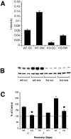
Cox-2 mRNA and protein induction are depressed in IL-1R1 null mice. A, 32P-labeled RT-PCR analysis of Cox-2 mRNA transcripts at 18 hr after a penetrating cortical injury in IL-1R1 null mice. Samples from three wild-type (WT) and three IL-1R1 null mice are depicted. For each mouse, tissue from the nondamaged hemisphere [contralateral cortex (CC)] as well as the lesioned hemisphere [stab wound (SW)] were analyzed by RT-PCR. Quantification of 32P-labeled PCR product was performed using the ImageQuant software program supplied with the PhosphorImager. Values of Cox-2 mRNA transcripts were normalized to cyclophilin from the same PCR. Comparisons between groups were statistically different from each other; p < 0.001 as determined by ANOVA followed by t test with Bonferroni correction.KO, Knock-out. B, Tissue from wild-type (wt) or IL-1R1 null mice at 2 d after stab wound (sw) analyzed by immunoblot for Cox-2 using chemiluminescence detection. Blots were reprobed for β-tubulin to establish equal protein loading. The top band in eachlane in B represents Cox-2 (Mr = 80 kDa), whereas the bottom band represents β-tubulin (Mr = 57 kDa). These immunoblots show that the level of Cox-2 was significantly lower in the IL-1R1 null mice under basal conditions, and that the levels did not increase after injury. cc, Contralateral cortex; ko, knock-out. C, Densitometric analysis of immunoblots at 2, 3, and 7 d after a penetrating brain injury. Measurements of the optical densities for Cox-2 for each sample were normalized to the level of β-tubulin; the level of Cox-2 in the injured hemisphere was expressed as the percentage of the level of Cox-2 in the contralateral cortex as indicated by the solid horizontal line.Asterisks indicate significant differences in the level of Cox-2 in the null mice compared with injured wild-type (WT) mice (p < 0.0001; n = 3 mice). The data for the 3 d recovery represent the average from two mice from each strain, whereas the other values are averaged from three mice per group. At 2 d after injury, the levels of Cox-2 in the null mice in the injured hemisphere were not different from the levels in the control hemisphere. By 7 d, however, there was a slight increase in Cox-2 protein levels. Measurements of the average optical densities between wild-type and knock-out (KO) samples and their respective controls showed no significant differences in β-tubulin levels.
Fig. 3.
Fig. 3.
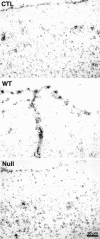
Cox-2 mRNA induction is abrogated 3 d after a penetrating cortical injury in IL-1R1 null mice compared with wild-type (WT) mice. Cryostat sections from brain-injured mice were processed for ISH using a 35S-labeled riboprobe for Cox-2. The control (CTL) panelillustrates the basal expression of Cox mRNA seen in the superficial layers of the mouse cerebral cortex. In a wild-type mouse at the level of the penetrating brain injury, increased hybridization was observed in cells along the pial surface as well as in labeled cells along the needle track. In contrast, in the IL-1R1 null mice the level of hybridization was equal to or less than that seen in the uninjured wild-type mouse cerebral cortex. Scale bar, 50 μm.
Fig. 4.
Fig. 4.
Microglia and macrophages produce Cox-2 adjacent to the injury site at 3 d after a penetrating brain injury. Cryostat sections from brain-injured mice were processed for Cox-2 immunoreactivity in combination with other cell type-specific markers. All sections were counterstained with 4′,6′-diamidino-2-phenylindole to reveal nuclear details of labeled cells. As illustrated in A, a subset of cells immediately adjacent to the injury site coexpressed tomato lectin-binding proteins (rhodamine) and Cox-2 (Alexa 488). Theinset depicts a higher-power view of coexpressing cells.B depicts a similar region adjacent to the injury site in the IL-1R1 null mouse. Cox-2-expressing cells were rarely observed. To confirm that the labeled cells were microglia or macrophages, adjacent sections were stained for CD11b (rhodamine) and Cox-2 (Alexa 488) (C) or for F4/80 (rhodamine) and Cox-2 (Alexa 488) (D). Scale bars: A–C, 40 μm; D, 20 μm.
Fig. 5.
Fig. 5.
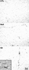
VCAM-1 induction is abrogated in IL-1R1 null mice at 3 d after a penetrating brain injury. Cryostat sections from brain-injured mice were processed for ISH using an35S-labeled riboprobe for VCAM-1. The control (CTL) panel illustrates the basal expression of VCAM-1 mRNA seen in the deep layers of the mouse cerebral cortex. In a wild-type (Wt) mouse at the level of the penetrating brain injury, increased hybridization was observed in cells along the needle track. The inset depicts hybridization for VCAM-1 in a cell adjacent to the lumen of a blood vessel. In contrast, in the IL-1R1 null mice the level of hybridization at the level of the penetrating brain injury was equal to or less than that seen in the uninjured wild-type mouse cerebral cortex. Scale bar, 50 μm.
Fig. 6.
Fig. 6.
GFAP immunoreactivity is reduced in IL-1R1 null mutant mice after a penetrating cortical injury. Adult IL-1R1 null or age-matched wild-type mice received a penetrating brain injury perpendicular to the pial surface using a sterile 25 gauge blunt needle. After 3 d, the animals were killed by perfusion and processed for GFAP immunohistochemistry. The stab wound is at thebottom of each panel. In the wild-type mice there is a robust increase in GFAP immunohistochemistry (A, C), whereas in the IL-1R1 null mice this response is blunted (B, D). Scale bars: A, B, 20 μm;C, D, 40 μm.
Fig. 7.
Fig. 7.
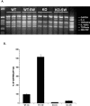
Induction of proinflammatory cytokine transcripts is abrogated in IL-1R null mutant mice after penetrating cortical injury. A, PCR cDNA products for IL-6, TNF-α, IL-1β, TGF-β, GM-CSF and glyceraldehyde-3-phosphate dehydrogenase (GAPDH) at the 18 hr time point after a penetrating cortical injury are shown. An ethidium-stained gel containing samples from three wild-type (WT) and three knock-out (KO) adult mice is depicted. Tissue from the nondamaged hemisphere as well as the lesioned hemisphere [stab wound (SW)] was analyzed by multiplex RT-PCR. This analysis revealed that the basal levels of IL-6 and IL-1 were much reduced in the IL-1R1 null mice compared with wild-type mice. After injury, the levels of these cytokine mRNAs increased in both strains of mice, although they did not increase to the same levels in the IL-1R1 null mice. The respective sizes of the PCR products are shown in Table 1. B,32P-labeled RT-PCR analysis of IL-6 mRNA transcripts was performed 18 hr after penetrating cortical injury. Tissue from the contralateral cortex (CC) as well as the stab wound (SW) from three wild-type and three IL-1R1 null adult mice was analyzed. Values of IL-6 mRNA transcripts were normalized to cyclophilin from the same PCR and then expressed as percentage of the value obtained for the wild-type unlesioned cortex. Comparisons between groups were statistically different from each other; p < 0.0001 as determined by ANOVA followed by a t test with Bonferroni correction, with the exception of the KO-CC versus the KO-SW, which was not statistically different.
Fig. 8.
Fig. 8.
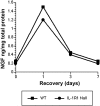
The increase in NGF protein is slightly decreased but not delayed in IL-1R1 null mutant mice after a penetrating cortical injury. NGF levels were measured by two-site ELISA at 1, 3, or 7 d after injury in wild-type (WT) or IL-1R1 null mice. NGF protein levels were normalized to the level of total protein in each sample. As shown, NGF increased both in wild-type and in the IL-1R1 null mice, reaching a peak at 1 d after injury. The levels in the receptor null mutants were 30% less than those in the wild-type animals; however, this difference was not statistically significant according to Student's t test. The increase in the IL-1R1 null also followed a normal time course.
Fig. 9.
Fig. 9.
IL-6-type cytokines activate astrocytes independent of IL-1R1 and TNF-αRp55. Adult IL-1R1/TNFα-Rp55 double null mice received an injection of 100 ng of rhCNTF into the neocortex. At 48 hr of survival, animals were perfused and processed for ISH with a 35S-labeled antisense probe to GFAP or vimentin (VIM). After hybridization, the slides were exposed to autoradiographic film and exposed for 2 (GFAP) or 5 (vimentin) d. Photographs were taken of the autoradiographic films using bright-field illumination. Hybridization on the contralateral hemisphere, which received heat-inactivated CNTF, was not above basal levels.

References

    1. Acarin L, Vela JM, González B, Castellano B. Demonstration of poly-N-acetyl lactosamine residues in ameboid and ramified microglial cells in rat brain by tomato lectin binding. J Histochem Cytochem. 1994;42:1033–1041. - PubMed
    1. Akiyama H, Barger S, Barnum S, Bradt B, Bauer J, Cole GM, Cooper NR, Eikelenboom P, Emmerling M, Fiebich BL, Finch CE, Frautschy S, Griffin WS, Hampel H, Hull M, Landreth G, Lue L, Mrak R, Mackenzie IR, McGeer PL. Inflammation and Alzheimer's disease. Neurobiol Aging. 2000;21:383–421. - PMC - PubMed
    1. Albrecht PJ, Dahl JP, Stoltzfus OK, Levenson R, Levison SW. Ciliary neurotrophic factor activates spinal cord astrocytes, stimulating their production and release of FGF-2, to increase motor neuron survival. Exp Neurol. 2002;173:46–62. - PubMed
    1. Aloisi F, Care A, Borsellino G, Gallo P, Rosa S, Bassani A, Cabibbo A, Testa U, Levi G, Peschle C. Production of hemolymphopoietic cytokines (IL-6, IL-8, colony-stimulating factors) by normal human astrocytes in response to IL-1β and tumor necrosis factor-α. J Immunol. 1992;149:2358–2366. - PubMed
    1. Araujo DM, Cotman CW. Basic FGF in astroglial, microglial, and neuronal cultures: characterization of binding sites and modulation of release by lymphokines and trophic factors. J Neurosci. 1992;12:1668–1678. - PMC - PubMed

Publication types

MeSH terms