. 2002 Jul;110(2):193-202.
doi: 10.1172/JCI15295.

Hepatocyte-specific inhibition of NF-kappaB leads to apoptosis after TNF treatment, but not after partial hepatectomy

Affiliations

Hepatocyte-specific inhibition of NF-kappaB leads to apoptosis after TNF treatment, but not after partial hepatectomy

Michelle L Chaisson et al. J Clin Invest. 2002 Jul.

Abstract

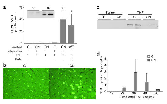
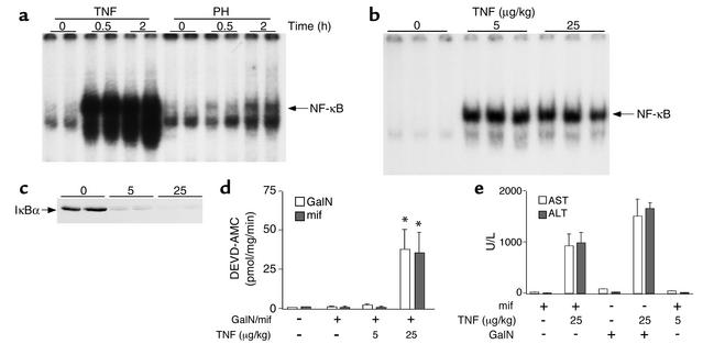
One of the earliest TNF-dependent events to occur during liver regeneration is the activation of the transcription factor NF-kappaB through TNF receptor type 1. NF-kappaB activation in the liver can have both antiapoptotic and proliferative effects, but it is unclear which liver cell types, hepatocytes or nonparenchymal cells (NPCs), contribute to these effects. To specifically evaluate the role of hepatocyte NF-kappaB, we created GLVP/DeltaN-IkappaB(alpha) transgenic mice, in which expression of a deletion mutant of IkappaB(alpha) (DeltaN-IkappaB(alpha)) was induced in hepatocytes after injection of mifepristone. In control mice, injection of 25 microg/kg TNF caused NF-kappaB nuclear translocation in virtually all hepatocytes by 30 minutes and no detectable apoptosis, while in mice expressing DeltaN-IkappaB(alpha), NF-kappaB nuclear translocation was blocked in 45% of hepatocytes, leading to apoptosis 4 hours after TNF injection. In contrast, expression of DeltaN-IkappaBalpha in hepatocytes during the first several hours after partial hepatectomy did not lead to apoptosis or decreased proliferation. As NF-kappaB activation was not inhibited in liver NPCs, it is likely that these cells are responsible for mediating the proliferative and antiapoptotic effects of NF-kappaB during liver regeneration.


Figures

Figure 1
Figure 1
Induced expression of ΔN-IκBα in double-transgenic mice injected with mifepristone. (a) Schematic of the inducible transgenic mouse system used. (b) Mifepristone dose–dependent expression. Mice were killed 3 hours after intraperitoneal injection of the indicated dose of mifepristone dissolved in sesame oil. Western blot analysis of liver protein lysates was performed using an antibody to either the C terminus of IκBα (top panel) or to the FLAG epitope (bottom panel). Lanes represent individual animals. (c) Timing of induced expression. Mice were killed at the indicated times after injection of 5 mg/kg mifepristone. (d) Liver-specific expression of ΔN-IκBα. Organs were harvested from a mouse killed 3 hours after injection with 5 mg/kg mifepristone. Western blot analysis was performed using an antibody to IκBα (top panel) or FLAG (bottom panel).
Figure 2
Figure 2
Inhibition of TNF-induced p65 nuclear translocation in hepatocytes of double-transgenic mice expressing ΔN-IκBα. Liver sections were stained with an antibody to the p65 subunit of NF-κB as described in Methods. (a) Negative control: no primary staining of a single-transgenic mouse killed 3 hours after injection with 5 mg/kg mifepristone. (b) Same tissue stained with p65 antibody. (c and d) p65 staining of a mifepristone-treated single-transgenic mouse (c) or double-transgenic mouse (d) killed 30 minutes after injection of 25 μg/kg TNF. Open arrow indicates an NPC with nuclear p65 staining. Filled arrow indicates a hepatocyte in which p65 nuclear translocation has been blocked. Original magnification, ×400.
Figure 3
Figure 3
TNF activates hepatocyte apoptosis in double-transgenic mice expressing ΔN-IκBα. (a) Caspase-3 activity. Single-transgenic (G) or double-transgenic (GN) mice were injected with 25 μg/kg TNF or saline 3 hours after injection with mifepristone or sesame oil. For comparison, C57BL6 wild-type (WT) mice were coinjected with TNF and D-galactosamine (GalN). Mice were killed 4 hours after TNF injection and liver protein lysates were used to measure caspase-3 activity by incubation with the fluorogenic substrate DEVD-AMC. Data represent average (± SEM) caspase-3 activity measured in each group. Three to five mice were used per group. *P < 0.05 compared with vehicle control. Inset: active caspase-3 Western blot analysis of mifepristone- and TNF-treated mice. (b) TUNEL assay. Liver sections from a single-transgenic (left) and a double-transgenic (right) mouse treated with mifepristone and TNF were incubated with FITC-dUTP and terminal deoxynucleotidyl transferase. Original magnification, ×400. (c) iNOS Western blot. (d) DNA synthesis. Mice were injected with BrdU prior to sacrifice at the indicated times after treatment with mifepristone and TNF. Liver sections were stained with an antibody to BrdU as described in Methods, and positive nuclei were scored for 30 fields (×400) per section. Data represent average (± SEM) percentage of BrdU-positive hepatocyte nuclei per group. Three mice were used per group.
Figure 4
Figure 4
PH does not cause hepatocyte apoptosis in double-transgenic mice expressing ΔN-IκBα. (a) IκBα Western blot. Liver lysates were prepared from mifepristone-treated double-transgenic mice killed 30 minutes or 4 hours after injection of 25 μg/kg TNF (left lanes) or PH (right lanes). (b) Caspase-3 activity measured using substrate DEVD-AMC. Liver protein lysates were prepared from mifepristone-treated single-transgenic and double-transgenic mice killed at the indicated times after PH. The last column (4 + TNF) represents mice injected with 25 μg/kg TNF 30 minutes prior to PH and killed 4 hours after surgery. Data represent average (± SEM) caspase activity measured in each group. Three mice were used per group. *P < 0.05 for double-transgenic compared with single-transgenic mice. (c) Bcl-xL Northern blot (top panel) and loading control cyclophilin (bottom panel).
Figure 5
Figure 5
Analysis of NF-κB activation in mifepristone-treated mice after PH. (a) EMSA analysis of NF-κB DNA binding. Nuclear extracts were prepared from livers of mifepristone-treated single- or double-transgenic mice killed at the indicated times after PH. Five micrograms of nuclear protein was incubated with labeled NF-κB probe as described in Methods. Lanes represent individual animals. (b) IκBα Western blot of liver nuclear protein. Ten micrograms of liver nuclear extract protein used for EMSA shown in a was used for IκBα immunoblot. (c) Coincubation assay. Nuclear extract protein from 2-hour PH samples used in a were incubated alone or together at the indicated ratios with labeled NF-κB probe. (d) Histological analysis of p65 nuclear translocation in mifepristone-treated double-transgenic mice killed without PH or 2 hours after PH. Original magnification, ×400.
Figure 6
Figure 6
Expression of ΔN-IκBα in hepatocytes does not inhibit liver regeneration or IL-6 or STAT3 activation after PH. (a) Hepatocyte DNA synthesis. Mifepristone-treated single-transgenic and double-transgenic mice were injected with BrdU 2 hours prior to killing at the indicated times after PH. Liver sections were stained with an antibody to BrdU as described in Methods, and BrdU-positive hepatocyte nuclei were scored for 30 fields (×400) per slide. Four mice were analyzed per group, and data represent average percentage (± SEM) of BrdU-positive nuclei per group. (b) Mitotic index. Mitotic hepatocytes were counted in mifepristone-treated mice killed 48 hours after PH. Four mice were analyzed per group, and data represent average total number ± SEM of mitotic hepatocytes counted per 30 fields (×400). (c) Liver/body weight ratio. Liver and whole body weights were determined for untreated mice (no PH) or mifepristone-treated mice killed 2 weeks after PH. Three mice were used per group, and data represent average (± SEM) liver weight expressed as a percentage of whole body weight. (d) DNA synthesis at 48 hours after PH in mice injected with mifepristone at 12-hour intervals. (e) IL-6 ELISA. Serum was obtained from mifepristone-treated single- or double-transgenic mice killed at the indicated times after PH. Data represent average (± SEM) IL-6 levels per group. Three mice were analyzed per group. (f) STAT3 EMSA. Nuclear extracts were prepared from livers of mice described in e. Five micrograms of nuclear protein were analyzed for STAT3 DNA binding activity as described in Methods. Each lane represents an individual animal.
Figure 7
Figure 7
TNF-induced apoptosis occurs at a higher dose of TNF than that required to activate NF-κB. (a) Comparison by EMSA of NF-κB activation after TNF injection or PH. Nuclear protein was prepared from livers of mifepristone-treated single-transgenic mice killed at the indicated times after injection of 25 μg/kg TNF (left lanes) or PH (right lanes). (b) NF-κB EMSA. Nuclear extracts were prepared from livers of C57BL6 mice killed 30 minutes after injection with the indicated dose of TNF. (c) IκBα Western blot. Liver protein lysates were prepared from mice described in b and immunoblotted for IκBα. (d) Caspase-3 activity. Mice were killed 4 hours after treatment with the indicated dose of TNF in combination with either mifepristone (mif) or D-galactosamine (GalN). For mifepristone, double-transgenic mice were used. For D-galactosamine, C57BL6 mice were used. One hundred micrograms of liver protein lysates were incubated with fluorogenic DEVD-AMC as described in Methods. *P < 0.05 compared with vehicle controls. (e) AST/ALT activity. Serum was collected from mice killed 7 hours after treatment with TNF in combination with mifepristone or D-galactosamine, as described in d.

References

    1. Beg AA, Sha WC, Bronson RT, Ghosh S, Baltimore D. Embryonic lethality and liver degeneration in mice lacking the RelA component of NF-κB. Nature. 1995;376:167–170. - PubMed
    1. Xu Y, et al. NF-kappaB inactivation converts a hepatocyte cell line TNF-alpha response from proliferation to apoptosis. Am J Physiol. 1998;275:C1058–C1066. - PubMed
    1. Kirillova I, Chaisson M, Fausto N. Tumor necrosis factor induces DNA replication in hepatic cells through nuclear factor κB activation. Cell Growth Differ. 1999;10:819–828. - PubMed
    1. Cressman DE, Greenbaum LE, Haber BA, Taub R. Rapid activation of post-hepatectomy factor/nuclear factor kappa B in hepatocytes, a primary response in the regenerating liver. J Biol Chem. 1994;269:30429–30435. - PubMed
    1. FitzGerald MJ, Webber EM, Donovan JR, Fausto N. Rapid DNA binding by nuclear factor kappa B in hepatocytes at the start of liver regeneration. Cell Growth Differ. 1995;6:417–427. - PubMed

Publication types

MeSH terms