. 2002 Jul;110(2):203-8.
doi: 10.1172/JCI15672.

Episodic coronary artery vasospasm and hypertension develop in the absence of Sur2 K(ATP) channels

Affiliations

Episodic coronary artery vasospasm and hypertension develop in the absence of Sur2 K(ATP) channels

William A Chutkow et al. J Clin Invest. 2002 Jul.

Abstract

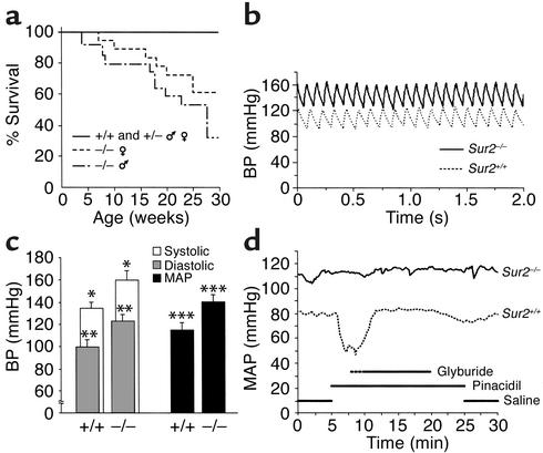
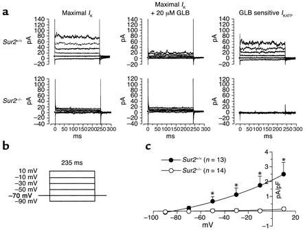
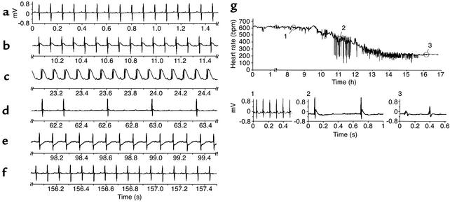
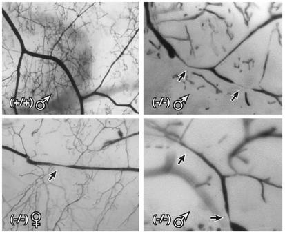
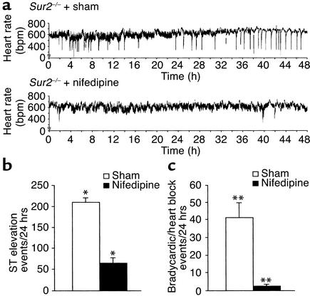
K(ATP) channels couple the intracellular energy state to membrane excitability and regulate a wide array of biologic activities. K(ATP) channels contain a pore-forming inwardly rectifying potassium channel and a sulfonylurea receptor regulatory subunit (SUR1 or SUR2). To clarify the role of K(ATP) channels in vascular smooth muscle, we studied Sur2 gene-targeted mice (Sur2(-/-)) and found significantly elevated resting blood pressures and sudden death. Using in vivo monitoring, we detected transient, repeated episodes of coronary artery vasospasm in Sur2(-/-) mice. Focal narrowings in the coronary arteries were present in Sur2(-/-) mice consistent with vascular spasm. We treated Sur2(-/-) mice with a calcium channel antagonist and successfully reduced vasospastic episodes. The intermittent coronary artery vasospasm seen in Sur2(-/-) mice provides a model for the human disorder Prinzmetal variant angina and demonstrates that the SUR2 K(ATP) channel is a critical regulator of episodic vasomotor activity.


Figures

Figure 1
Figure 1
Cardiovascular physiology in Sur2–/– mice. (a) Sur2–/– mice exhibit decreased survival. Survival of littermate control and heterozygous mice (Sur2+/+ n = 15, 5 males and 10 females, and Sur2+/–, n = 41, 12 males and 29 females), homozygous mutant females (Sur2–/–, n = 18), and homozygous mutant males (Sur2–/–, n = 17) as a function of age. (b) Telemetric blood pressure recordings of Sur2–/– and normal littermate control mice. Representative blood pressure tracings from conscious, untethered control (Sur2+/+) and homozygous mutant (Sur2–/–) mice. (c) Systolic (white bars), diastolic (gray bars), and MAP (black bars) for littermate control and mutant mice (n = 6 and 4, respectively), recorded between days 2 and 4 after implantation. *P < 0.04, **P < 0.03, ***P < 0.02 versus normal littermate controls by two-tailed, unpaired Student t test. (d) Sur2–/– mice do not respond to KATP vasoactive agents. Representative MAP tracings from a littermate control mouse (dotted line) and a Sur2–/– mouse (solid line) during infusions of the agents indicated. Pinacidil was administered at 5 μg/kg/min and glibenclamide at 100 μg/kg/min. The interrupted line for glyburide infusion indicates the beginning of infusion for the normal control mouse (Sur2+/+), while the mutant mouse (Sur2–/–) infusion begins with the solid line.
Figure 2
Figure 2
Absence of KATP current in aortic smooth muscle cells isolated from Sur2–/– mice. (a) Examples of original whole cell current traces recorded in a Sur2+/+ and a Sur2–/– aortic smooth muscle cell 2 minutes after cell rupture (maximal potassium channel current [IK], left), after 20 μM of glibenclamide (GLB, middle), and the GLB-sensitive IKATP obtained by subtraction (right). The glibenclamide-sensitive current was interpreted as KATP current and was normalized by cell capacitance to obtain the whole cell current density. The GLB-sensitive current was absent from the Sur2–/– cell. (b) Whole cell current protocol. The cell was held at –70 mV, and 235 ms test pulses from –90 to 10 mV were delivered to induce a whole cell current. (c) Mean summary data of the current voltage plot for the GLB-sensitive IKATP density at all voltages tested (n = 13 cells from five Sur2+/+ mice vs. n = 14 cells from four Sur2–/– mice, *P < 0.05).
Figure 3
Figure 3
In vivo evidence of coronary artery vasospasm in Sur2–/– mice. (af) Representative telemetric ECG recording of a 12-week-old male Sur2–/– mouse. Each of the serial tracings reflects 1.5 seconds of the entire 160-second episode. The timing of each 1.5-second segment with respect to the duration of the entire episode is indicated in seconds below the tracing. (a) A baseline tracing at the beginning of the event. (b and c) Marked ST segment elevation that occurs within 30 seconds. ST segment elevation leads to atrioventricular heart block seen in d. Following the acute injury pattern of ST segment elevation, ST segment depression was present during recovery (e). (f) A return to baseline ECG readings. (g) Continuous ECG recording capturing sudden death. Upper tracing depicts heart rate and bradycardia events (downward spikes) over a 17-hour period. Individually, numbered regions are magnified in the lower three sets of ECG waveform tracings indicating: 1, normal rate and rhythm; 2, post-ST elevation atrioventricular heart block with bradycardia; 3, agonal rhythm with a widened QRS.
Figure 4
Figure 4
Coronary artery vasospasm. The coronary vasculature was perfused with Microfil, a liquid latex medium, as described previously (20). Microvascular perfusion was visualized by transillumination under low-power magnification. (upper left panel) Normal microvasculature of a littermate control male mouse. (upper right panel and lower panels) Examples of focal artery stenoses in three Sur2–/– mice. Arrows indicate significant coronary artery stenoses.
Figure 5
Figure 5
Successful treatment of coronary artery vasospasm in Sur2–/– mice with nifedipine. (a) Representative continuous ECG recordings obtained during subcutaneous infusion of a sham-infusate (saline or DMSO) versus nifedipine at 3 mg/kg/d. Tracings depict heart rate, with sharp downward deflections representing deceleration in the heart rate or episodic bradycardia. Graphic summary of quantitative decrease of (b) ST segment elevation events per 24-hour period, and (c) heart block episodes per 24-hour period (n = 5 each). White bars represent sham-infusate group. Black bars represent nifedipine group. *,**P < 0.0001 by two-tailed, unpaired Student t test.

Comment in

References

    1. Noma A. ATP-regulated K+ channels in cardiac muscle. Nature. 1983;305:147–148. - PubMed
    1. Ashcroft SJ, Ashcroft FM. Properties and functions of ATP-sensitive K-channels. Cell Signal. 1990;2:197–214. - PubMed
    1. Inagaki N, et al. Reconstitution of IKATP: an inward rectifier subunit plus the sulfonylurea receptor. Science. 1995;270:1166–1170. - PubMed
    1. Inagaki N, et al. Cloning and functional characterization of a novel ATP-sensitive potassium channel ubiquitously expressed in rat tissues, including pancreatic islets, pituitary, skeletal muscle, and heart. J Biol Chem. 1995;270:5691–5694. - PubMed
    1. Aguilar-Bryan L, et al. Cloning of the beta cell high-affinity sulfonylurea receptor: a regulator of insulin secretion. Science. 1995;268:423–426. - PubMed

Publication types

MeSH terms

Substances