. 2002 Jul;110(2):209-17.
doi: 10.1172/JCI15745.

Thyroid-stimulating autoantibodies in Graves disease preferentially recognize the free A subunit, not the thyrotropin holoreceptor

Affiliations

Thyroid-stimulating autoantibodies in Graves disease preferentially recognize the free A subunit, not the thyrotropin holoreceptor

Gregorio D Chazenbalk et al. J Clin Invest. 2002 Jul.

Abstract

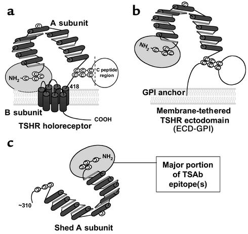
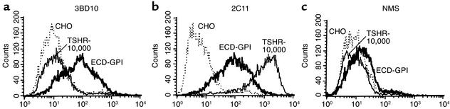
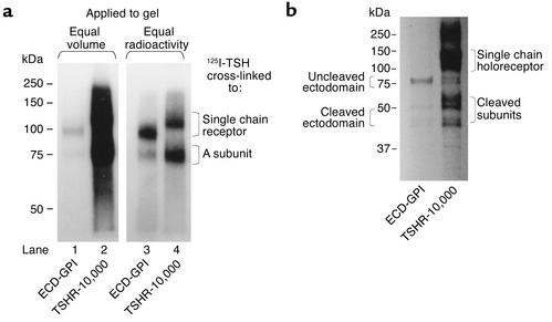
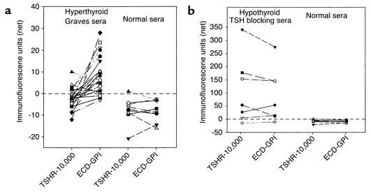
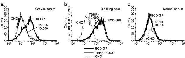
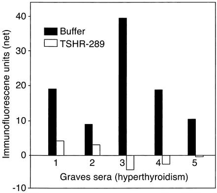
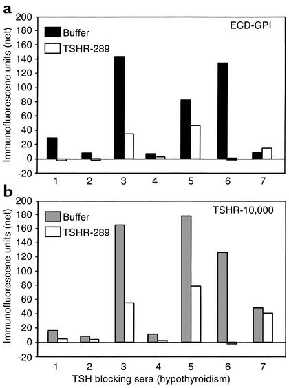
Graves disease is directly caused by thyroid-stimulating autoantibodies (TSAb's) that activate the thyrotropin receptor (TSHR). We observed upon flow cytometry using intact cells that a mouse mAb (3BD10) recognized the TSHR ectodomain with a glycosidylphosphatidylinositol (ECD-GPI) anchor approximately tenfold better than the same ectodomain on the wild-type TSHR, despite the far higher level of expression of the latter. The 3BD10 epitope contains the N-terminal cysteine cluster critical for TSAb action. Consequently, we hypothesized and confirmed that TSAb (but not thyrotropin-blocking autoantibodies [TBAb's]) also poorly recognize the wild-type TSHR relative to the ECD-GPI. Despite poor recognition by TSAb of the holoreceptor, soluble TSHR A subunits (known to be shed from surface TSHR) fully neutralized autoantibody-binding activity. These data indicate that the epitope(s) for TSAb's, but not for TBAb's, are partially sterically hindered on the holoreceptor by the plasma membrane, the serpentine region of the TSHR, or by TSHR dimerization. However, the TSAb epitope on the soluble A subunit is freely accessible. This observation, as well as other evidence, supports the concept that A subunit shedding either initiates or amplifies the autoimmune response to the TSHR, thereby causing Graves disease in genetically susceptible individuals.


Figures

Figure 1
Figure 1
Schematic representation of different forms of the TSHR. (a) TSH holoreceptor. Intramolecular cleavage of the single polypeptide chain is followed by removal of the C peptide region, with the A subunit remaining tethered to the membrane-spanning B subunit by disulfide bonds. The cylinders depict the α helices in the nine leucine-rich repeats in the A subunit, as well as the seven transmembrane segments of the B subunit. The ectodomain comprises the entire A subunit and the extracellular region of the B subunit. The ectodomain enters the plasma membrane at approximately amino acid residue 418. A critical domain in the TSAb epitope(s) involves the cysteine-rich area at the extreme N terminus of the TSHR and is shown by the gray oval. (b) TSHR ectodomain tethered to the plasma membrane by a GPI anchor (ECD-GPI). This construct involved the attachment after TSHR codon 412 of a 39–amino acid sequence containing a signal for GPI attachment (31). The putative GPI attachment site of the anchor is at codon 425. Because of the additional length of the ectodomain prior to insertion into the membrane, and because the GPI anchor only traverses the outer leaf of the plasma membrane, the TSHR ectodomain is shown in a more “open” orientation relative to the wild-type TSHR. (c) Shed A subunit. The exact site of cleavage has not been established, but is approximately at amino acid residue 310. The purified TSHR-289 preparation used in this study is truncated at TSHR amino acid residue 289 and therefore comprises nearly the entire A subunit.
Figure 2
Figure 2
Differential recognition by murine TSHR mAb of the wild-type TSHR ectodomain and the GPI-anchored ectodomain. Flow cytometry was performed with the following murine mAb’s: (a) mAb 3BD10, epitope including the cysteine-rich segment at the extreme N terminus of the TSHR ectodomain; (b) mAb 2C11, epitope in the C peptide region of the TSHR ectodomain, present only in the uncleaved, but not in the cleaved, TSHR; (c) normal mouse serum (NMS) included as a control. The assays used the following cell lines: TSHR-10,000, ECD-GPI, and CHO. TSHR-10,000 is the CHO cell line stably expressing the wild-type TSH holoreceptor. The transgenome has been amplified using the dihydrofolate reductase minigene approach (adaptation to final methotrexate concentration of 10,000 nM), leading to very high TSHR expression on the cell surface (∼2 × 10⁶/cell) (30). ECD-GPI is the CHO cell line stably expressing the TSHR ectodomain anchored to the plasma membrane by a GPI moiety. CHO are untransfected CHO cells. Note the approximately 100-fold difference in 3BD10 versus 2C11 recognition of the TSHR holoreceptor, despite their similar recognition of the ECD-GPI cell line.
Figure 3
Figure 3
(a) TSH covalent cross-linking to intact cells reveals greater level of cell surface TSHR expression in TSHR-10,000 cells than in ECD-GPI cells. After simultaneous 125I-TSH cross-linking (same number of cells, same amount of 125I-TSH), cell extracts were subjected to polyacrylamide gel electrophoresis (10%) under reducing conditions (see Methods). Autoradiography (representative of three experiments) was for 16 hours. In lanes 1 and 2, equal volumes of cell extract were applied. Lanes 3 and 4 were loaded with equal amounts of radioactivity. TSH cross-linking to intact cells, unlike immunoprecipitation (b), visualizes only mature TSHR expressed on the cell surface. Two forms of TSHR are seen: single-chain uncleaved TSHR and the ligand binding A subunit in the cleaved TSHR released from the serpentine region (TSHR-10,000 cells) or from the GPI-anchored portion (ECD-GPI) by disulfide bond reduction. Note the lesser proportion of cleaved versus uncleaved receptors in the ectodomain with the GPI anchor than in the wild-type TSHR. (b) Immunoprecipitation confirms higher level of TSHR expression in TSHR-10,000 cells than in ECD-GPI cells. Similar numbers of TSHR-10,000 cells and ECD-GPI cells were precursor labeled simultaneously with the same quantity of 35S-methionine and 35S-cysteine (see Methods). Detergent-solubilized particulate preparations were immunoprecipitated with murine mAb A9 (epitope in the midregion of the TSHR ectodomain), the most effective mAb available to us for immunoprecipitating all forms of the TSHR. After polyacrylamide gel electrophoresis (10%), autoradiography (representative of duplicate experiments) was performed for 16 hours.
Figure 4
Figure 4
(a) Stimulating TSHR autoantibodies recognize the wild-type TSHR less well than the TSHR ectodomain with a GPI anchor. Sera (diluted 1:50) from hyperthyroid Graves patients, all with high levels of TSHR autoantibodies, were tested by flow cytometry on cells expressing the wild-type TSH holoreceptor (TSHR-10,000) and the TSHR ectodomain with a GPI anchor (see Methods). Net mean fluorescence values for the Graves sera (and for control sera from normal individuals) are plotted after subtraction of background fluorescence obtained with untransfected CHO cells. Statistical significance of differences between individual Graves serum recognition of the TSHR-10,000 and the ECD-GPI cells was determined by the paired t test; P < 0.001. (b) Sera from rare hypothyroid patients with TBAb’s show no difference in their recognition of the wild-type TSHR and the GPI-anchored TSHR ectodomain. Sera (diluted 1:50) were tested by flow cytometry on cells expressing the wild-type TSH holoreceptor (TSHR-10,000) and the TSHR ectodomain with a GPI anchor (see Methods). Net mean fluorescence values for the sera (and for control sera from normal individuals) are indicated after subtraction of background fluorescence obtained with untransfected CHO cells. Note the difference scales for the Y axes in a and b, reflecting the higher titers of TBAb’s versus TSAb’s.
Figure 5
Figure 5
Examples of flow cytometry data obtained with a Graves hyperthyroid serum and serum from a hypothyroid patient with TBAb activity. Note that despite giving an excellent fluorescent signal on the ECD-GPI cell line, the same Graves serum does not recognize the TSHR-10,000 cells overexpressing the TSH holoreceptor on their surface (a). In contrast, the serum from a hypothyroid patient with TBAb activity recognizes both ECD-GPI and TSHR-10,000 cells equally well (b). Serum from a normal individual is included as a control (c).
Figure 6
Figure 6
Neutralization by purified TSHR A subunits of TSHR autoantibodies from Graves patients with hyperthyroidism. Flow cytometry was performed using sera from five Graves patients whose signal upon flow cytometry gave the largest differential between recognition of the wild-type TSHR and GPI-anchored ectodomain (Figure 4a). Sera were preincubated in either buffer alone or in buffer containing a large excess of TSHR-289 (0.1 μg/0.1 ml of serum diluted 1:50) before addition to CHO cells expressing ECD-GPI. TSHR-289 is a secreted, soluble, heavily glycosylated protein that comprises amino acid residues 22–289 of the TSHR, essentially the A subunit (residues 1–21 being the signal peptide). Net fluorescence values for the sera are indicated after subtraction of background fluorescence obtained with untransfected CHO cells.
Figure 7
Figure 7
Variable neutralization by purified TSHR A subunits of TSHR autoantibodies from hypothyroid patients with TBAb’s. Flow cytometry was performed using sera from seven patients. Sera were preincubated in either buffer alone or in buffer containing TSHR-289 (0.1 μg/0.1 ml of serum diluted 1:50) before addition to CHO cells expressing the wild-type (TSHR-10,000 cells) or the GPI-anchored TSHR ectodomain (ECD-GPI cells). Net fluorescence values for the sera are indicated after subtraction of background fluorescence obtained with untransfected CHO cells. Note the difference scales for the Yaxes here and in Figure 6, reflecting the higher titers of TBAb versus TSAb.

Comment in

  • The TSH receptor reveals itself.
    Davies T, Marians R, Latif R. Davies T, et al. J Clin Invest. 2002 Jul;110(2):161-4. doi: 10.1172/JCI16234. J Clin Invest. 2002. PMID: 12122107 Free PMC article. Review. No abstract available.

References

    1. Rapoport B, McLachlan SM. Thyroid autoimmunity. J Clin Invest. 2001;108:1253–1259. doi:10.1172/JCI200114321. - PMC - PubMed
    1. Zakarija M. Immunochemical characterization of the thyroid-stimulating antibody (TSAb) of Graves’ disease: evidence for restricted heterogeneity. J Clin Lab Immunol. 1983;10:77–85. - PubMed
    1. Knight J, Laing P, Knight A, Adams D, Ling N. Thyroid stimulating autoantibodies usually contain only lambda-light chains: evidence for the “forbidden clone” theory. J Clin Endocrinol Metab. 1986;62:342–347. - PubMed
    1. Williams RC, Jr, et al. Kappa/lambda immunoglobulin distribution in Graves’ thyroid-stimulating antibodies. Simultaneous analysis of C lambda gene polymorphisms. J Clin Invest. 1988;82:1306–1312. - PMC - PubMed
    1. Weetman AP, et al. Thyroid-stimulating antibody activity between different immunoglobulin G subclasses. J Clin Invest. 1990;86:723–727. - PMC - PubMed

Publication types

MeSH terms