. 2002 Jul;110(2):247-57.
doi: 10.1172/JCI15058.

Functional redundancy of Rab27 proteins and the pathogenesis of Griscelli syndrome

Affiliations

Functional redundancy of Rab27 proteins and the pathogenesis of Griscelli syndrome

Duarte C Barral et al. J Clin Invest. 2002 Jul.

Erratum in

  • J Clin Invest 2002 Oct;110(8):1213

Abstract

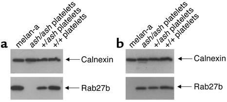
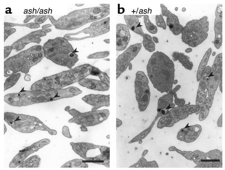
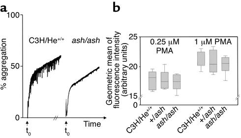
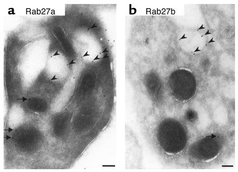
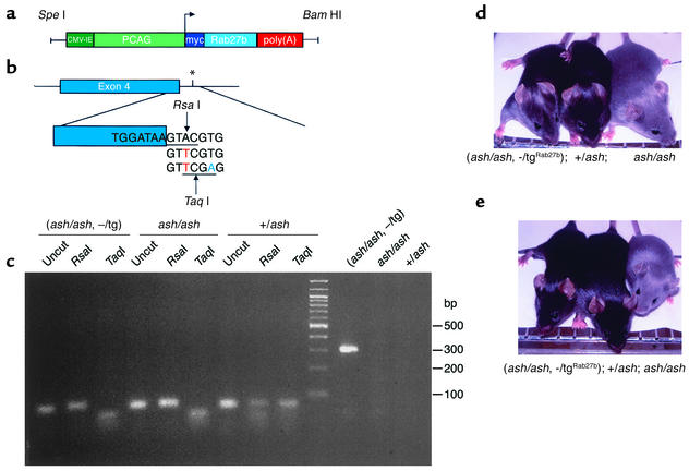
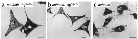
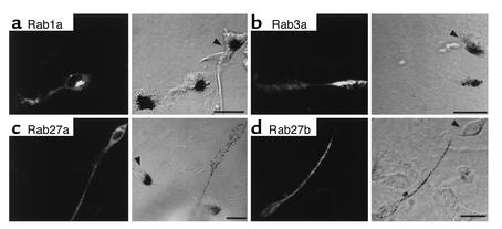
Griscelli syndrome (GS) patients and the corresponding mouse model ashen exhibit defects mainly in two types of lysosome-related organelles, melanosomes in melanocytes and lytic granules in CTLs. This disease is caused by loss-of-function mutations in RAB27A, which encodes 1 of the 60 known Rab GTPases, critical regulators of vesicular transport. Here we present evidence that Rab27a function can be compensated by a closely related protein, Rab27b. Rab27b is expressed in platelets and other tissues but not in melanocytes or CTLs. Morphological and functional tests in platelets derived from ashen mice are all within normal limits. Both Rab27a and Rab27b are found associated with the limiting membrane of platelet-dense granules and to a lesser degree with alpha-granules. Ubiquitous transgenic expression of Rab27a or Rab27b rescues ashen coat color, and melanocytes derived from transgenic mice exhibit widespread peripheral distribution of melanosomes instead of the perinuclear clumping observed in ashen melanocytes. Finally, transient expression in ashen melanocytes of Rab27a or Rab27b, but not other Rab's, restores peripheral distribution of melanosomes. Our data suggest that Rab27b is functionally redundant with Rab27a and that the pathogenesis of GS is determined by the relative expression of Rab27a and Rab27b in specialized cell types.


Figures

Figure 1
Figure 1
Tissue distribution of Rab27a and Rab27b. Tissues from perfused mice were lysed, the postnuclear supernatant was ultracentrifuged, and identical amounts of protein (35 μg) from the pellet fractions were subjected to SDS-PAGE and immunoblot analysis as described in Methods. Monoclonal anti-Rab27a Ab 4B12 was used to probe Rab27a (a) and affinity-purified polyclonal anti-Rab27b Ab S086 to probe Rab27b (b). Anti-calnexin Ab recognizing a ubiquitous endoplasmic reticulum-membrane protein (ER-membrane protein) was used as a loading control. Recombinant his6Rab27a and his6Rab27b (25 ng) were used as controls for Ab specificity.
Figure 2
Figure 2
Expression of Rab27a and Rab27b in melanocytes and platelets. Platelets from wild-type (C3H/He+/+), homozygous (ash/ash), and heterozygous (+/ash) ashen mice and a melanocytic cell line (melan-a) were lysed, and identical amounts of protein (13 μg) from total lysates (for platelets) or postnuclear supernatants (for melan-a cells) were subjected to SDS-PAGE and immunoblotting as described in Methods. Monoclonal anti-Rab27a Ab, 4B12 was used to probe Rab27a (a) and affinity-purified polyclonal anti-Rab27b Ab S086 to probe Rab27b (b). Anti-calnexin Ab recognizing a ubiquitous ER-membrane protein was used as a loading control.
Figure 3
Figure 3
EM of ashen platelets. Platelets were isolated, fixed, and processed for EM as described under Methods. Ashen (ash/ash) platelets (a) appear normal when compared with heterozygous (+/ash) controls (b). Typical dense granules (arrowheads) are shown. The granules are electron dense due to their high calcium content. Bars, 1 μm.
Figure 4
Figure 4
ELISA detection of endogenous vWF. Platelets from ashen homozygous (ash/ash) and heterozygous mice controls (+/ash) (a) and from gunmetal homozygous (gm/gm) and heterozygous mice controls (+/gm) (b) were lysed and the indicated amount of total protein incubated with 100 μl of buffer in a sandwich ELISA (see Methods). Each step corresponds to a 1:2 dilution, and duplicates were made for each point. The results are representative of three independent experiments. The OD450nm value from the blank reaction (buffer only) was subtracted.
Figure 5
Figure 5
Platelet aggregation and expression of P-selectin at cell surface upon activation. (a) The aggregation capacity of homozygous ashen mice (ash/ash) and wild-type C3H/He mice (C3H/He+/+) was tested, measuring the increasing light transmission through a platelet suspension. The agonist (thrombin at 2 U/ml) was added at t0, and the traces were recorded for 5 minutes. The experiment was repeated independently, and the results were not significantly different (see text). The 0% corresponds to the light transmission of the resting platelet suspension and the 100% to the transmission of light in suspension buffer (or totally aggregated platelets). (b) The expression of P-selectin (mainly localized in α-granule membrane) at the surface of platelets upon stimulation with PMA was used to test α-granule release capacity. Platelets in whole blood were identified with an anti–CD41 (gpIIb) Ab. The geometric mean of the population of platelets labeled with anti–P-selectin Ab was plotted. Triplicates were done for each mouse, and six mice were tested in each case. Boxes represent the 25th and 75th percentiles, and median is represented by horizontal line inside the boxes. The error bars represent the 5th and 95th percentiles. The statistical analysis was performed using an ANOVA test, and the were results found not to be significantly different. The negative controls (without anti–P-selectin Ab) showed less that 2% activated platelets.
Figure 6
Figure 6
Immuno-EM of wild-type platelets. Platelets from C3H/He+/+ mice were processed for immuno-EM as described in Methods. Affinity-purified polyclonal anti-Rab27a Ab N688 was used to probe for Rab27a (a) and affinity-purified polyclonal anti-Rab27b S086 to probe for Rab27b (b). The labeling was found mainly in dense granules (arrowheads). Some labeling was also detected in α-granules (arrows). Bars, 100 nm.
Figure 7
Figure 7
(a) Organization of the transgenic construct encoding Rab27b under the control of β-actin promoter. The PCAG/mycRab27/β-globin construct contains the CMV enhancer (dark green) and the chicken β-actin promoter sequence (light green) upstream of Rab27a or Rab27b cDNA (light blue), followed by the rabbit β-globin poly(A) sequence (red). (b) Strategy to detect ashen mutation (*). This is an A→T transversion (red) that is present on the third base of the splice donor site downstream of exon 4 (blue). Primers ASH2 (see Methods), introduces a T→A transversion (green). RsaI, which recognizes the sequence GTAC (underlined), cuts the DNA where indicated only in the absence of the ashen mutation. TaqI, which recognizes the sequence TCGA (underlined), cuts the DNA where indicated only if the ashen mutation is present. (c) Mouse genotyping. After PCR amplification the 80 bp product was digested with RsaI and TaqI, which originates one fragment of 50 bp and one fragment of 30 bp. The uncut amplified product is shown as a control. The screening for the presence of the transgene was done as described in Methods, generating a product of 324 bp. The PCR and digestion products were resolved on a 3% agarose gel. The molecular weight of the standards is indicated on the right. (d and e) Photography of representative mice for Rab27b (d) or Rab27a (e) rescue experiment. Littermates resulting from crosses between a homozygous ashen mice with heterozygous transgenic mice were genotyped as above.
Figure 8
Figure 8
Melanocytes from rescued mice show normal melanosome distribution. Primary melanocytes from ash/ash, –/tgRab27a (a), ash/ash, –/tgRab27b (b), or ash/ash (c) were cultured and subjected to phase-contrast light microscopy as described in Methods. The ash/ash cells in c were derived from a littermate of ash/ash, –/tgRab27b. Bars, 20 μM.
Figure 9
Figure 9
Transient expression of Rab proteins in ashen melanocytes. Melan-ash melanocytes derived from ashen mice were transiently transfected with pEGFP-Rab1a (a), pEGFP-Rab3a (b), pEGFP-Rab27a (c), and pEGFP-Rab27b (d) and subjected to fluorescence and phase-contrast light microscopy as described under Methods. Nontransfected cells are indicated by arrowheads. Bars, 20 μM.

References

    1. Pereira-Leal JB, Seabra MC. The mammalian Rab family of small GTPases: definition of family and subfamily sequence motifs suggests a mechanism for functional specificity in the Ras superfamily. J Mol Biol. 2000;301:1077–1087. - PubMed
    1. Zerial M, McBride H. Rab proteins as membrane organizers. Nat Rev Mol Cell Biol. 2001;2:107–117. - PubMed
    1. Segev N. Ypt and Rab GTPases: insight into functions through novel interactions. Curr Opin Cell Biol. 2001;13:500–511. - PubMed
    1. Pfeffer SR. Rab GTPases: specifying and deciphering organelle identity and function. Trends Cell Biol. 2001;11:487–491. - PubMed
    1. Seabra MC, Mules EH, Hume AN. Rab GTPases, intracellular traffic and disease. Trends Mol Med. 2002;8:23–30. - PubMed

Publication types

MeSH terms