Physical mobilization of secretory vesicles facilitates neuropeptide release by nerve growth factor-differentiated PC12 cells
- PMID: 12122140
- PMCID: PMC2290425
- DOI: 10.1113/jphysiol.2002.021733
Physical mobilization of secretory vesicles facilitates neuropeptide release by nerve growth factor-differentiated PC12 cells
Abstract
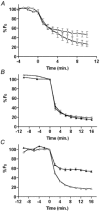
It has been speculated that neurosecretion can be enhanced by increasing the motion, and hence, the availability of cytoplasmic secretory vesicles. However, facilitator-induced physical mobilization of secretory vesicles has not been observed directly in living cells, and recent experimental results call this hypothesis into question. Here, high resolution green fluorescent protein (GFP)-based measurements in nerve growth factor-differentiated PC12 cells are used to test whether altering dense core vesicle (DCV) motion affects neuropeptide release. Experiments with mycalolide B and jasplakinolide demonstrate that neuropeptidergic DCV motion at the ends of processes is proportional to F-actin. Furthermore, Ba2+ increases DCV mobility without detectably modifying F-actin. Finally, we show that altering DCV motion by changing F-actin or stimulating with Ba2+ proportionally changes sustained neuropeptide release. Therefore, increasing DCV mobility facilitates prolonged neuropeptide release.
Figures


Similar articles
-
Unexpected mobility variation among individual secretory vesicles produces an apparent refractory neuropeptide pool.Biophys J. 2003 Jun;84(6):4127-34. doi: 10.1016/S0006-3495(03)75137-6. Biophys J. 2003. PMID: 12770915 Free PMC article.
-
Mycalolide B dissociates dynactin and abolishes retrograde axonal transport of dense-core vesicles.Mol Biol Cell. 2015 Jul 15;26(14):2664-72. doi: 10.1091/mbc.E14-11-1564. Epub 2015 May 28. Mol Biol Cell. 2015. PMID: 26023088 Free PMC article.
-
Nerve growth factor-induced differentiation changes the cellular organization of regulated Peptide release by PC12 cells.J Neurosci. 2002 May 15;22(10):3890-7. doi: 10.1523/JNEUROSCI.22-10-03890.2002. J Neurosci. 2002. PMID: 12019308 Free PMC article.
-
The secretory response through electric stimulation of differentiated PC12 rat pheochromocytoma cells transfected with neuropeptide Y fused with enhanced green fluorescent protein.Biotechnol Lett. 2003 Apr;25(7):547-52. doi: 10.1023/a:1022894220755. Biotechnol Lett. 2003. PMID: 12882143
-
Sorting of neuropeptides and neuropeptide receptors into secretory pathways.Prog Neurobiol. 2010 Feb 9;90(2):276-83. doi: 10.1016/j.pneurobio.2009.10.011. Epub 2009 Oct 22. Prog Neurobiol. 2010. PMID: 19853638 Review.
Cited by
-
Astroglial excitability and gliotransmission: an appraisal of Ca2+ as a signalling route.ASN Neuro. 2012 Mar 22;4(2):e00080. doi: 10.1042/AN20110061. ASN Neuro. 2012. PMID: 22313347 Free PMC article. Review.
-
Unexpected mobility variation among individual secretory vesicles produces an apparent refractory neuropeptide pool.Biophys J. 2003 Jun;84(6):4127-34. doi: 10.1016/S0006-3495(03)75137-6. Biophys J. 2003. PMID: 12770915 Free PMC article.
-
Dynamics of peptidergic secretory granule transport are regulated by neuronal stimulation.BMC Neurosci. 2010 Mar 4;11:32. doi: 10.1186/1471-2202-11-32. BMC Neurosci. 2010. PMID: 20202202 Free PMC article.
-
The role of actin remodeling in the trafficking of intracellular vesicles, transporters, and channels: focusing on aquaporin-2.Pflugers Arch. 2008 Jul;456(4):737-45. doi: 10.1007/s00424-007-0404-2. Epub 2007 Dec 8. Pflugers Arch. 2008. PMID: 18066585 Review.
-
Mycalolide B dissociates dynactin and abolishes retrograde axonal transport of dense-core vesicles.Mol Biol Cell. 2015 Jul 15;26(14):2664-72. doi: 10.1091/mbc.E14-11-1564. Epub 2015 May 28. Mol Biol Cell. 2015. PMID: 26023088 Free PMC article.
References
-
- Bubb MR, Senderowicz AM, Sausville EA, Duncan KL, Korn ED. Jasplakinolide, a cytotoxic natural product, induces actin polymerization and competitively inhibits the binding of phalloidin to F-actin. Journal of Biological Chemistry. 1994;269:14869–14871. - PubMed
-
- Burke N, Han W, Li D, Takimoto K, Watkins SC, Levitan ES. Neuronal peptide release is limited by secretory granule mobility. Neuron. 1997;19:1095–1102. - PubMed
Publication types
MeSH terms
Substances
Grants and funding
LinkOut - more resources
Full Text Sources
Miscellaneous

