. 2002 Jul 15;542(Pt 2):453-76.
doi: 10.1113/jphysiol.2002.018630.

Stimulation-dependent regulation of the pH, volume and quantal size of bovine and rodent secretory vesicles

Affiliations

Stimulation-dependent regulation of the pH, volume and quantal size of bovine and rodent secretory vesicles

Emmanuel N Pothos et al. J Physiol. .

Abstract

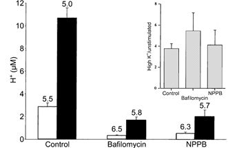
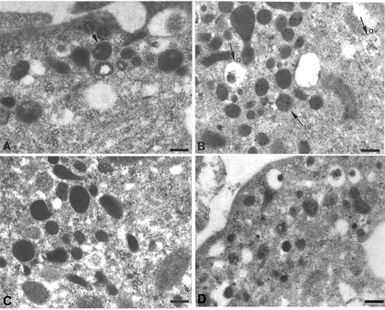
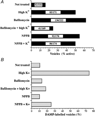
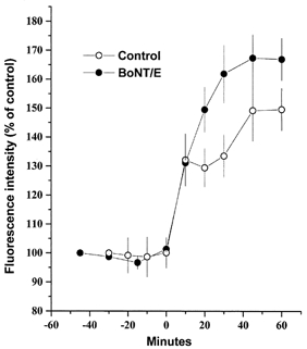
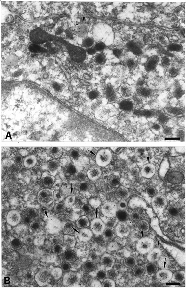
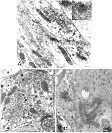
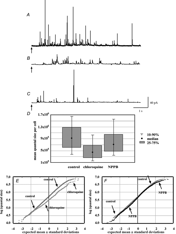
Trapping of weak bases was utilized to evaluate stimulus-induced changes in the internal pH of the secretory vesicles of chromaffin cells and enteric neurons. The internal acidity of chromaffin vesicles was increased by the nicotinic agonist 1,1-dimethyl-4-phenyl-piperazinium iodide (DMPP; in vivo and in vitro) and by high K+ (in vitro); and in enteric nerve terminals by exposure to veratridine or a plasmalemmal [Ca2+]o receptor agonist (Gd3+). Stimulation-induced acidification of chromaffin vesicles was [Ca2+]o-dependent and blocked by agents that inhibit the vacuolar proton pump (vH+-ATPase) or flux through Cl- channels. Stimulation also increased the average volume of chromaffin vesicles and the proportion that displayed a clear halo around their dense cores (called active vesicles). Stimulation-induced increases in internal acidity and size were greatest in active vesicles. Stimulation of chromaffin cells in the presence of a plasma membrane marker revealed that membrane was internalized in endosomes but not in chromaffin vesicles. The stable expression of botulinum toxin E to prevent exocytosis did not affect the stimulation-induced acidification of the secretory vesicles of mouse neuroblastoma Neuro2A cells. Stimulation-induced acidification thus occurs independently of exocytosis. The quantal size of secreted catecholamines, measured by amperometry in cultured chromaffin cells, was found to be increased either by prior exposure to L-DOPA or stimulation by high K+, and decreased by inhibition of vH+-ATPase or flux through Cl- channels. These observations are consistent with the hypothesis that the content of releasable small molecules in secretory vesicles is increased when the driving force for their uptake is enhanced, either by increasing the transmembrane concentration or pH gradients.


Figures

Figure 4
Figure 4
Mean calculated dense-core vesicle H+ concentration The bars represent [H+ ] (mean ± s.e.m.; μm) determined from immunogold labelling of vesicles to detect DAMP, as reported in Fig. 2 and Table 1. The corresponding pH values (for which s.e.m.s are inappropriate; see Methods) for each unstimulated (□) and stimulated (▪) pair are written above the bars. While bafilomycin and NPPB produce more alkaline vesicles, for each pair the stimulated preparations display greater acidity. The inset shows the same data as a ratio of H+ concentrations in resting and high-K+-treated cells in the absence and in the presence of bafilomycin or NPPB.
Figure 1
Figure 1
Acidification of intracellular organelles in rat chromaffin cells exposed to high K+ or DMPP Acidification of intracellular sites as observed by acridine orange accumulation was apparent after exposure to the 50 mm K+ (A, before; B, 40 min after 50 mm K+). Scale bar: 10 μm. C, mean increase in fluorescence (± s.e.m.) for cytoplasmic regions of interest in 16 cells in 16 separate micrographs. D, increase in punctate fluorescence emission over time for a single chromaffin cell. E, the increased fluorescence due to exposure to 10 μm DMPP was blocked by the vH+-ATPase inhibitor bafilomycin (500 nm), in Ca2+-free medium, and by the Cl channel inhibitor NPPB (30 μm; mean ± s.e.m. of controls, >10 regions of interest per condition).
Figure 2
Figure 2
Effects of bafilomycin and NPPB on K+-induced changes in the acidification and morphology of vesicles in cultured chromaffin cells The acidity of vesicles in rat chromaffin cultures was evaluated by using immunogold labelling to detect the trapping of the weak base DAMP. A, control. A number of dense-cored vesicles can be seen in the field. None has become labelled by incubation with DAMP. Note that most of the vesicles lack electron-lucent halos and are thus stable vesicles (sv). B, 50 mm K+ for 40 min. Several of the dense-cored vesicles in the field have trapped DAMP. Some of these vesicles possess halos around the dense cores and are thus active vesicles (av). Note that the dense cores of the active vesicles tend to be less electron-dense than those of neighbouring stable vesicles. C, 50 mm K+ plus bafilomycin (500 nm for 40 min). The K+-induced trapping of DAMP within dense-cored vesicles is abolished. Almost all vesicles are stable in morphology. D, 50 mm K+ plus NPPB (30 μm) for 40 min. The K+-induced trapping of DAMP within dense-cored vesicle is again prevented. Many active vesicles, however, can be seen in the field. Scale bars: 200 nm.
Figure 3
Figure 3
Fraction of active and DAMP-labelled dense-cored chromaffin vesicles The percentage of active vesicles (A) and the percentage of DAMP-labelled vesicles (B) for each condition are indicated. Cultured bovine chromaffin cells were exposed to high K+ in the absence or presence of inhibitors of vH+-ATPase (bafilomycin) or flux through chloride channels (NPPB). The numbers of dense-cored vesicles assayed in each condition are indicated within the bars in A, as number of active vesicles/total number of vesicles. The data are non-parametric.
Figure 5
Figure 5
Cationic ferritin and DAMP colocalization in cultured chromaffin cells A, non-stimulated cells. Cationic ferritin, used as a tracer for endosomes, has a stippled, electron-dense appearance and is labelled with 10 nm diameter immunogold particles. DAMP, used to indicated acidic organelles, is immunolabelled with 5 nm immunogold particles. The plasma membrane (arrowheads) and examples of cationic ferritin-labelled endosomal structures (larger arrows and inset) are indicated. DAMP-containing active dense-core vesicles (small arrows) do not display cationic ferritin. B, in stimulated chromaffin cells, cationic ferritin also does not appear in active dense-cored vesicles (arrows as in A). Scale bar: 200 nm.
Figure 6
Figure 6
Effect of botulinum toxin E expression on stimulation-dependent vesicle acidification Stimulation-dependent acidification of intracellular sites in native Neuro2A cells (○) and cells expressing BoNT/E-LC (•), as observed by acridine orange accumulation after exposure to 50 mm K+ (added at time = 0 min). Each data point represents n = 3 experiments (± s.e.m.), with >30 cells imaged per experiment.
Figure 7
Figure 7
Effects of the nicotinic receptor agonist DMPP on the acidification and morphology of vesicles of chromaffin cells in vivo A, vehicle-injected control. Both active and stable dense-cored vesicles are present. Although few are labelled with DAMP, a highly labelled lysosome (L) is present. The trapping of DAMP within the lysosome indicates that the injected DAMP has entered the chromaffin cell and that its distribution reflects the ΔH+ gradient across the membrane of this organelle. B, DMPP treated. More chromaffin vesicles exhibit an active morphology (arrows) with halo around the dense cores. Note also that many of the vesicles are now labelled with DAMP (see also Table 3). Scale bars: 200 nm.
Figure 8
Figure 8
Effects of stimulation on the acidification of small and large synaptic vesicles of rat myenteric neurons A, in control preparations of rat myenteric neurons in which spontaneous activity of neurons was blocked by the addition of tetrodotoxin, synaptic vesicles contained very little DAMP. In contrast, lysosomes, which are abundant in the enteric perikarya, were highly labelled (inset). B, stimulation of myenteric neurons with Ca2+ receptor agonist, Gd3+ (500 μm for 30 min) in the presence of DAMP. Arrows indicate small and large synaptic vesicles labelled with DAMP. C, rat myenteric neurons were stimulated with the Na+ channel activator veratridine (1 μm for 30 min) in the presence of DAMP. Both small clear and large dense-cored synaptic vesicles were labelled (arrows). Scale bars: 100 nm.
Figure 9
Figure 9
Effects of high-K+ stimulation and bafilomycin on the size of quanta recorded from bovine chromaffin cells Sample amperometric recordings from control cells (A), cells stimulated to release with high K+ (B; 50 mm for 40 min) and bafilomycin-exposed cells (C; 0.5 μm for 40 min). The arrows indicate application of a 6 s 80 mm K+ puff as a secretagogue. D, box-and-whiskers plot of the distribution of the median quantal size from each cell recorded. Each distribution is significantly different from the other two (see text). E and F, normal probability distributions of the population of logs of each of the quantal size data points from each group. Histograms of the untransformed distribution of quantal sizes resulting from each treatment are shown as supplemental material.
Figure 10
Figure 10
Effects of chloroquine and NPPB on the size of quanta recorded from rat chromaffin cells Sample amperometric recordings from control cells (A), cells treated with chloroquine (B; 100 μm for 30 min) and NPPB-exposed cells (C; 30 μm for 30 min). D, box-and-whiskers plot of the distribution of the median quantal size recorded from each cell recorded. Each distribution is significantly different from the other two (see text). E and F, normal probability distributions of the population of logs of each of the quantal size data points from each group. Histograms of the untransformed distribution of quantal sizes resulting from each treatment are shown as supplemental material.

References

    1. Aguado F, Gombau L, Majo G, Marsal J, Blanco J, Blasi J. Regulated secretion is impaired in AtT-20 endocrine cells stably transfected with botulinum neurotoxin type A light chain. Journal of Biological Chemistry. 1997;272:26005–26008. - PubMed
    1. Anderson RGW, Falck JR, Goldstein JL, Brown MS. Visualization of acidic organelles in intact cells by electron microscopy. Proceeding of the National Academy of Sciences of the USA. 1984;81:4838–4842. - PMC - PubMed
    1. Anderson RGW, Pathak RK. Vesicles and cisternae in the trans Golgi apparatus of human fibroblasts are acidic compartments. Cell. 1985;40:635–643. - PubMed
    1. Aspinwall CA, Brooks SA, Kennedy RT, Lakey JR. Effects of intravesicular H+ and extracellular H+ and Zn2+ on insulin secretion in pancreatic beta cells. Journal of Biological Chemistry. 1997;272:31308–31314. - PubMed
    1. Bae H-R, Verkman AS. Protein kinase A regulates chloride conductance in endocytic vesicles from proximal tubule. Nature. 1990;348:637–639. - PubMed

Publication types

MeSH terms

LinkOut - more resources