

Break excitation alone does not explain the delay and amplitude of anodal current-induced vasodilatation in human skin
- PMID: 12122152
- PMCID: PMC2290427
- DOI: 10.1113/jphysiol.2002.022731
Break excitation alone does not explain the delay and amplitude of anodal current-induced vasodilatation in human skin
Abstract
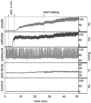
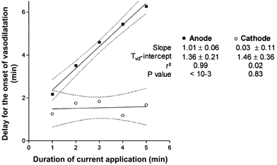
In iontophoresis experiments, a 'non-specific' current-induced vasodilatation interferes with the effects of the diffused drugs. This current-induced vasodilatation is assumed to rely on an axon reflex due to excitation of cutaneous nociceptors and is weaker and delayed at the anode as compared to the cathode. We analysed whether these anodal specificities could result from a break excitation of nociceptors. Break excitation is the generation of action potentials at the end of a square anodal DC current application, which are generally weaker than those observed at the onset of a same application at the cathode. In eight healthy volunteers, we studied forearm cutaneous laser Doppler flow (LDF) responses to: (1) anodal and cathodal 100 microA current applications of 1, 2, 3, 4 or 5 min; (2) 100 microA anodal applications of 3 min with a progressive ending over 100 s (total charge 23 mC); these were compared to square-ended 100 microA anodal applications of the same total charge (23 mC) or duration (3 min); (3) a 4 min 100 microA anodal application with a 333 msec break at half time. Results (mean +/- S.D.) are expressed as percentage of heat-induced maximal vasodilatation (%MVD). Onset (T(vd)) and amplitude (LDF(peak)) of vasodilatation were determined. We observed that: T(vd) was linearly related to the duration of current application at the anode (slope = 1.01, r(2) = 0.99, P < 0.0001) but not at the cathode (slope = 0.03, r(2) = 0.02, n.s.). Progressive ending of anodal current did not decrease LDF(peak) (63.3 +/- 24.6 %MVD) as compared to square-ending of current application of the same duration (36.9 +/- 22.2 %MVD) or the same total charge (57.1 +/- 23.5 %MVD). A transient break of anodal current did not allow for the vasodilatation to develop until current was permanently stopped. We conclude that, during iontophoresis, anodal break excitation alone cannot account for the delay and amplitude of the vascular response.
Figures
Similar articles
-
Anodal current intensities above 40 microA interfere with current-induced axon-reflex vasodilatation in human skin.J Vasc Res. 2004 May-Jun;41(3):261-7. doi: 10.1159/000078665. Epub 2004 May 19. J Vasc Res. 2004. PMID: 15153776
-
Vasodilatation in response to repeated anodal current application in the human skin relies on aspirin-sensitive mechanisms.J Physiol. 2002 Apr 1;540(Pt 1):261-9. doi: 10.1113/jphysiol.2001.013364. J Physiol. 2002. PMID: 11927685 Free PMC article.
-
Early vasodilator response to anodal current application in human is not impaired by cyclooxygenase-2 blockade.Am J Physiol Heart Circ Physiol. 2005 Apr;288(4):H1668-73. doi: 10.1152/ajpheart.00415.2004. Epub 2004 Nov 24. Am J Physiol Heart Circ Physiol. 2005. PMID: 15563538 Clinical Trial.
-
Cathodal current-induced vasodilation to single application and the amplified response to repeated application in humans rely on aspirin-sensitive mechanisms.J Appl Physiol (1985). 2005 Oct;99(4):1538-44. doi: 10.1152/japplphysiol.00258.2005. Epub 2005 Jun 23. J Appl Physiol (1985). 2005. PMID: 15976365
-
Current-induced vasodilation during water iontophoresis (5 min, 0.10 mA) is delayed from current onset and involves aspirin sensitive mechanisms.J Vasc Res. 2002 Jan-Feb;39(1):59-71. doi: 10.1159/000048994. J Vasc Res. 2002. PMID: 11844938
Cited by
-
Oral single high-dose aspirin results in a long-lived inhibition of anodal current-induced vasodilatation.Br J Pharmacol. 2002 Oct;137(3):384-90. doi: 10.1038/sj.bjp.0704868. Br J Pharmacol. 2002. PMID: 12237259 Free PMC article.
-
Fundamentals of transcranial electric and magnetic stimulation dose: definition, selection, and reporting practices.Brain Stimul. 2012 Oct;5(4):435-53. doi: 10.1016/j.brs.2011.10.001. Epub 2011 Nov 1. Brain Stimul. 2012. PMID: 22305345 Free PMC article. Review.
References
-
- Asberg A, Holm T, Vassbotn T, Andreassen AK, Hartmann A. Nonspecific microvascular vasodilation during iontophoresis is attenuated by application of hyperosmolar saline. Microvascular Research. 1999;58:41–48. - PubMed
-
- Berliner MN. Skin microcirculation during tapwater iontophoresis in humans: Cathode stimulates more than anode. Microvascular Research. 1997;54:74–80. - PubMed
Publication types
MeSH terms
LinkOut - more resources
Full Text Sources
Medical
Miscellaneous