. 2002 Jul 15;542(Pt 2):643-63.
doi: 10.1113/jphysiol.2001.015644.

Age-dependent variations in the directional sensitivity of balance corrections and compensatory arm movements in man

Affiliations

Age-dependent variations in the directional sensitivity of balance corrections and compensatory arm movements in man

J H J Allum et al. J Physiol. .

Abstract

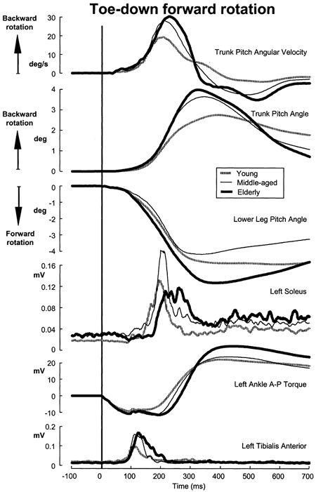
We investigated the effects of ageing on balance corrections induced by sudden stance perturbations in different directions. Effects were examined in biomechanical and electromyographic (EMG) recordings from a total of 36 healthy subjects divided equally into three age groups (20-34, 35-55 and 60-75 years old). Perturbations consisted of six combinations of support-surface roll (laterally) and pitch (forward-backward) each with 7.5 deg amplitude (2 pure pitch, and 4 roll and pitch) delivered randomly. To reduce stimulus predictability further and to investigate scaling effects, perturbations were at either 30 or 60 deg s(-1). In the legs, trunk and arms we observed age-related changes in balance corrections. The changes that appeared in the lower leg responses included smaller stretch reflexes in soleus and larger reflexes in tibialis anterior of the elderly compared with the young. For all perturbation directions, onsets of balance correcting responses in these ankle muscles were delayed by 20-30 ms and initially had smaller amplitudes (between 120-220 ms) in the elderly. This reduced early activity was compensated by increased lower leg activity after 240 ms. These EMG changes were paralleled by comparable differences in ankle torque responses, which were initially (after 160 ms) smaller in the elderly, but subsequently greater (after 280 ms). Findings in the middle-aged group were generally intermediate between the young and the elderly groups. Comparable results were obtained for the two different stimulus velocities. Stimulus-induced trunk roll, but not trunk pitch, changed dramatically with increasing age. Young subjects responded with early large roll movements of the trunk in the opposite direction to platform roll. A similarly directed but reduced amplitude of trunk roll was observed in the middle-aged. The elderly had very little initial roll modulation and also had smaller stretch reflexes in paraspinals. Balance-correcting responses (over 120-220 ms) in gluteus medius and paraspinals were equally well tuned to roll in the elderly, as in the young, but were reduced in amplitude. Onset latencies were delayed with age in gluteus medius muscles. Following the onset of trunk and hip balance corrections, trunk roll was in the same direction as support-surface motion for all age groups and resulted in overall trunk roll towards the fall side in the elderly, but not in the young. Protective arm movements also changed with age. Initial arm roll movements were largest in the young, smaller in the middle aged, and smallest in the elderly. Initial arm roll movements were in the same direction as initial trunk motion in the young and middle aged. Thus initial roll arm movements in the elderly were directed oppositely to those in the young. Initial pitch motion of the arms was similar across age groups. Subsequent arm movements were related to the amplitude of deltoid muscle responses which commenced at 100 ms in the young and 20-30 ms later in the elderly. These deltoid muscle responses preceded additional arm roll motion which left the arms directed 'downhill' (in the direction of the fall) in the elderly, but 'uphill' (to counterbalance motion of the pelvis) in the young. We conclude that increased trunk roll stiffness is a key biomechanical change with age. This interferes with early compensatory trunk movements and leads to trunk displacements in the direction of the impending fall. The reversal of protective arm movements in the elderly may reflect an adaptive strategy to cushion the fall. The uniform delay and amplitude reduction of balance-correcting responses across many segments (legs, hips and arms) suggests a neurally based alteration in processing times and response modulation with age. Interestingly, the elderly compensated for these 'early abnormalities' with enlarged later responses in the legs, but no similar adaptation was noted in the arms and trunk. These changes with age provide an insight into possible mechanisms underlying falls in the elderly.


Figures

Figure 1
Figure 1
Schematic diagram of the perturbation directions The angular notation indicates the direction of the stimulus for the polar plots of the other figures.
Figure 2
Figure 2
Population responses to a pure toe-up backward pitch rotation (7.5 deg at 60 deg s−1) of the support surface Each of the traces shown is the average of 12 subjects’ responses to seven randomized repetitions of the stimulus yielding a total of 84 responses to the average curve. The thick vertical line at 0 ms represents the onset of the support-surface rotation and is at the first inflexion of the A-P ankle torque trace. A positive deflection of the ankle torque trace represents increased ankle torque imposed on the support surface. The initial increase of ankle torque lasts ≈120 ms, the duration of the stimulus. The movement directions for lower leg pitch and trunk pitch are opposite as indicated in the figure. Note the decreased response amplitudes in soleus and delayed onsets of tibialis anterior in the elderly.
Figure 3
Figure 3
Population responses to a forward toe-down rotation of the support surface Details of the responses are provided in the legend to Fig. 2. Note the increased early response in tibialis anterior and the delayed decreased response initially in soleus for the elderly compared with the young.
Figure 4
Figure 4
Polar plots for areas of tibialis anterior and soleus EMG responses averaged over two time intervals consisting of balance-correcting (120–220 ms) and secondary balance-correcting (240-340 ms) reactions Each radial line represents one of the six directions of support-surface rotation (0, 45, 135, 180, 225 and 315 deg). For each direction, mean population values are plotted with amplitude represented as distance from the centre. The standard error of the mean (s.e.m.) has been added to the mean value of the young. The response amplitude is scaled according to the different vertical scales between the set of plots for the two recording sites. Note the slightly off-pitch plane orientation of soleus responses and the smaller, compared with the young, early responses but larger, later response amplitudes in the elderly.
Figure 5
Figure 5
Polar plots for A-P ankle torque changes between 160–260 and 280–380 ms for the left leg The upper plots are arranged similarly to those of Fig. 4. The two lower plots indicate the vector direction of torque obtained when the lateral torque is taken into account. The resulting torque vector is always oriented just off the pitch direction for each direction and age group except for 45 deg due to a small, sign-reversed, lateral torque for the elderly. Note smaller initial torque amplitude in the left polar plot, but later larger torque for the elderly compared with the young.
Figure 7
Figure 7
Polar plots of trunk angular velocity with corresponding vector plots The plotted amplitude of roll velocity is the average between 90–130 ms, and for pitch velocity the average between 180–220 ms. The vector directions of trunk angular velocity at these two times are indicated by the diagrams below to the left and right of the respective polar plots. Note the differences in roll velocities between the two populations and the effect this has on the early direction of trunk roll movement.
Figure 6
Figure 6
Trunk muscle and velocity population responses to a backward left rotation of 60 deg s−1 Details of the responses have been provided in the legend to Fig. 2. The trunk roll angle traces were obtained by numerically integrating the trunk angular velocity recordings. Note the negligible initial trunk roll velocity in the direction opposite to the support surface tilt and the final trunk roll displacement in the same direction as the tilt in the elderly.
Figure 8
Figure 8
Trunk mean angular changes from stimulus onset to 700 ms later when trunk velocities have stabilized Mean and standard errors of the mean are shown for each age group. Trunk roll and pitch angles are shown. The direction of support-surface rotation causing the angle changes is indicated by the plot abscissa, leftward directions on the left, rightward on the right. Note the opposite directions of trunk roll changes between the young and elderly populations, but similar directions of trunk pitch rotations.
Figure 9
Figure 9
Polar plots for right paraspinals and left gluteus medius mean EMG response areas over the 120–220 ms interval for each direction The format of the polar plots is identical to that of Fig. 4. Note how the maximum activity directions are the same for right paraspinals and left gluteus medius. These are oriented towards backwards roll. For the backwards roll direction the response amplitudes of the young are clearly larger than those of the elderly.
Figure 11
Figure 11
A, polar plots for right deltoid medius mean EMG response areas over the intervals 120–220 and 350–700 ms. B, left arm angular displacements with respect to the trunk. The format of the polar plots is identical to that of Figs 4 and 9. Note how the maximum activity is clearly oriented towards back left in the young over the interval 120–220 ms but not in the elderly. In the lower part of the plot angular changes of the left arm relative to the trunk from stimulus onset to 700 ms later are shown for all roll stimuli. The format of the plot is similar to that of Fig. 8. Note the different directions of arm roll between the young and elderly.
Figure 10
Figure 10
Arm muscle and velocity population responses to a backward left rotation of 60 deg s−1 The relative angular velocity of the arm shown in the roll direction was computed from the difference of the measured angular velocities of the left upper arm and that of the trunk. Left roll angular position of the arm is the numerical integral of this trace. The relative pitch angle of the arm was obtained using the same technique as for roll. Note the different directions of the roll arm movements and the delayed and smaller deltoid responses for the elderly compared with the young.
Figure 12
Figure 12
Schematic illustration of body segment movements for leftwards support surface roll tilt as viewed from behind young and elderly subjects The arrows on the body segments indicate the direction roll velocity at ca 100 ms after stimulus onset.

References

    1. Accornero N, Capozza M, Rinalduzzi S, Manfredi GW. Clinical multisegmental posturography: age-related changes in stance control. Electroencephalography and Clinical Neurophysiology. 1997;105:213–219. - PubMed
    1. Adkin AL, Frank JS, Peysar GW, Carpenter MG. Fear of falling modifies anticipatory postural control. Experimental Brain Research. 2002;143:160–170. - PubMed
    1. Alexander NB, Shepard N, Gu MJ, Schultz A. Postural control in young and elderly adults when stance is perturbed: Kinematics. Journal of Gerontology. 1992;47:M79. - PubMed
    1. Allum JHJ, Honegger F, Schicks H. Vestibular and proprioceptive modulation of postural synergies in normal subjects. Journal of Vestibular Research. 1993;3:59–85. - PubMed
    1. Allum JHJ, Mauritz K-H. Compensation for intrinsic muscle stiffness by short-latency reflexes in human triceps surae. Journal of Neurophysiology. 1984;52:797–818. - PubMed

Publication types

LinkOut - more resources