. 2002 Aug 6;99(16):10813-8.
doi: 10.1073/pnas.152339799. Epub 2002 Jul 16.

alpha -Synucleinopathy and selective dopaminergic neuron loss in a rat lentiviral-based model of Parkinson's disease

Affiliations

alpha -Synucleinopathy and selective dopaminergic neuron loss in a rat lentiviral-based model of Parkinson's disease

C Lo Bianco et al. Proc Natl Acad Sci U S A. .

Abstract

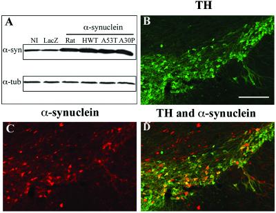
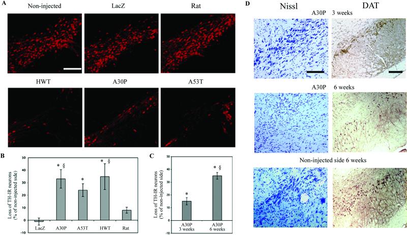
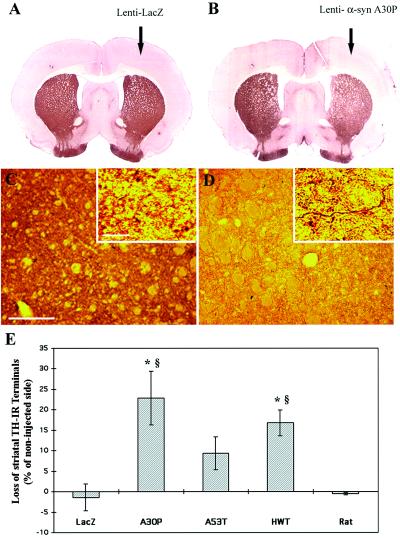
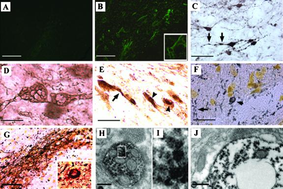
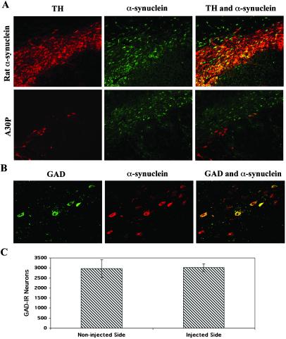
Parkinson's disease (PD) is characterized by the progressive loss of substantia nigra dopaminergic neurons and the presence of cytoplasmic inclusions named Lewy bodies. Two missense mutations of the alpha-synuclein (alpha-syn; A30P and A53T) have been described in several families with an autosomal dominant form of PD. alpha-Syn also constitutes one of the main components of Lewy bodies in sporadic cases of PD. To develop an animal model of PD, lentiviral vectors expressing different human or rat forms of alpha-syn were injected into the substantia nigra of rats. In contrast to transgenic mice models, a selective loss of nigral dopaminergic neurons associated with a dopaminergic denervation of the striatum was observed in animals expressing either wild-type or mutant forms of human alpha-syn. This neuronal degeneration correlates with the appearance of abundant alpha-syn-positive inclusions and extensive neuritic pathology detected with both alpha-syn and silver staining. Lentiviral-mediated expression of wild-type or mutated forms of human alpha-syn recapitulates the essential neuropathological features of PD. Rat alpha-syn similarly leads to protein aggregation but without cell loss, suggesting that inclusions are not the primary cause of cell degeneration in PD. Viral-mediated genetic models may contribute to elucidate the mechanism of alpha-syn-induced cell death and allow the screening of candidate therapeutic molecules.


Figures

Fig 1.
Fig 1.
Lentiviral-mediated overexpression of α-syn. (A) Western blot analysis shows the overexpression of normal and mutated (A53T and A30P) human and rat α-syn in the SH-SY5Y human neuroblastoma cell line. All α-syn forms are expressed at similar levels for the same amount of viral particles. Protein (25 μg per lane) were loaded for the noninfected cells (NI) and cells transduced with lentiviral vectors encoding for cytoplasmic LacZ, rat α-syn, wild-type (HWT), and mutated forms of human α-syn. The 19-kDa α-syn bands (α-syn) were detected with a polyclonal rabbit Ab generated against the 101- to 124-aa sequence of human α-syn. This Ab recognizes both human and rat α-syn on Western blot. The amount of protein loaded was checked by reprobing the same membrane with an α-tubulin Ab (α-tub). (BD) Lentiviral vectors encoding for wild-type and mutated human α-syn were stereotactically injected in the substantia nigra of rats. The nigral dopaminergic neurons were specifically labeled with a TH Ab (B). Detection with an α-syn polyclonal Ab revealed a significant overexpression of A30P α-syn (C) in the injected hemisphere. No α-syn staining was observed on the contralateral side. Double staining (D, yellow-orange color) shows a large proportion of TH-IR neurons overexpressing α-syn. (Scale bars = 200 μm.)
Fig 2.
Fig 2.
Dopaminergic-specific marker (TH, DAT) expression in the substantia nigra of rats injected with lentiviral vectors encoding for wild-type (HWT) or mutated (A30P, A53T) human and wild-type rat α-syn. (A) Overexpression of normal or mutant human α-syn significantly decreases the number of TH-IR neurons. No significant loss of TH expression was observed with the lentivirus encoding for rat α-syn or the reporter protein β-galactosidase (LacZ). (B) Histograms representing the loss of TH-IR nigral neurons at 5 mo relative to the contralateral side in rats unilaterally injected with the different lentiviral constructs. (C) The loss of dopaminergic neurons was quantified at 3 and 6 weeks for the A30P mutant and compared to Lenti-LacZ- or Lenti-rat α-syn-injected animals at 5 mo. The A30P lentiviral-mediated lesion represents a time-dependant process occurring within 6 weeks. (D) Nissl and DAT staining for A30P expressing animals at 3 and 6 weeks confirming the loss of dopaminergic cells over time. Values refer to means ± SEM; n = five animals per group; *, P < 0.005; §, P < 0.05; *, compared to lenti-lacZ-injected animals; §, compared to lenti-rat α-syn-injected animals at 5 mo. (Scale bars = 200 μm.)
Fig 3.
Fig 3.
Dopaminergic innervation in the striatum of rats expressing the different α-syn forms after 5 mo after lentiviral injection. (A and B) Striatal sections from rats that received intra-nigral injection of either lenti-LacZ (A) or lenti-A30P (B) in the right side of the substantia nigra. Lenti-A30P-injected animals show a significant reduction of TH staining on the ipsilateral side of the striatum whereas lenti-LacZ has no effect. (C and D) Double staining for TH and silver in the striatum of an animal injected with lenti-LacZ (C) or lenti-A30P (D). Higher magnification reveals that the reduction for TH staining in A30P-injected animals is associated with the presence of degenerating fibers containing silver deposits. (E) Quantification of the density of TH-IR terminals in the striatum of rats injected with the different α-syn forms. The histogram shows a significant reduction in TH-positive terminal density for the wild-type and A30P mutant α-syn forms. Values refer to means ± SEM; n = 5 animals per groups; *, P < 0.05; §, P < 0.01; *, compared to lenti-LacZ-injected animals; §, compared to lenti-rat α-syn-injected animals. [Scale bars = 200 μm (C and D) and 10 μm for higher magnification of (C and D).]
Fig 4.
Fig 4.
Neuropathology induced by lentiviral-mediated expression of α-syn. (A) No fluoroJade B labeled neurons were detected in both Lenti-LacZ-injected animals and noninjected side of animals expressing human α-syn. (B) Scattered FluoroJade B-positive neurons detected in the substantia nigra of rats expressing A30P α-syn. Higher magnification shows a degenerating neuron with FluoroJade B labeling in their perikaria and neurites. (C and D) α-Syn staining revealed that transduced neurons develop dystrophic neurites (C, arrows) and abnormal α-syn immunopositive swollen structures (D) with axonal and cell body distribution. (E) Nigral neurons abnormally accumulate cytoplasmic α-syn-immunopositive aggregates in cell bodies and neurites in rats overexpressing A30P α-syn. To confirm the presence of inclusions rather than a particular cellular sublocalization of α-syn, a silver staining (dark deposits) was performed alone (F) or in combination with α-syn staining (red-brown color) (G). Cytoplasmic inclusions (arrowheads) and extensive neuritic pathology (arrows) are detected with both methods. Higher magnification of a double-stained aggregate reveals that the inclusions contain abundant α-syn. (HJ) Ultrastructural analysis of A30P α-syn expressing nigral neurons. Abundant α-syn immunoreactive aggregation was detected in both the axon (H and I) and cell body (J) of nigral neurons expressing human α-syn. These cytoplasmic structures are mainly present as scattered granular microaggregates, which are occasionally associated with mitochondria or synaptic vesicles (H) and nuclear or cytoplasmic membrane (J). A degenerating cell with clustered aggregates in its perikaria reveals an important loss of organelles in its cytoplasm and a disorganized nuclear membrane (J). The size of the cell body and the presence of a varicosity full of synaptic vesicules observed (on the top) in close apposition with α-syn-positive structures support that this degenerating cell is a neuron. [Scale bars = 50 μm (A, B, and G); 25 μm (C, E, and F); 8 μm (D); and 0.3 μm (H and J).]
Fig 5.
Fig 5.
Specificity of the lesion induced by human A30P α-syn expression. (A) Double staining for TH (red) and α-syn (green) in rat α-syn or A30P expressing animals at 5 mo after lentiviral injection. TH-IR nigral neurons still overexpress rat α-syn (yellow-orange) whereas a significant loss of TH-IR neurons is observed in animals overexpressing the A30P human mutant. However, nondopaminergic neurons overexpressing human α-syn (green) survived inside the lesioned area of the substantia nigra. (B) GABAergic neurons are preserved in rats expressing A30P mutant α-syn. Double staining showing spared GAD-positive neurons (green) transduced and expressing high level of A30P α-syn (red). GABAergic neurons expressing human α-syn appear yellow-orange. (C) The number of GAD-positive neurons in the substantia nigra was determined for the noninjected and lenti-A30P-injected side. Overexpression of A30P α-syn does not induce a significant loss of GABAergic neurons. Values refer to means ± SEM. [Scale bars = 200 μm (A) and 50 μm (B).]

Similar articles

Cited by

References

    1. Polymeropoulos M. H., Lavedan, C., Leroy, E., Ide, S. E., Dehejia, A., Dutra, A., Pike, B., Root, H., Rubenstein, J., Boyer, R., et al. (1997) Science 276, 2045-2047. - PubMed
    1. Kruger R., Kuhn, W., Muller, T., Woitalla, D., Graeber, M., Kosel, S., Przuntek, H., Epplen, J. T., Schols, L. & Riess, O. (1998) Nat. Genet. 18, 106-108. - PubMed
    1. Spillantini M. G., Schmidt, M. L., Lee, V. M., Trojanowski, J. Q., Jakes, R. & Goedert, M. (1997) Nature (London) 388, 839-840. - PubMed
    1. Spillantini M. G., Crowther, R. A., Jakes, R., Hasegawa, M. & Goedert, M. (1998) Proc. Natl. Acad. Sci. USA 95, 6469-6473. - PMC - PubMed
    1. Iwai A., Masliah, E., Yoshimoto, M., Ge, N., Flanagan, L., de Silva, H. A., Kittel, A. & Saitoh, T. (1995) Neuron 14, 467-475. - PubMed

Publication types

MeSH terms

LinkOut - more resources