Ventricular expression of natriuretic peptides in Npr1(-/-) mice with cardiac hypertrophy and fibrosis
- PMID: 12124219
- PMCID: PMC4321891
- DOI: 10.1152/ajpheart.00677.2001
Ventricular expression of natriuretic peptides in Npr1(-/-) mice with cardiac hypertrophy and fibrosis
Abstract
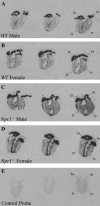
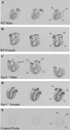
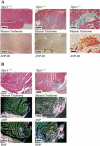
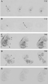
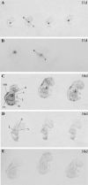
Atrial natriuretic peptide (ANP) and brain natriuretic peptide (BNP) are cardiac hormones that regulate blood pressure and volume, and exert their biological actions via the natriuretic peptide receptor-A gene (Npr1). Mice lacking Npr1 (Npr(-/-)) have marked cardiac hypertrophy and fibrosis disproportionate to their increased blood pressure. This study examined the relationships between ANP and BNP gene expression, immunoreactivity and fibrosis in cardiac tissue, circulating ANP levels, and ANP and BNP mRNA during embryogenesis in Npr1(-/-) mice. Disruption of the Npr1 signaling pathway resulted in augmented ANP and BNP gene and ANP protein expression in the cardiac ventricles, most pronounced for ANP mRNA in females [414 +/- 57 in Npr1(-/-) ng/mg and 124 +/- 25 ng/mg in wild-type (WT) by Taqman assay, P < 0.001]. This increased expression was highly correlated to the degree of cardiac hypertrophy and was localized to the left ventricle (LV) inner free wall and to areas of ventricular fibrosis. In contrast, plasma ANP was significantly greater than WT in male but not female Npr1(-/-) mice. Increased ANP and BNP gene expression was observed in Npr1(-/-) embryos from 16 days of gestation. Our study suggests that cardiac ventricular expression of ANP and BNP is more closely associated with local hypertrophy and fibrosis than either systemic blood pressure or circulating ANP levels.
Figures
Similar articles
-
Influence of natriuretic peptide receptor-1 on survival and cardiac hypertrophy during development.Biochim Biophys Acta. 2009 Dec;1792(12):1175-84. doi: 10.1016/j.bbadis.2009.09.009. Epub 2009 Sep 24. Biochim Biophys Acta. 2009. PMID: 19782130 Free PMC article.
-
Npr1-regulated gene pathways contributing to cardiac hypertrophy and fibrosis.J Mol Endocrinol. 2007 Feb;38(1-2):245-57. doi: 10.1677/jme.1.02138. J Mol Endocrinol. 2007. PMID: 17293444
-
Gestational hypertension in atrial natriuretic peptide knockout mice and the developmental origins of salt-sensitivity and cardiac hypertrophy.Regul Pept. 2013 Sep 10;186:108-15. doi: 10.1016/j.regpep.2013.08.006. Epub 2013 Aug 25. Regul Pept. 2013. PMID: 23981445
-
The role of natriuretic peptides in cardioprotection.Cardiovasc Res. 2006 Feb 1;69(2):318-28. doi: 10.1016/j.cardiores.2005.10.001. Epub 2005 Nov 10. Cardiovasc Res. 2006. PMID: 16289003 Review.
-
Minireview: natriuretic peptides during development of the fetal heart and circulation.Endocrinology. 2003 Jun;144(6):2191-4. doi: 10.1210/en.2003-0127. Endocrinology. 2003. PMID: 12746273 Review.
Cited by
-
Relaxin and atrial natriuretic peptide pathways participate in the anti-fibrotic effect of a melon concentrate in spontaneously hypertensive rats.Food Nutr Res. 2016 Apr 12;60:30985. doi: 10.3402/fnr.v60.30985. eCollection 2016. Food Nutr Res. 2016. PMID: 27079780 Free PMC article.
-
SGLT2 inhibitor dapagliflozin prevents atherosclerotic and cardiac complications in experimental type 1 diabetes.PLoS One. 2022 Feb 17;17(2):e0263285. doi: 10.1371/journal.pone.0263285. eCollection 2022. PLoS One. 2022. PMID: 35176041 Free PMC article.
-
C-type natriuretic peptide activates a non-selective cation current in acutely isolated rat cardiac fibroblasts via natriuretic peptide C receptor-mediated signalling.J Physiol. 2007 Apr 1;580(Pt 1):255-74. doi: 10.1113/jphysiol.2006.120832. Epub 2007 Jan 4. J Physiol. 2007. PMID: 17204501 Free PMC article.
-
Myocardial infarction-induced microRNA-enriched exosomes contribute to cardiac Nrf2 dysregulation in chronic heart failure.Am J Physiol Heart Circ Physiol. 2018 May 1;314(5):H928-H939. doi: 10.1152/ajpheart.00602.2017. Epub 2018 Jan 26. Am J Physiol Heart Circ Physiol. 2018. PMID: 29373037 Free PMC article.
-
Molecular Signaling Mechanisms and Function of Natriuretic Peptide Receptor-A in the Pathophysiology of Cardiovascular Homeostasis.Front Physiol. 2021 Aug 19;12:693099. doi: 10.3389/fphys.2021.693099. eCollection 2021. Front Physiol. 2021. PMID: 34489721 Free PMC article. Review.
References
-
- Cameron V, Rademaker M, Ellmers L, Espiner E, Nicholls M, Richards A. Atrial (ANP) and brain natriuretic peptide (BNP) expression after myocardial infarction in sheep: ANP is synthesized by fibroblasts infiltrating the infarct. Endocrinology. 2000;141:4690–4697. - PubMed
-
- Cameron VA, Aitken GD, Ellmers LJ, Kennedy MA, Espiner EA. The sites of gene expression of atrial, brain, and c-type natriuretic peptide in mouse fetal development: temporal changes in embryos and placenta. Endocrinology. 1996;137:817–824. - PubMed
-
- Cameron VA, Nishimura E, Mathews LS, Lewis KA, Sawchenko PE, Vale WW. Hybridization histochemical localization of activin receptor subtypes in rat brain, pituitary, ovary and testis. Endocrinology. 1994;134:799–808. - PubMed
-
- Cao L, Gardner D. Natriuretic peptides inhibit DNA synthesis in cardiac fibroblasts. Hypertension. 1995;25:227–234. - PubMed
Publication types
MeSH terms
Substances
Grants and funding
LinkOut - more resources
Full Text Sources
Medical
Molecular Biology Databases
Miscellaneous

