

doi: 10.1101/gad.978002.
Brn-1 and Brn-2 share crucial roles in the production and positioning of mouse neocortical neurons
Affiliations
- PMID: 12130536
- PMCID: PMC186401
- DOI: 10.1101/gad.978002
Item in Clipboard
Brn-1 and Brn-2 share crucial roles in the production and positioning of mouse neocortical neurons
Genes Dev.
.
Abstract
Formation of highly organized neocortical structure depends on the production and correct placement of the appropriate number and types of neurons. POU homeodomain proteins Brn-1 and Brn-2 are coexpressed in the developing neocortex, both in the late precursor cells and in the migrating neurons. Here we show that double disruption of both Brn-1 and Brn-2 genes in mice leads to abnormal formation of the neocortex with dramatically reduced production of layer IV-II neurons and defective migration of neurons unable to express mDab1. These data indicate that Brn-1 and Brn-2 share roles in the production and positioning of neocortical neuron development.
Figures
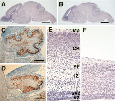
Morphological alterations in Brn-1/Brn-2 double mutant P0 brains. Sagittal sections of whole brain (HE stain) (A,B), cerebellum (brown, anti-Calbindin; blue, hematoxylin) (C,D), and neocortex (HE stain)(E,F) of wild-type (A,C,E) and Brn-1/Brn-2 double mutant (B,D,F) mice. Normally laminated structure consisting of the ventricular zone (VZ), subventricular zone (SVZ), intermediate zone (IZ), subplate (SP), cortical plate (CP), and marginal zone (MZ) is observed in wild-type neocortex (E), whereas, in Brn-1/Brn-2 mutant neocortex, the overall thickness is markedly reduced and the IZ is not clearly distinguishable from the CP (F). Scale bar: (A,B) 1 mm, (C,D) 200 μm, (E,F) 100 μm.
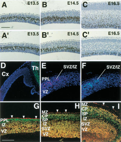
Reduced cell proliferation in Brn-1/Brn-2 mutant neocortex and expression of Brn-1 and Brn-2 in developing neocortex. BrdU labeling (brown) in sagittal sections of wild (A–C) and Brn-1/Brn-2 mutant (A‘–C‘) neocortex at indicated stages. Fluorescent micrographs of sagittal sections of E12.5 (D), E13.5 (E,F), E14.5 (G), E15.5 (H), and E16.5 (I) cortices doubly stained with anti-Brn-1 (red) and anti-Brn-2 (green). Cells expressing both Brn-1 and Brn-2 appear yellow (G–I). All nuclei were stained with DAPI (blue) in D–F. Arrowheads indicate background staining by secondary antibodies in pia matter (G–I). From E13.5, Brn-1-expressing and Brn-2-expressing cells are clearly seen outside of the VZ in rostral and lateral cortex (E,F). From E14.5, Brn-1/Brn-2 coexpression also becomes prominent in the VZ/SVZ as well as in the IZ and the CP (G–I). Brn-1 or Brn-2 singularly expressing cells are also found within the MZ (H,I) or in the PPL (G) and the presumptive SP (H,I), respectively. (Cx) Neocortex, (Th) thalamus, (PPL) preplate. For other abbreviations, see Fig. 1. Scale bar: (A–C,A‘–C‘) 100 μm, (D–I) 200 μm.
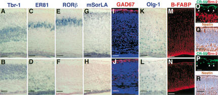
Loss of upper-layer neurons and altered positioning of cortical neurons in Brn-1/Brn-2 mutant neocortex. In situ hybridization using Tbr-1 (A,B), ER81 (C,D), RORβ (E,F), mSorLA (G,H), Olg-1 (K,L) riboprobes on coronal sections of E19.0 wild-type (A,C,E,G,K) and Brn-1/Brn-2 mutant (B,D,F,H,L) cortices. In wild-type cortex, Tbr-1-positive layer VI, ER81-positive layer V, RORβ-positive layer IV, and mSorLA-positive layer II/III neurons are ordered from deep to superficial (A,C,E,G). In Brn-1/Brn-2 mutant cortex, however, the majority of the ER81-positive neurons are found beneath the Tbr-1-positive layer, with a few ER81-positive neurons detected in the superficial region within the cortical plate (B,D), and the numbers of layer IV or layer II/III neurons positive for RORβ or mSorLA are drastically reduced (F,H), although mSorLA expression is found in the SVZ with a similar pattern of Tbr-1 expression in the SVZ (H,B). Immunostaining against GAD67 (red) (I,J), B-FABP (red) (M,N), Reelin (green) and Brn-2 (red) (O,P), and Nestin (brown) (Q,R) on sagittal (M,N,Q,R) and coronal (I,J,O,P) sections of E19.0 (I,J), E18.5 (M–P) and E16.5 (Q,R) cortices of wild-type (I,M,O,Q) and Brn-1/Brn-2 mutant (J,N,P,R). Scale bar: (A–N) 100 μm, (O,P) 20 μm, (Q,R) 50 μm.
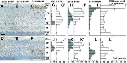
Abnormal migration of Brn-1/Brn-2 mutant neurons. (Left panels) Distribution of cells labeled with BrdU during E12.5 (A,D), E13.5 (B,E), or E14.5 (C,F) in E19.0 wild-type (A–C) and Brn-1/Brn-2 mutant (D–F) sagittal cortical sections. BrdU-positive nuclei (brown) were detected by immunohistochemistry. (Right panels) Bar graphs showing the radial distribution of heavily labeled cells (first generation at time of BrdU injection) (G–L) and lightly labeled cells (the majority of them are second and perhaps third generation cells from subsequent progenitor cell divisions) (G‘–L‘) in E19.0 wild-type (G–I,G‘–I‘) and Brn-1/Brn-2 mutant (J–L,J‘–L‘) neocortex. In Brn-1/Brn-2 mutants, most E14.5 BrdU-labeled cells (L,L‘) occupy the deepest positions. The subpopulation of E13.5 lightly labeled cells is also shifted to deeper positions (K‘). (2–4) Layer II–VI, (5) Layer V, (6) Layer VI. For other abbreviations, see Fig. 1. Scale bar: 80 μm.
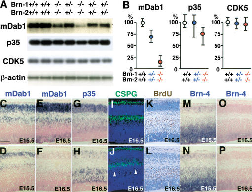
Reduced mDab1 expression level and congestion of migrating neurons just beneath the subplate in Brn-1/Brn-2 mutant cortex. (A) RT-PCR analysis for mDab1, p35, CDK5, and β-actin mRNA expression in the E16.5 dorsal cortex of wild-type (+/+), Brn-1/Brn-2 double heterozygotes (+/−), and Brn-1/Brn-2 double homozygotes (−/−), and (B) quantitation of their mRNA levels in Brn-1/Brn-2 double heterozygotes (n = 5) and Brn-1/Brn-2 double homozygotes (n = 5) relative to those of wild-type embryos (n = 4). In situ hybridization using mDab1 (C–F), p35 (G,H), and Brn-4 (M–P) riboprobes (blue) and immunostaining against CSPGs (green) (I,J) and BrdU labeled at E14.5 (brown) (K,L) on sagittal sections of E15.5 (C,D,M,N) and E16.5 (E–L,O,P) wild-type (C,E,G,I,K,M,O) and Brn-1/Brn-2 mutant (D,F, H,J,L,N,P) cortices. All nuclei were stained with DAPI (blue) in I and J. Although mDab1 expression is detected in wild-type and Brn-1/Brn-2 mutant cortices (C,D) at E15.5 when Brn-4 is expressed (M,N), it is hardly detectable in E16.5 mutant cortex (F) when and where Brn-4 expression is decreased (P). The p35-expressing neurons in Brn-1/Brn-2 mutant cortex are less abundant in the CP and more so in the IZ beneath the SP (H) compared with wild-type (G). Immunolabeling for CSPGs, which is intense in the MZ and the SP (I,J), shows proper splitting of the preplate into the MZ and SP and abnormal cell congestion just beneath the CSPG-positive SP (white arrowheads) in Brn-1/Brn-2 mutant cortex (J). In the wild-type cortex, the majority of E14.5-born neurons are found in the IZ, and some of them have already entered into the CP (K), whereas in Brn-1/Brn-2 mutant cortex, none of the E14.5-born cells are found in the CP, and all of them stay beneath the presumptive SP (L). Scale bar: (C–P) 100 μm.
References
-
- Anderson SA, Eisenstat DD, Shi L, Rubenstein JL. Interneuron migration from basal forebrain to neocortex: Dependence on Dlx genes. Science. 1997;278:474–476. - PubMed
-
- Angevine JB, Sidman RL. Autoradiographic study of cell migration during histogenesis of the cerebral cortex in the mouse. Nature. 1961;192:766–768. - PubMed
-
- Berman NE, Johnson JK, Klein RM. Early generation of glia in the intermediate zone of the developing cerebral cortex. Brain Res Dev Brain Res. 1997;101:149–164. - PubMed
-
- The Boulder Committee. Embryonic vertebrate central nervous system: Revised terminology. Anat Rec. 1970;166:257–261. - PubMed
MeSH terms
Substances
LinkOut - more resources
Full Text Sources
Other Literature Sources
Molecular Biology Databases