. 2002 Aug 1;169(3):1535-41.
doi: 10.4049/jimmunol.169.3.1535.

Activation of toll-like receptor 2 in acne triggers inflammatory cytokine responses

Affiliations

Activation of toll-like receptor 2 in acne triggers inflammatory cytokine responses

Jenny Kim et al. J Immunol. .

Abstract

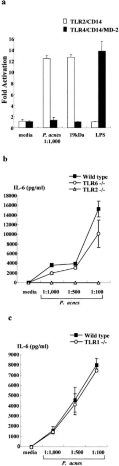
One of the factors that contributes to the pathogenesis of acne is Propionibacterium acnes; yet, the molecular mechanism by which P. acnes induces inflammation is not known. Recent studies have demonstrated that microbial agents trigger cytokine responses via Toll-like receptors (TLRs). We investigated whether TLR2 mediates P. acnes-induced cytokine production in acne. Transfection of TLR2 into a nonresponsive cell line was sufficient for NF-kappa B activation in response to P. acnes. In addition, peritoneal macrophages from wild-type, TLR6 knockout, and TLR1 knockout mice, but not TLR2 knockout mice, produced IL-6 in response to P. acnes. P. acnes also induced activation of IL-12 p40 promoter activity via TLR2. Furthermore, P. acnes induced IL-12 and IL-8 protein production by primary human monocytes and this cytokine production was inhibited by anti-TLR2 blocking Ab. Finally, in acne lesions, TLR2 was expressed on the cell surface of macrophages surrounding pilosebaceous follicles. These data suggest that P. acnes triggers inflammatory cytokine responses in acne by activation of TLR2. As such, TLR2 may provide a novel target for treatment of this common skin disease.


Figures

FIGURE 1
FIGURE 1
TLR2, but not TLR4, TLR6, or TLR1, is sufficient for induction by P. acnes of monocyte activation. a, HEK 293 cells transfected with TLR2, CD14, and the NF-κB responsive E-selectin (ELAM) enhancer luciferase reporter gene and BaF3 cells stably expressing TLR4, CD14, MD-2, and an ELAM luciferase reporter gene were activated with P. acnes, M. tuberculosis 19-kDa lipoprotein, or LPS for 6 h. NF-κB activity was measured by luciferase assay. Data reflect at least two independent experiments. b, Peritoneal macrophages were obtained from wild-type, TLR2−/−, and TLR6−/− mice, activated with P. acnes, and supernatants were assayed for the presence of IL-6. c, Peritoneal macrophages were obtained from wild-type and TLR1−/− mice, activated with P. acnes, and supernatants were assayed for the presence of IL-6.
FIGURE 2
FIGURE 2
P. acnes induces IL-12 p40 promoter activity via TLR2. RAW 264.7 cells were transiently transfected with a murine IL-12 p40 promoter CAT reporter. Cells were also cotransfected with TLR2 dn1 (■) or with a vector control ( formula image). Transfected cells were stimulated with P. acnes sonicate or M. tuberculosis 19-kDa lipoprotein, or left unstimulated (□) for 24 h. Activation of IL-12 p40 promoter activity was measured according to CAT activity (percent chloramphenicol acetylation) with a phosphor imager. Data reflects at least two independent experiments and are reported as a percentage of Ag-stimulated IL-12 p40 promoter activity cotransfected with a vector control. Media controls were comparable between vector control and TLR2 dn1 transfectants.
FIGURE 3
FIGURE 3
P. acnes induces IL-12 and IL-8 production in human adherent monocytes via TLR2. Human adherent monocytes were stimulated with P. acnes and the production of IL-12 p40 and IL-8 was determined by ELISA. A dose-response curve demonstrates the ability of P. acnes to stimulate monocytes to release IL-12 p40 (a) and IL-8 (b). Cells were coincubated with anti-TLR2, anti-TLR4, or isotype control mAbs. The IL-12 p40 (c) and IL-8 (d) release into the supernatants was determined by ELISA. The results from one representative experiment of three are shown.
FIGURE 4
FIGURE 4
TLR2 expression in acne lesions. Representative sections from skin biopsy specimens from two acne patients stained by the immunoperoxidase method with mAbs specific for TLR2, CD14, CD3, and CD1a. Multiple TLR2+ cells were detected primarily in the inflammatory infiltrate around the perifollicular/peribulbar region. Numerous CD14+ and CD3+ cells were detected in the similar area. CD1a+ cells were found within the follicular wall and in the epidermis. A total of 19 different lesions from acne patients were examined. Original magnification, ×40.
FIGURE 5
FIGURE 5
Kinetics of TLR2 expression in acne according to the evolution of the lesion. Representative sections from skin biopsy specimens of acne lesions stained by the immunoperoxidase method with mAbs specific for TLR2. In early acne lesions, up to 6 h, few rare TLR2+ cells were detected. In lesions obtained between 12 and 24 h, TLR2+ cells were much more numerous around the pilosebaceous follicles. In later older lesions obtained between 48 and 72 h, greater numbers of TLR2+ cells were detected.
FIGURE 6
FIGURE 6
Phenotype of cells expressing TLR2 in acne lesions. Two-color immunofluorescence confocal images were obtained for (a) CD14, (b) CD3 (green, first panel of each row), and TLR2 (red, second panel of each row). The two images were then superimposed (third panel of each row). Double-positive cells are shown in yellow. TLR2-positive cells near the follicular bulbar region colocalized with CD14+ macrophages but not with CD3+ T cells. Original magnification, ×630.

References

    1. Leyden JJ, McGinley KJ, Mills OH, Kligman AM. Propionibacterium levels in patients with and without acne vulgaris. J Invest Dermatol. 1975;65:382. - PubMed
    1. Kamisango K, Saiki I, Tanio Y, Okumura H, Araki Y, Sekikawa I, Azuma I, Yamamura Y. Structures and biological activities of peptidoglycans of Listeria monocytogenes and Propionibacterium acnes. J Biochem. 1982;92:23. - PubMed
    1. Vowels BR, Yang S, Leyden JJ. Induction of proinflammatory cytokines by a soluble factor of Propionibacterium acnes: implications for chronic inflammatory acne. Infect Immun. 1995;63:3158. - PMC - PubMed
    1. Hoeffler U. Enzymatic and hemolytic properties of Propionibacterium acnes and related bacteria. J Clin Microbiol. 1977;6:555. - PMC - PubMed
    1. Hoeffler U, Ko HL, Pulverer G. Antimicrobiol susceptibility of Propionibacterium acnes and related microbial species. Antimicrob Agents Che-mother. 1976;10:387. - PMC - PubMed

Publication types

MeSH terms