Comparative Study
. 2002 Aug;76(16):7932-41.
doi: 10.1128/jvi.76.16.7932-7941.2002.

Avian reoviruses cause apoptosis in cultured cells: viral uncoating, but not viral gene expression, is required for apoptosis induction

Affiliations
Comparative Study

Avian reoviruses cause apoptosis in cultured cells: viral uncoating, but not viral gene expression, is required for apoptosis induction

Lucía Labrada et al. J Virol. 2002 Aug.

Abstract

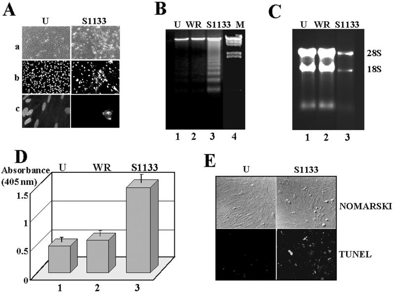
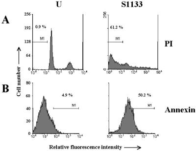
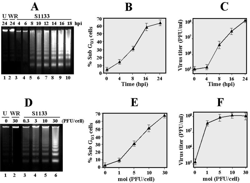
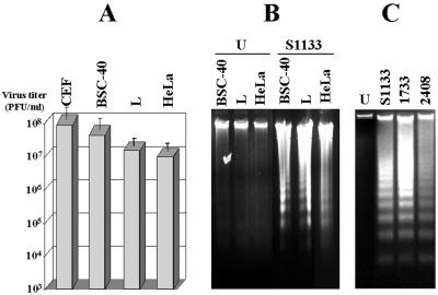
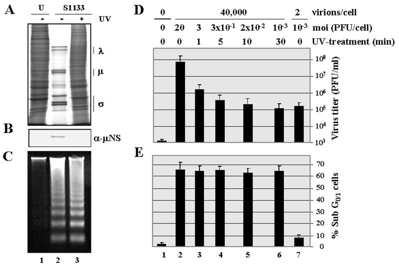
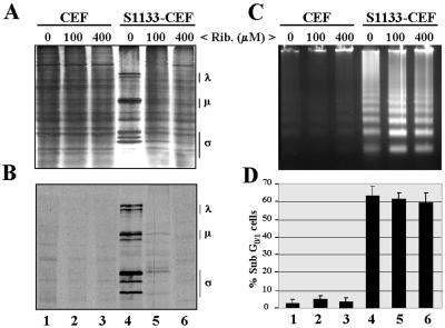
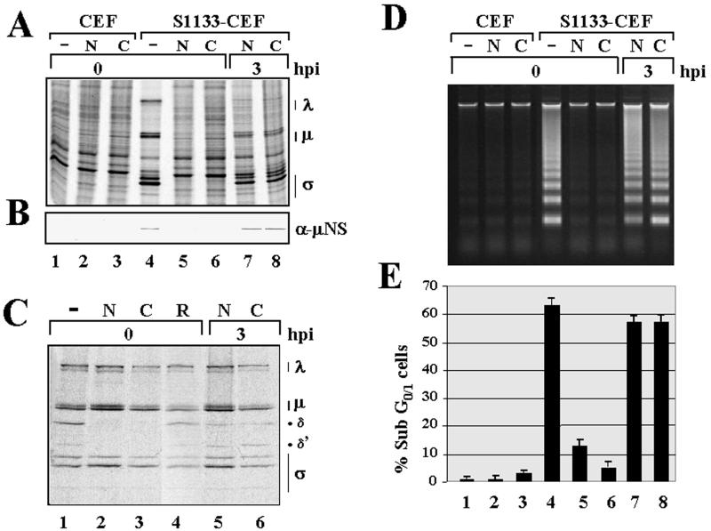
The cytopathic effect evidenced by cells infected with avian reovirus S1133 suggests that this virus may induce apoptosis in primary cultures of chicken embryo fibroblasts. In this report we present evidence that avian reovirus infection of cultured cells causes activation of the intracellular apoptotic program and that this activation takes place during an early stage of the viral life cycle. The ability of avian reoviruses to induce apoptosis is not restricted to a particular virus strain or to a specific cell type, since different avian reovirus isolates were able to induce apoptosis in several avian and mammalian cell lines. Apoptosis was also provoked in ribavirin-treated avian reovirus-infected cells and in cells infected with UV-irradiated reovirions, indicating that viral mRNA synthesis and subsequent steps in viral replication are not needed for apoptosis induction in avian reovirus-infected cells and that the number of inoculated virus particles, not their infectivity, is the critical factor for apoptosis induction by avian reovirus. Our finding that apoptosis is no longer induced when intracellular viral uncoating is blocked indicates that intraendosomal virion disassembly is required for apoptosis induction and that attachment and uptake of parental reovirions are not sufficient to cause apoptosis. Taken together, our results suggest that apoptosis is triggered from within the infected cell by viral products generated after intraendosomal uncoating of parental reovirions.


Figures

FIG. 1.
FIG. 1.
Morphological and biochemical analysis. CEF cells were mock infected (U) or infected with 20 PFU of either vaccinia virus (WR) or the avian reovirus S1133 (S1133) per cell for 24 h. (A) Cells were either visualized with a light microscope (a) or stained with Hoechst and visualized with a fluorescent microscope at 450 nm after the samples had been excited at 330 nm (b and c). Magnification: (a and b) ×10; (c) ×60. (B) DNA extracted from these cells and λ DNA digested with both HindIII and EcoRI (M) were analyzed by electrophoresis on 1.5% agarose gels and stained with ethidium bromide. (C) Electrophoretic analysis of total cytoplasmic RNA isolated from these cells. Positions of 28S and 18S rRNAs are shown on the right. (D) ELISA detection of histone-associated DNA fragments from cell extracts. The values shown are means of four independent experiments, and error bars indicate standard deviations of the means. (E) Cells were fixed and permeabilized and subsequently stained with a Boehringer Mannheim TUNEL/FITC kit according to the manufacturer's instructions. TUNEL-positive cells were observed by fluorescence (TUNEL) or by visible light (Nomarski) at a magnification of ×20.
FIG. 2.
FIG. 2.
Cytometric analysis. CEF cells were mock infected (U) or infected at 50 PFU/cell with the avian reovirus S1133 (S1133). Cells were harvested at 20 h postinfection, permeabilized, and subsequently stained with either propidium iodide (PI) or annexin V-FITC conjugate (annexin). One million cells were then analyzed by flow cytometry, and cell number was plotted against fluorescence. M1 indicates the percentage of apoptotic cells.
FIG. 3.
FIG. 3.
Analysis of apoptosis dependence and viral growth on time and multiplicity of infection. (A to C) Mock-infected cells (U) and cells infected with 20 PFU of either vaccinia virus (WR) or the avian reovirus S1133 (S1133) per cell were incubated for the indicated times. Cells were then either subjected to flow cytometric analysis after propidium iodide staining (B) or lysed for subsequent electrophoretic DNA analysis (A) and determination of virus titers (C). (D to F) Similar analyses were performed with cells infected for 12 h with the indicated S1133 multiplicities. The values shown in panels B, C, E, and F are means of four different experiments, and error bars indicate standard deviations of the means.
FIG. 4.
FIG. 4.
Infections of different cell lines with different avian reovirus strains. (A) The cell lines indicated at the top of the figure were infected at 20 PFU/cell with the avian reovirus S1133. At 20 h postinfection, cells were lysed, and the virus concentration of the cell extracts was determined by plaque assay on fresh CEF monolayers. The values shown are means of three independent experiments, and error bars indicate standard deviations of the means. (B) The cell lines indicated at the top of the figure were mock infected (U) or infected with 20 PFU of the avian reovirus S1133 (S1133) per cell. At 20 h postinfection, DNA was extracted and analyzed by electrophoresis. (C) Electrophoretic analysis of DNA extracted from CEF that were either mock infected (U) or infected with 20 PFU of the avian reovirus isolates per cell as indicated at the top of the figure.
FIG. 5.
FIG. 5.
Effect of UV treatment on avian reovirus replication and apoptosis induction. Monolayers of CEF were mock infected (U) or infected with purified avian S1133 reovirions (S1133) either before (−UV) or after (+UV) treatment with UV light. For the experiments shown in panels A to D, the virus input contained approximately 40,000 particles/cell, which corresponds to a multiplicity of infection of 20 PFU/cell in the sample of untreated virus and to a multiplicity of 10−3 PFU/cell in the sample of UV-treated virus. (A) At 16 h postinfection, cells were labeled with [35S]methionine-cysteine, and 1 h later cytoplasmic extracts were prepared and analyzed by SDS-PAGE. Positions of the three size classes of avian reovirus polypeptides are shown on the right. (B) Proteins from extracts of nonradiolabeled cells were analyzed by Western blotting with polyclonal antibodies raised against the nonstructural protein μNS. (C) At 18 h postinfection, cellular DNA was extracted and analyzed by electrophoresis. (D and E) Cells were mock infected (lane 1) or infected with purified avian reovirions for 18 h, either before (lanes 2 and 7) or after UV treatment, with the number of viral particles and multiplicities of infection (moi) indicated on the top. (D) Cytoplasmic extracts were then prepared, and their virus concentration was determined on fresh monolayers of CEF. (E) Cells were then analyzed by flow cytometry after propidium iodide staining. Values in panels D and E are means of three independent experiments, and error bars indicate standard deviations of the means.
FIG. 6.
FIG. 6.
Effect of ribavirin on protein synthesis and apoptosis induction. Monolayers of either mock-infected CEF (U) or CEF infected with 20 PFU of avian reovirus S1133 (S1133) per cell were treated, from the onset of the infection, with the concentrations of ribavirin (Rib.) indicated at the top of the figure. (A and B) At 16 h postinfection, cells were labeled with [35S]methionine-cysteine, and 1 h later cytoplasmic extracts were prepared and analyzed by SDS-PAGE and autoradiography, either before (A) or after (B) immunoprecipitation with anti-S1133 rabbit antiserum. Positions of the three size classes of avian reovirus polypeptides are shown on the right. (C) At 18 h postinfection, cellular DNA was extracted and analyzed by electrophoresis. (D) At 18 h postinfection, cells were analyzed by flow cytometry after propidium iodide staining. The values shown are the means of three independent experiments, and error bars indicate standard deviations of the means.
FIG. 7.
FIG. 7.
Effect of viral inhibitors on protein synthesis, virus uncoating, and apoptosis induction. (A, B, D, and E) Monolayers of either mock-infected CEF (U) or CEF infected with 20 PFU of avian reovirus S1133 (S1133) per cell were treated at the times indicated with 10 mM ammonium chloride (N) or 100 μM chloroquine (C) or untreated (−). (A) At 16 h postinfection (hpi), cells were labeled for 1 h with [35S]methionine-cysteine, and cytoplasmic extracts were prepared and analyzed by SDS-PAGE and autoradiography. (B) Cytoplasmic extracts of nonradiolabeled cells were analyzed by Western blotting with anti-μNS polyclonal antibodies. (D) Cellular DNA was isolated at 17 h postinfection and analyzed by electrophoresis. (E) Cells were analyzed by flow cytometry after propidium iodide staining. The values shown are means of three independent experiments, and error bars indicate standard deviations of the means. (C) Monolayers of CEF infected with 35S-labeled purified reovirions were treated at the times indicated with the two viral inhibitors and with 200 μM ribavirin (R), and at 5 h postinfection cytoplasmic extracts were prepared and analyzed by SDS-PAGE and autoradiography. Positions of the three size classes of avian reovirus polypeptides are indicated at the right of panels A and C.

Similar articles

Cited by

References

    1. Arends, M. J., R. G. Morris, and A. H. Willey. 1990. Apoptosis: the role of the endonuclease. Am. J. Pathol. 136:593-608. - PMC - PubMed
    1. Baer, G. S., and T. S. Dermody. 1997. Mutations in reovirus outer-capsid protein σ3 selected during persistent infections of L cells confer resistance to protease inhibitor E64. J. Virol. 71:4921-4928. - PMC - PubMed
    1. Barrett, A. J., A. A. Kembhavi, M. A. Brown, H. Kirschke, C. G. Knight, M. Tamai, and K. Hanada. 1982. l-trans-Epoxysuccinyl-leucylamido(4-guanidino)butane (E-64) and its analogues as inhibitors of cysteine proteinases including cathepsins B, H, and L. Biochem. J. 201:189-198. - PMC - PubMed
    1. Barton, E. S., J. C. Forrest, J. L. Connolly, J. D. Chappell, Y. Liu, F. J. Schnell, A. Nusrat, C. A. Parkos, and T. S. Dermody. 2001. Junction adhesion molecule is a receptor for reovirus. Cell 104:441-451. - PubMed
    1. Bartz, S. R., and M. Emerman. 1999. Human immunodeficiency virus type 1 Tat induces apoptosis and increases sensitivity to apoptotic signals by up-regulating FLICE/caspase-8. J. Virol. 73:1956-1963. - PMC - PubMed

Publication types

MeSH terms

LinkOut - more resources