. 2002 Aug;76(16):7956-67.
doi: 10.1128/jvi.76.16.7956-7967.2002.

Bovine leukemia virus SU protein interacts with zinc, and mutations within two interacting regions differently affect viral fusion and infectivity in vivo

Affiliations

Bovine leukemia virus SU protein interacts with zinc, and mutations within two interacting regions differently affect viral fusion and infectivity in vivo

Jean-Stéphane Gatot et al. J Virol. 2002 Aug.

Abstract

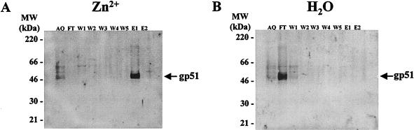
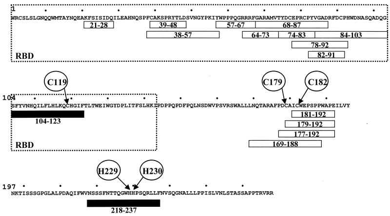
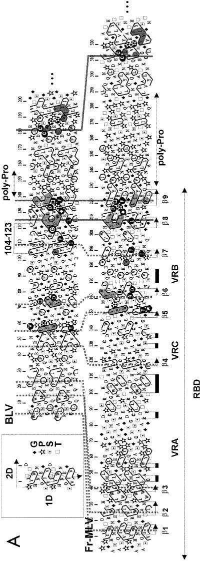
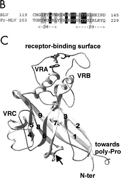
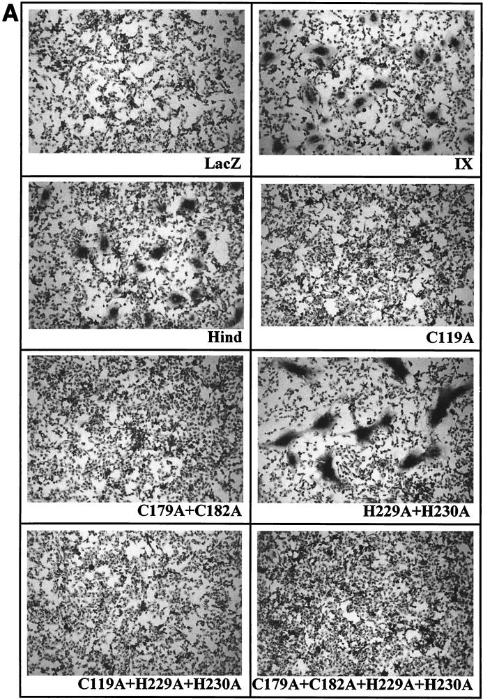
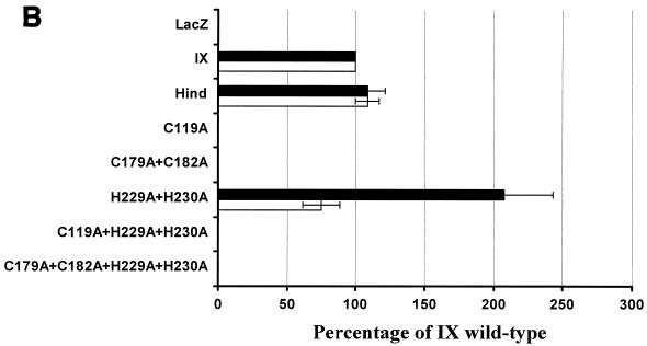
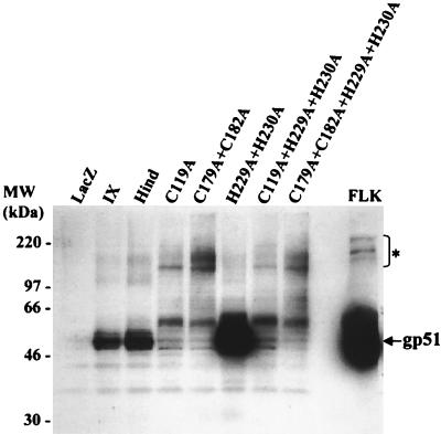
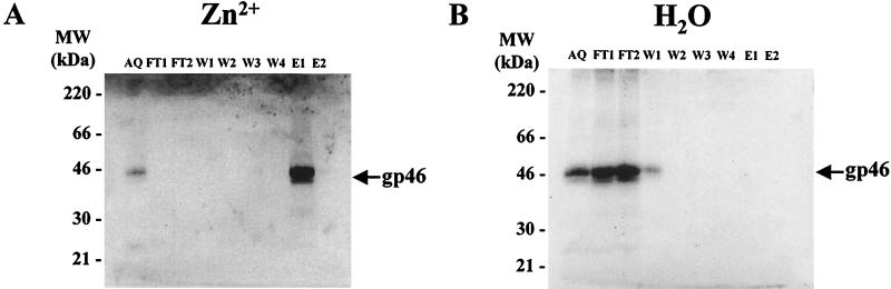
Bovine leukemia virus (BLV) and human T-cell lymphotropic virus type 1 (HTLV-1) belong to the genus of deltaretroviruses. Their entry into the host cell is supposed to be mediated by interactions of the extracellular (SU) envelope glycoproteins with cellular receptors. To gain insight into the mechanisms governing this process, we investigated the ability of SU proteins to interact with specific ligands. In particular, by affinity chromatography, we have shown that BLV SU protein specifically interacted with zinc ions. To identify the protein domains involved in binding, 16 peptides distributed along the sequence were tested. Two of them appeared to be able to interact with zinc. To unravel the role of these SU regions in the biology of the virus, mutations were introduced into the env gene of a BLV molecular clone in order to modify residues potentially interacting with zinc. The fusogenic capacity of envelope mutated within the first zinc-binding region (104 to 123) was completely abolished. Furthermore, the integrity of this domain was also required for in vivo infectivity. In contrast, mutations within the second zinc-binding region (218 to 237) did not hamper the fusogenic capacity; indeed, the syncytia were even larger. In sheep, mutations in region 218 to 237 did not alter infectivity or viral spread. Finally, we demonstrated that the envelope of the related HTLV-1 was also able to bind zinc. Interestingly, zinc ions were found to be associated with the receptor-binding domain (RBD) of Friend murine leukemia virus (Fr-MLV) SU glycoprotein, further supporting their relevance in SU structure. Based on the sequence similarities shared with the Fr-MLV RBD, whose three-dimensional structure has been experimentally determined, we located the BLV zinc-binding peptide 104-123 on the opposite side of the potential receptor-binding surface. This observation supports the hypothesis that zinc ions could mediate interactions of the SU RBD either with the C-terminal part of SU, thereby contributing to the SU structural integrity, or with a partner(s) different from the receptor.


Figures

FIG. 1.
FIG. 1.
BLV envelope SU glycoprotein interacts with zinc. (A) Chelating Sepharose beads charged with Zn2+ ions were incubated with cell culture supernatant from BLV-producing FLK cells. An aliquot (AQ) was taken before loading the column. The flowthrough fraction (FT) was harvested, and the column was washed five times with load and wash buffer (20 mM sodium phosphate, 500 mM NaCl, 0.5% Tween 20, pH 7.5). Wash fractions W1 to W5 were harvested. Finally, elution fractions (E1 and E2) were collected after addition of elution buffer (the same composition as load and wash buffer except that EDTA was added at a concentration of 250 mM). The material was concentrated by PEG precipitation and migrated onto an SDS-polyacrylamide gel. After electroblotting of the proteins, the nitrocellulose membrane was first incubated with monoclonal antibodies directed against BLV gp51 and subsequently with a secondary antibody conjugated to alkaline phosphatase. The presence of the BLV envelope SU protein (gp51) was then visualized by chemiluminescence. (B) As a control, the chelating Sepharose fast flow gel was incubated with water and the procedure described for panel A was followed.
FIG. 2.
FIG. 2.
Two peptides within BLV SU glycoprotein specifically bind zinc. The sequence of BLV SU protein is presented without considering the signal peptide (residue 1 corresponding to the precursor cleavage site) (15). A series of 16 peptides encompassing the SU protein (peptides 21-28, 38-57, 39-48, 57-67, 64-73, 68-87, 74-83, 78-92, 82-91, 84-103, 104-123, 169-188, 177-192, 179-192, 181-192, and 218-237) (16, 59) were tested for their ability to bind chelating Sepharose charged with Zn2+ ions. The concentration of the peptides in the different fractions was determined by an ELISA procedure with specific antisera (13, 16, 59). Peptides 104-123 and 218-237 were able to specifically interact with zinc ions (black boxes), while other peptides exhibited no zinc-binding activity (open boxes). Within peptides 104-123 and 218-237, residues C119, H229, and H230, potentially interacting with zinc, are indicated. Conserved amino acids C179 and C182 are also circled. Localization of the BLV receptor-binding domain (RBD), deduced from the sequence alignment with the Fr-MLV RBD, is surrounded.
FIG. 3.
FIG. 3.
Sequence-structure relationships of the BLV gp51 protein. (A) Hydrophobic cluster analysis of the SU N-terminal ends. Sequences are shown on a duplicated α-helical net, where hydrophobic amino acids (V, I, L, F, M, Y, and W) are contoured (14). These residues form clusters that statistically correspond to the internal faces of regular secondary structures (α-helices and β-strands) (82). The way to read sequences (1D) and secondary structures (2D) and the symbols used for particular amino acids are indicated in the upper left inset. The Fr-MLV receptor-binding domain (RBD) and its secondary structures (arrows for the β-sheets and black boxes for the α-helices) are indicated below the Fr-MLV sequence. The position of the BLV 104-123 peptide is shown, and the C119 residue is circled. Both Fr-MLV and BLV RBD sequences end with a region rich in proline residues (poly-Pro), indicating the presence of a hinge separating distinct domains. A region located just before the hinge is particularly well conserved between Fr-MLV and BLV and encompasses strands β8 and β9 of the Fr-MLV RBD. Identities are indicated in white on a black background, and cluster similarities are shaded in gray. Sequences upstream of β8 and β9 are less conserved, and the variable regions (VRA to VRC), poor in hydrophobic residues, are reduced in BLV gp51. Proposed correspondences between the different strands of the immunoglobulin-like domain are indicated with stippled lines. (B) Alignment of the BLV and Fr-MLV SU sequences encompassing strands β8 and β9 of the Fr-MLV RBD structure. Cysteine 119 is located just upstream of strand β8. Identities and cluster similarities are indicated as in panel A. (C) Ribbon representation of the three-dimensional structure of the Fr-MLV RBD, corresponding to the N-terminal part of the SU protein (PDB 1AOL [31]). Strands are labeled 1 to 9, and variable regions VRA to VRC are indicated. Following the alignment presented in A, the cysteine present in peptide 104-123 (circled in panel A) should be located between strands β7 and β8 (see arrow). The position of the receptor-binding surface is shown, as well as three amino acids (S84, D86, and W102) which were shown to be critical for receptor binding and infection (24).
FIG. 3.
FIG. 3.
Sequence-structure relationships of the BLV gp51 protein. (A) Hydrophobic cluster analysis of the SU N-terminal ends. Sequences are shown on a duplicated α-helical net, where hydrophobic amino acids (V, I, L, F, M, Y, and W) are contoured (14). These residues form clusters that statistically correspond to the internal faces of regular secondary structures (α-helices and β-strands) (82). The way to read sequences (1D) and secondary structures (2D) and the symbols used for particular amino acids are indicated in the upper left inset. The Fr-MLV receptor-binding domain (RBD) and its secondary structures (arrows for the β-sheets and black boxes for the α-helices) are indicated below the Fr-MLV sequence. The position of the BLV 104-123 peptide is shown, and the C119 residue is circled. Both Fr-MLV and BLV RBD sequences end with a region rich in proline residues (poly-Pro), indicating the presence of a hinge separating distinct domains. A region located just before the hinge is particularly well conserved between Fr-MLV and BLV and encompasses strands β8 and β9 of the Fr-MLV RBD. Identities are indicated in white on a black background, and cluster similarities are shaded in gray. Sequences upstream of β8 and β9 are less conserved, and the variable regions (VRA to VRC), poor in hydrophobic residues, are reduced in BLV gp51. Proposed correspondences between the different strands of the immunoglobulin-like domain are indicated with stippled lines. (B) Alignment of the BLV and Fr-MLV SU sequences encompassing strands β8 and β9 of the Fr-MLV RBD structure. Cysteine 119 is located just upstream of strand β8. Identities and cluster similarities are indicated as in panel A. (C) Ribbon representation of the three-dimensional structure of the Fr-MLV RBD, corresponding to the N-terminal part of the SU protein (PDB 1AOL [31]). Strands are labeled 1 to 9, and variable regions VRA to VRC are indicated. Following the alignment presented in A, the cysteine present in peptide 104-123 (circled in panel A) should be located between strands β7 and β8 (see arrow). The position of the receptor-binding surface is shown, as well as three amino acids (S84, D86, and W102) which were shown to be critical for receptor binding and infection (24).
FIG. 4.
FIG. 4.
Fusogenic capacity of recombinant BLV envelope proteins. (A) The fusogenic capacity of the different mutants was tested by cocultivation of CC81 indicator cells with BHK cells expressing the BLV envelope proteins. The wild-type (IX and Hind) and mutated (C119A, C179A+C182A, H229A+H230A, C119A+H229A+H230A, and C179A+C182A+H229A+H230A) envelope genes were cloned into the pSFV expression vector (pSFVenv plasmids). BHK cells were transfected with RNAs transcribed from the different pSFVenv vectors and cocultivated for 20 h with CC81 indicator cells. As a negative control, cells were also transfected with a pSFV vector expressing the lacZ gene. After fixation, the nuclei were colored with Giemsa, and the syncytia were visualized microscopically at ×100 magnification. The diameter of the syncytia induced by the wild-type envelope proteins was in the range of 100 to 200 μm. (B) The numbers of syncytia counted in 10 different microscope fields (open bars) were arbitrarily normalized to the wild-type levels (IX = 100%). In addition, the mean number of nuclei within 20 syncytia was evaluated for each mutant envelope protein (black bars). The data represent mean values of three independent experiments.
FIG. 4.
FIG. 4.
Fusogenic capacity of recombinant BLV envelope proteins. (A) The fusogenic capacity of the different mutants was tested by cocultivation of CC81 indicator cells with BHK cells expressing the BLV envelope proteins. The wild-type (IX and Hind) and mutated (C119A, C179A+C182A, H229A+H230A, C119A+H229A+H230A, and C179A+C182A+H229A+H230A) envelope genes were cloned into the pSFV expression vector (pSFVenv plasmids). BHK cells were transfected with RNAs transcribed from the different pSFVenv vectors and cocultivated for 20 h with CC81 indicator cells. As a negative control, cells were also transfected with a pSFV vector expressing the lacZ gene. After fixation, the nuclei were colored with Giemsa, and the syncytia were visualized microscopically at ×100 magnification. The diameter of the syncytia induced by the wild-type envelope proteins was in the range of 100 to 200 μm. (B) The numbers of syncytia counted in 10 different microscope fields (open bars) were arbitrarily normalized to the wild-type levels (IX = 100%). In addition, the mean number of nuclei within 20 syncytia was evaluated for each mutant envelope protein (black bars). The data represent mean values of three independent experiments.
FIG. 5.
FIG. 5.
Expression of wild-type and mutant gp51 proteins with the SFV system. After in vitro transcription of the different pSFVenv vectors (wild-type and mutants), the corresponding purified RNAs were transfected into BHK cells by electroporation. The cells were cultivated for 60 h and harvested, and cell lysates corresponding to 1.5 million cells were added per well of an SDS-polyacrylamide gel. The proteins were transferred onto a nitrocellulose membrane and incubated in the presence of a mixture of monoclonal antibodies directed against the BLV gp51 protein. After incubation with an anti-mouse immunoglobulin antibody coupled to peroxidase, the presence of the gp51 proteins was visualized by chemiluminescence. Supernatant from BLV-producing FLK cells was used as a positive control. The unprocessed precursors are indicated (*).
FIG. 6.
FIG. 6.
In contrast to histidines 229 and 230, cysteine 119 is essential for infectivity in vivo. (A) To assess infectivity, recombinant proviruses carrying mutated gp51 genes were constructed, mixed with cationic liposomes, and injected intradermally into sheep. The infectious potential was evaluated by two criteria: the presence of antibodies directed towards the gp51 protein (as measured by ELISA and immunodiffusion) and PCR amplification of viral sequences. (B) To evaluate the proviral loads in sheep 2674 and 2675 infected with the H229A+H230A mutant, DNA was extracted from 500-μl aliquots of peripheral blood at 6 months postseroconversion and amplified by 29 cycles of PCR. Of note, the lymphocyte counts for the different animals were all in the normal range (3,000 to 5,000 per mm3). The samples were then analyzed by Southern blot hybridization with a BLV probe. DNAs from sheep 114 and 117 (uninfected [N.I.] animals) and 292 and 293 (infected with the wild-type virus) were used as negative and positive controls, respectively. As a standard for quantification, serial dilutions of the lysates (1:10 and 1:100) were amplified in parallel.
FIG. 7.
FIG. 7.
Binding of HTLV-1 envelope SU glycoprotein to zinc. Chelating Sepharose fast flow gel was charged with Zn2+ ions (A) or water (B) and packed into a column. The columns were loaded with MT2 cell culture supernatant mixed with load and wash buffer, and the procedure described for Fig. 1 was followed. The HTLV-1 gp46 glycoprotein was detected by incubation with the MF2 monoclonal antibody before incubation with a secondary antibody conjugated to alkaline phosphatase. AQ, aliquot; FT, flowthrough; W, wash; E, eluate.

References

    1. Agadjanyan, M. G., K. E. Ugen, B. Wang, W. V. Williams, and D. B. Weiner. 1994. Identification of an 80-kilodalton membrane glycoprotein important for human T-cell leukemia virus type I and type II syncytium formation and infection. J. Virol. 68:485-493. - PMC - PubMed
    1. Bae, Y., S. M. Kingsman, and A. J. Kingsman. 1997. Functional dissection of the Moloney murine leukemia virus envelope protein gp70. J. Virol. 71:2092-2099. - PMC - PubMed
    1. Ban, J., D. Portetelle, C. Altaner, B. Horion, D. Milan, V. Krchnak, A. Burny, and R. Kettmann. 1993. Isolation and characterization of a 2.3-kilobase-pair cDNA fragment encoding the binding domain of the bovine leukemia virus cell receptor. J. Virol. 67:1050-1057. - PMC - PubMed
    1. Barnett, A. L., and J. M. Cunningham. 2001. Receptor binding transforms the surface subunit of the mammalian C-type retrovirus envelope protein from an inhibitor to an activator of fusion. J. Virol. 75:9096-9105. - PMC - PubMed
    1. Barnett, A. L., R. A. Davey, and J. M. Cunningham. 2001. Modular organization of the Friend murine leukemia virus envelope protein underlies the mechanism of infection. Proc. Natl. Acad. Sci. USA 98:4113-4118. - PMC - PubMed

Publication types

MeSH terms

LinkOut - more resources