. 2002 Aug;76(16):7976-86.
doi: 10.1128/jvi.76.16.7976-7986.2002.

Multiple transmembrane amino acid requirements suggest a highly specific interaction between the bovine papillomavirus E5 oncoprotein and the platelet-derived growth factor beta receptor

Affiliations

Multiple transmembrane amino acid requirements suggest a highly specific interaction between the bovine papillomavirus E5 oncoprotein and the platelet-derived growth factor beta receptor

Valerie M Nappi et al. J Virol. 2002 Aug.

Abstract

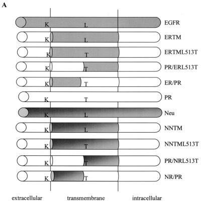
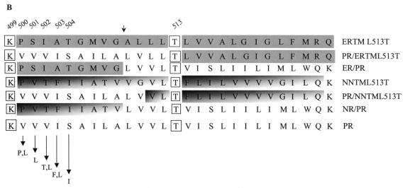
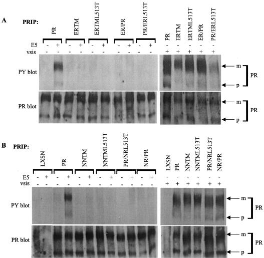
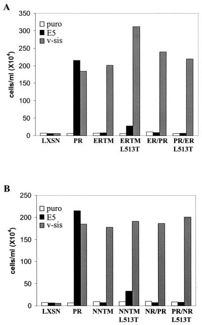
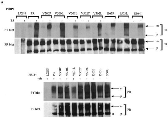
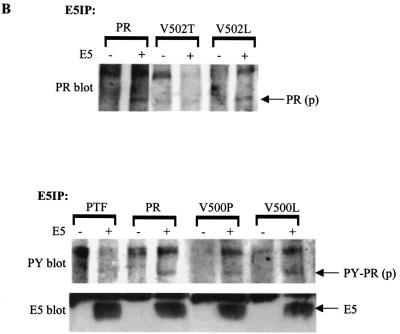
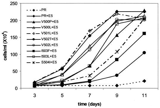
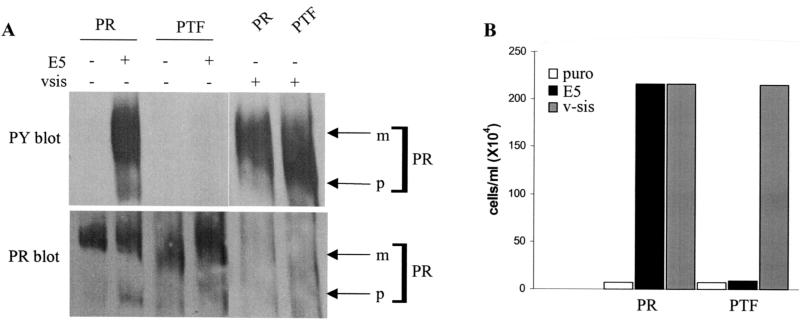
The bovine papillomavirus E5 protein activates the cellular platelet-derived growth factor beta receptor (PDGFbetaR) tyrosine kinase in a ligand-independent manner. Evidence suggests that the small transmembrane E5 protein homodimerizes and physically interacts with the transmembrane domain of the PDGFbetaR, thereby inducing constitutive dimerization and activation of this receptor. Amino acids in the receptor previously found to be required for the PDGFbetaR-E5 interaction are a transmembrane Thr513 and a juxtamembrane Lys499. Here, we sought to determine if these are the only two receptor amino acids required for an interaction with the E5 protein. Substitution of large portions of the PDGFbetaR transmembrane domain indicated that additional amino acids in both the amino and carboxyl halves of the receptor transmembrane domain are required for a productive interaction with the E5 protein. Indeed, individual amino acid substitutions in the receptor transmembrane domain identified roles for the extracellular proximal transmembrane residues in the interaction. These data suggest that multiple amino acids within the transmembrane domain of the PDGFbetaR are required for a stable interaction with the E5 protein. These may be involved in direct protein-protein contacts or may support the proper transmembrane alpha-helical conformation for optimal positioning of the primary amino acid requirements.


Figures

FIG. 1.
FIG. 1.
Structure of the mutant receptors. (A) Schematic representations of the chimeric mutant receptors generated and characterized in these studies. Chimeric receptor mutants were derived from ERTM or NNTM, which contain the transmembrane domain of the EGF receptor and Neu, respectively. Included are the ERTML513T and NNTML513T mutants, which contain a Thr at position 513 in ERTM and NNTM, respectively. Also included are the split transmembrane domain receptor mutants PR/ERL513T, ER/PR, PR/NRL513T, and NR/PR. The required Lys (K) located at position 499 and Thr (T) at position 513 are indicated. Shaded areas are derived from EGF receptor or Neu, while unshaded areas are derived from the PDGFβR. (B) Amino acid sequence of the transmembrane domain of each receptor examined in this study compared to the wild-type murine PDGFβR (PR). Fully shaded sequences were derived from the EGF receptor transmembrane domain, differentially shaded sequences were derived from the Neu receptor transmembrane domain, and unshaded sequences were derived from the wild-type PDGFβR transmembrane domain. The small arrow above the sequences denotes where a SpeI site was introduced into the PDGFβR, ERTML513T, and NNTML513T receptor constructs. Arrows at the bottom depict amino acid changes made in the wild-type PDGFβR at positions 500 to 504. The essential Lys (K) 499 and Thr (T) 513 of the PDGFβR required for an interaction with the E5 protein are boxed. Specific residue numbers are indicated above the sequences.
FIG. 1.
FIG. 1.
Structure of the mutant receptors. (A) Schematic representations of the chimeric mutant receptors generated and characterized in these studies. Chimeric receptor mutants were derived from ERTM or NNTM, which contain the transmembrane domain of the EGF receptor and Neu, respectively. Included are the ERTML513T and NNTML513T mutants, which contain a Thr at position 513 in ERTM and NNTM, respectively. Also included are the split transmembrane domain receptor mutants PR/ERL513T, ER/PR, PR/NRL513T, and NR/PR. The required Lys (K) located at position 499 and Thr (T) at position 513 are indicated. Shaded areas are derived from EGF receptor or Neu, while unshaded areas are derived from the PDGFβR. (B) Amino acid sequence of the transmembrane domain of each receptor examined in this study compared to the wild-type murine PDGFβR (PR). Fully shaded sequences were derived from the EGF receptor transmembrane domain, differentially shaded sequences were derived from the Neu receptor transmembrane domain, and unshaded sequences were derived from the wild-type PDGFβR transmembrane domain. The small arrow above the sequences denotes where a SpeI site was introduced into the PDGFβR, ERTML513T, and NNTML513T receptor constructs. Arrows at the bottom depict amino acid changes made in the wild-type PDGFβR at positions 500 to 504. The essential Lys (K) 499 and Thr (T) 513 of the PDGFβR required for an interaction with the E5 protein are boxed. Specific residue numbers are indicated above the sequences.
FIG. 2.
FIG. 2.
Biochemical and functional analysis of the split transmembrane PDGFβR chimeras. Ba/F3 cell lines expressing the wild-type PDGFβR (PR), the ERTM, ERTML513T, NNTM, or NNTML513T chimeric receptors, or the PR/ERL513T, ER/PR, PR/NRL513T, or NR/PR split transmembrane receptor chimeras with (+) or without (−) E5 or v-sis were generated as described in Materials and Methods. (A and B) PDGFβ receptor (PR) was immunoprecipitated (IP) from cell extracts and immunoblotted with an antiphosphotyrosine antibody (PY) for detection of activated receptor (upper panels). The blots then were stripped (Materials and Methods) and incubated with anti-PDGFβR antiserum (PR) to assess receptor expression levels (lower panels). Each lane represents approximately 300 μg of extracted protein. Arrows on the right point to the mature (m) and precursor (p) forms of the receptor.
FIG. 3.
FIG. 3.
IL-3 independence assay of Ba/F3 cells expressing the split transmembrane chimeric receptors. Cells expressing the split transmembrane chimeric receptors with or without E5 or v-sis were seeded in medium lacking IL-3 and counted 10 days later. The densities of the cell lines expressing the ERTM-derived chimeras are shown in panel A, and the densities of the cell lines expressing the NNTM-derived chimeras are shown in panel B. These data are representative of experiments listed in Table 1.
FIG. 4.
FIG. 4.
Biochemical and functional analysis of PDGFβR mutants containing extracellular proximal transmembrane amino acid substitutions. Each of the PDGFβR mutants containing an extracellular proximal transmembrane amino acid substitution, the wild-type PDGFβR (PR), or no exogenous receptor (LXSN) was expressed in Ba/F3 cells with (+) or without (−) E5 or v-sis as described in Materials and Methods. (A) PDGFβR was immunoprecipitated (PRIP) from cell extracts and subjected to antiphosphotyrosine (PY) or anti-PDGFβR (PR) immunoblotting to detect receptor activation and total receptor levels, respectively. (B) The E5 protein with any associated proteins was immunoprecipitated (E5IP) from cell extracts and subjected to anti-PDGFβR (PR) or antiphosphotyrosine (PY) immunoblotting to assess physical complex formation between the E5 protein and the PDGFβR. Anti-E5 (E5) immunoblotting was performed to detect E5 protein expression levels. In these experiments, the presence of the 165-kDa precursor form of the PDGFβR in E5 immunoprecipitates is indicative of complex formation between the E5 protein and the PDGFβR. Arrows to the right mark the mature (m) and precursor (p) isoforms of each receptor as well as the E5 protein. Each lane represents approximately 585 μg (A) or 675 μg (B) of extracted protein.
FIG. 4.
FIG. 4.
Biochemical and functional analysis of PDGFβR mutants containing extracellular proximal transmembrane amino acid substitutions. Each of the PDGFβR mutants containing an extracellular proximal transmembrane amino acid substitution, the wild-type PDGFβR (PR), or no exogenous receptor (LXSN) was expressed in Ba/F3 cells with (+) or without (−) E5 or v-sis as described in Materials and Methods. (A) PDGFβR was immunoprecipitated (PRIP) from cell extracts and subjected to antiphosphotyrosine (PY) or anti-PDGFβR (PR) immunoblotting to detect receptor activation and total receptor levels, respectively. (B) The E5 protein with any associated proteins was immunoprecipitated (E5IP) from cell extracts and subjected to anti-PDGFβR (PR) or antiphosphotyrosine (PY) immunoblotting to assess physical complex formation between the E5 protein and the PDGFβR. Anti-E5 (E5) immunoblotting was performed to detect E5 protein expression levels. In these experiments, the presence of the 165-kDa precursor form of the PDGFβR in E5 immunoprecipitates is indicative of complex formation between the E5 protein and the PDGFβR. Arrows to the right mark the mature (m) and precursor (p) isoforms of each receptor as well as the E5 protein. Each lane represents approximately 585 μg (A) or 675 μg (B) of extracted protein.
FIG. 5.
FIG. 5.
IL-3 independence assay of Ba/F3 cells expressing PDGFβR mutants with the extracellular proximal transmembrane substitutions. The Ba/F3 cell lines described in the Fig. 4 legend were seeded into medium lacking IL-3 and counted at the indicated intervals. These growth curves are representative of multiple experiments listed in Table 1.
FIG. 6.
FIG. 6.
Biochemical and functional analysis of the triple substitution PTF mutant. Ba/F3 cells expressing the wild-type PDGFβR (PR) or the PTF mutant receptor with (+) or without (−) E5 or v-sis were established as described in Materials and Methods. (A) PDGFβR immunoprecipitates from cell extracts were immunoblotted with an antiphosphotyrosine antibody to assess receptor activation levels (top panel). The phosphotyrosine immunoblot was then stripped (see Materials and Methods) and reprobed with the PDGFβR antiserum to detect receptor expression levels (bottom panel). Each lane represents approximately 300 μg of extracted protein. Arrows on the right point to the mature (m) and precursor (p) forms of the receptors. (B) IL-3 independence assay of cells expressing the PTF mutant receptor. Ba/F3 cells expressing the wild-type PDGFβR (PR) or the PTF mutant receptor without (puro) or with E5 or v-sis were incubated in the absence of IL-3 for 11 days and then counted. The data are representative of multiple experiments listed in Table 1.

Similar articles

Cited by

References

    1. Bergman, P., M. Ustav, J. Sedman, J. Moreno-Lopez, B. Vennstrom, and U. Pettersson. 1988. The E5 gene of bovine papillomavirus type 1 is sufficient for complete oncogenic transformation of mouse fibroblasts. Oncogene 2:453-459. - PubMed
    1. Burkhardt, A., D. DiMaio, and R. Schlegel. 1987. Genetic and biochemical definition of the bovine papillomavirus E5 transforming protein. EMBO J. 6:2381-2385. - PMC - PubMed
    1. Burkhardt, A., M. Willingham, C. Gay, K. T. Jeang, and R. Schlegel. 1989. The E5 oncoprotein of bovine papillomavirus is oriented asymmetrically in Golgi and plasma membranes. Virology 170:334-339. - PubMed
    1. Claesson-Welsh, L. 1994. Platelet-derived growth factor receptor signals. J. Biol. Chem. 269:32023-32026. - PubMed
    1. Cohen, B. D., D. R. Lowy, and J. T. Schiller. 1993. The conserved C-terminal domain of the bovine papillomavirus E5 oncoprotein can associate with an alpha-adaptin-like molecule: a possible link between growth factor receptors and viral transformation. Mol. Cell. Biol. 13:6462-6468. - PMC - PubMed

Publication types

MeSH terms

Substances

LinkOut - more resources