. 2002 Aug;76(16):7987-95.
doi: 10.1128/jvi.76.16.7987-7995.2002.

Given the opportunity, the Sendai virus RNA-dependent RNA polymerase could as well enter its template internally

Affiliations

Given the opportunity, the Sendai virus RNA-dependent RNA polymerase could as well enter its template internally

Diane Vulliémoz et al. J Virol. 2002 Aug.

Abstract

The negative-stranded RNA viral genome is an RNA-protein complex of helicoidal symmetry, resistant to nonionic detergent and high salt, in which the RNA is protected from RNase digestion. The 15,384 nucleotides of the Sendai virus genome are bound to 2,564 subunits of the N protein, each interacting with six nucleotides so tightly that the bases are poorly accessible to soluble reagents. With such a uniform structure, the question of template recognition by the viral RNA polymerase has been raised. In a previous study, the N-phase context has been proposed to be crucial for this recognition, a notion referring to the importance of the position in which the nucleotides interact with the N protein. The N-phase context ruled out the role of the template 3'-OH congruence, a feature resulting from the obedience to the rule of six that implies the precise interaction of the last six 3'-OH nucleotides with the last N protein. The N-phase context then allows prediction of the recognition by the RNA polymerase of a replication promoter sequence even if internally positioned, a promoter which normally lies at the template extremity. In this study, with template minireplicons bearing tandem replication promoters separated by intervening sequences, we present data that indeed show that initiation of RNA synthesis takes place at the internal promoter. This internal initiation can best be interpreted as the result of the polymerase entering the template at the internal promoter. In this way, the data are consistent with the importance of the N-phase context in template recognition. Moreover, by introducing between the two promoters a stretch of 10 A residues which represent a barrier for RNA synthesis, we found that the ability of the RNA polymerase to cross this barrier depends on the type of replication promoter, strong or weak, that the RNA polymerase starts on, a sign that the RNA polymerase may be somehow imprinted in its activity by the nature of the promoter on which it starts synthesis.


Figures

FIG. 1.
FIG. 1.
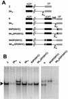
Replication of the AGPint-GPext constructs. (A) Schematic representation of the replication system. For a detailed description, see Results. NoP and PE refer, respectively, to the Northern blot riboprobe and to the primer extension oligonucleotide, both of positive polarity, complementary to the replicated RNA, depicted in diagram e. (B) Schematic representation of the AGPint-GPext series constructs. 0, Partial scheme of the pSP65 plasmid harboring the basic copyback minireplicon pSV-DI-H4Δ96 flanked by the T7 RNA polymerase promoter (T7p) and the hepatitis delta virus ribozyme (Rbz). 1, T7 RNA transcript produced from 0, carrying at its 5′ and 3′ ends the complementary sequence of the antigenomic promoter (AGPLC) and the antigenomic promoter sequence (AGPR), respectively. 2, Minireplicon T7 transcript harboring at its 3′ end the genomic promoter (GP) instead of the AGP. 3, Minireplicon T7 transcript harboring at its 3′ end an internal AGP sequence adjacent to an external GP. 4 to 9, Series of minireplicon T7 transcripts harboring at their 3′ ends a double promoter, as in construct 3, but separated by increasing intervening sequences as indicated (nt, nucleotides). Note that all the constructs harbor the same 5′ end sequence (AGPLC). (C) Northern blot analysis (see Materials and Methods) of the encapsidated minireplicon RNAs of negative polarity produced upon transfection of A549 cells with the various plasmids described for panel B in replication assays made in the absence of the C proteins (P/Cstop conditions; see Materials and Methods). Open and solid arrowheads, external and internal initiation, respectively. (D) As for panel C, but the replication assays were performed in HeLa cells in the presence of the C proteins (Cwt conditions; see Materials and Methods).
FIG. 2.
FIG. 2.
Effect of the 10A stretch on minireplicon replication. (A) Schematic representation of the single-promoter minireplicons used. 0, as described in the legend to Fig. 1B, with further features related to the present experiment: the restriction sites [DraIII, BamHI (BH1), and MunI], the sequencing primer (SE), and the Sp6 promoter. 1 and 2, as for Fig. 1B. 1A10 and 2A10, Derivatives of 1 and 2, respectively, containing a 10A stretch inserted at the DraIII site. (B) Northern blot analysis of the in vivo-replicated minireplicons. Lanes −L, replication assays performed in the absence of the L-expressing plasmid. (C) Northern blot analysis of in vitro T7 RNA polymerase transcriptions of pSp65 plasmids carrying the indicated minireplicons. The plasmids were linearized at the BamHI (BH1) site (see panel A). The probe of negative polarity resulting from an Sp6 RNA polymerase transcription of plasmid 1 or 2 linearized at the DraIII site (indicated as probe − in panel A) was used. Open arrowhead, plasmids. Solid arrowhead, T7 transcripts. −T7p, in vitro transcription without T7 RNA polymerase. (D) The plasmid-carrying construct 1A10 and the RT-PCR product amplified (see text) from the in vivo-replicated minireplicon 1A10 were sequenced with a primer situated as indicated in panel A (SE). Only the relevant portions of the sequence are shown. ≫ indicates further extension of A’s past the 10A stretch, ∗ indicates longer replication products, and <<< indicates the large accumulation of heterogeneous shorter products.
FIG. 3.
FIG. 3.
Replication of the AGPint-GPext constructs harboring a 10A stretch in the intervening sequence. (A) Schematic representation of the minireplicons used. 2 and 9 are as described in the legend to Fig. 1. 2A10, 9A10, 9A10GP(G91C), and 9AGPA10(G91C) harbor a 10A stretch (positive polarity sequence) adjacent to the internal border of GP (see Materials and Methods). 9GP(G91C), 9A10GP(G91C), 9AGP(G91C), and 9A10AGP(G91C) harbor a G-to-C substitution at position 91 of GP or AGP. (B) Northern blot analysis of the replicated minireplicons. When two bands are visible, the upper and lower one correspond to initiation at GP and AGP, respectively. Open and solid arrowheads, as for Fig. 1.
FIG. 4.
FIG. 4.
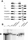
Replication of the AGPint-AGPext constructs. (A) Schematic representation of AGPint-AGPext series constructs. Construct 1, as described in the legend to Fig. 1. Constructs 10 to 13, minireplicons harboring two AGP promoters, adjacent (construct 10) or separated by increasing intervening sequence (nucleotides [nt]) as indicated. (B) Northern blot analysis (see the encapsidated minireplicon RNAs produced from the plasmids described for panel A). The open rectangle is meant to help show the different migration of the bands. Note the presence of a single band regardless. (C) Primer extension (PE) analysis (see Materials and Methods) on construct 10 to better demonstrate the preponderant presence of the replicated product initiating at AGPext. The end position of the product elongated on the RNA is given by comparison with the sequence performed on the respective plasmid with the same primer. 3′-end sequence (Seq) of AGP, 5′…TTGTCTGGT-3′. Adjacent ribozyme sequence, 5′…CCGGCCGA…3′. Only portions of the sequencing gel relevant for the 3′ ends of the replicated RNAs are shown.
FIG. 5.
FIG. 5.
Gradually diminishing the template availability. (A) Schematic representation of the minireplicons used. Constructs 1 and 13, as described in the legend to Fig. 4. 13A10 harbors a 10A stretch (positive polarity sequence) adjacent to the internal border of external AGP. (B) Northern blot analysis of the replicated products. μg, micrograms of the template plasmid added. Note the presence of a faint signal below the main band for lanes 13A10, 5 and 0.5 μg (see text). Open and solid arrowheads, as for Fig. 1.
FIG. 6.
FIG. 6.
Replication of the AGPint-AGPext constructs harboring a 10A stretch in the intervening sequence. (A) Schematic representation of the constructs. Constructs 1, 13, and 13A10, as described in the legend to Fig. 5. 13-Ex(G91C) and 13-In(G91C), as for construct 13 but harboring the G91C mutation in the external or the internal AGP, respectively. 13-A10Ex(G91C) and 13-A10In(G91C), as for 13-Ex(G91C) and 13-In(G91C) but with the 10A stretch in the intervening sequence. (B) Northern blot analysis of the replicated products. Construct 9 (Fig. 3A) was used a size marker for the replication products initiating at both promoters. Open and solid arrowheads, as for Fig. 1.
FIG. 7.
FIG. 7.
Replication of the GPint-GPext constructs. (A) Schematic representation of GPint-GPext series constructs. Construct 2, as described in the legend to Fig. 1. Construct 2A10, as for Fig. 2. Constructs 9 and 9A10, as for Fig. 3. Constructs 14 and 14A10, minireplicons harboring two GP promoters, adjacent or separated by 240 nucleotides, respectively. (B) Northern blot analysis of the replicated constructs. 2, 2A10, 9, and 9A10 are shown for comparison and as size markers. 14 and 14A10 constitute the newly analyzed constructs GPint-GPext. Open and solid arrowheads, as for Fig. 1. ∗, residual non-fully denatured RNA, occasionally seen upon poor solubilization. Note its position close to the main band.
FIG. 8.
FIG. 8.
Schematic summary of the data. The explanations of the symbols are presented at the right of the figure. The two promoters are shown, in their wild-type and mutated configurations, as well as the intervening sequence and the 10A stretch. Below, the curved arrows portray external or internal initiations of RNA synthesis, as well as interdicted initiations. The sizes of the arrows reflect the inherent activity of the promoter, AGP≫GP. This activity is further modulated by the relative use of the promoter, as indicated by the width of the straight arrows (in the left part of the figure) depicting the importance of the replicated products. In the left part of the figure, only the two promoters at the 3′ end of the encapsidated T7 RNA transcripts are shown. (A) AGPint-GPext series. (B) AGPint-GPext series. (C) GPint-GPext series.

Similar articles

Cited by

References

    1. Baron, M. D., and T. Barrett. 1997. Rescue of rinderpest virus from cloned cDNA. J. Virol. 71:1265-1271. - PMC - PubMed
    1. Baudin, F., C. Bach, S. Cusack, and R. W. H. Ruigrok. 1994. Structure of influenza virus RNP. I. Influenza virus nucleoprotein melts secondary structure in panhandle RNA and exposes the bases to the solvent. EMBO J. 13:3158-3165. - PMC - PubMed
    1. Boyer, J.-C., and A.-L. Haenni. 1994. Infectious transcripts and cDNA clones of RNA viruses. Virology 198:415-426. - PubMed
    1. Bridgen, A., and R. M. Elliott. 1996. Rescue of a segmented negative-strand RNA virus entirely from cloned complementary DNAs. Proc. Natl. Acad. Sci. USA 93:15400-15404. - PMC - PubMed
    1. Cadd, T., D. Garcin, C. Tapparel, M. Itoh, M. Homma, L. Roux, J. Curran, and D. Kolakofsky. 1996. The Sendai paramyxovirus accessory C proteins inhibit viral genome amplification in a promoter-specific fashion. J. Virol. 70:5067-5074. - PMC - PubMed

Publication types

LinkOut - more resources