. 2002 Aug;76(16):8090-100.
doi: 10.1128/jvi.76.16.8090-8100.2002.

The herpes simplex virus type 1 US11 protein binds the coterminal UL12, UL13, and UL14 RNAs and regulates UL13 expression in vivo

Affiliations

The herpes simplex virus type 1 US11 protein binds the coterminal UL12, UL13, and UL14 RNAs and regulates UL13 expression in vivo

Helen L Attrill et al. J Virol. 2002 Aug.

Abstract

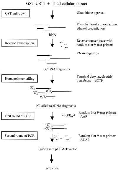
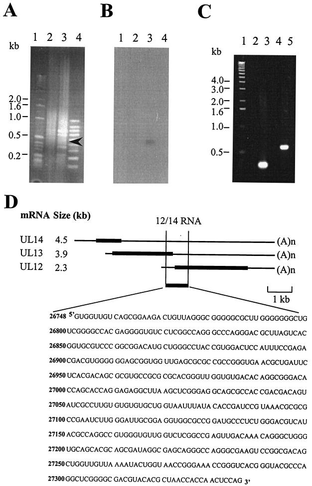
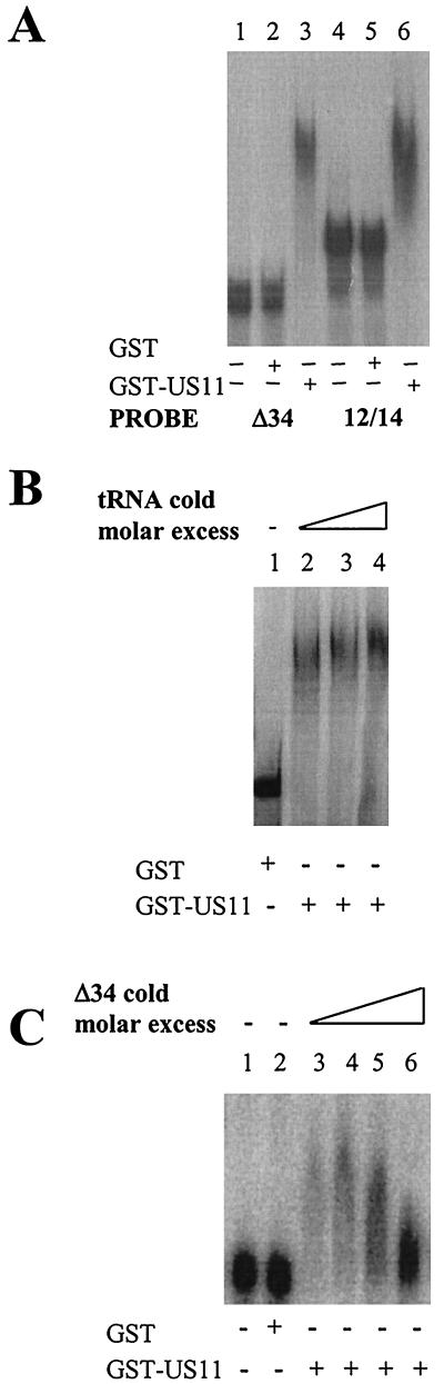
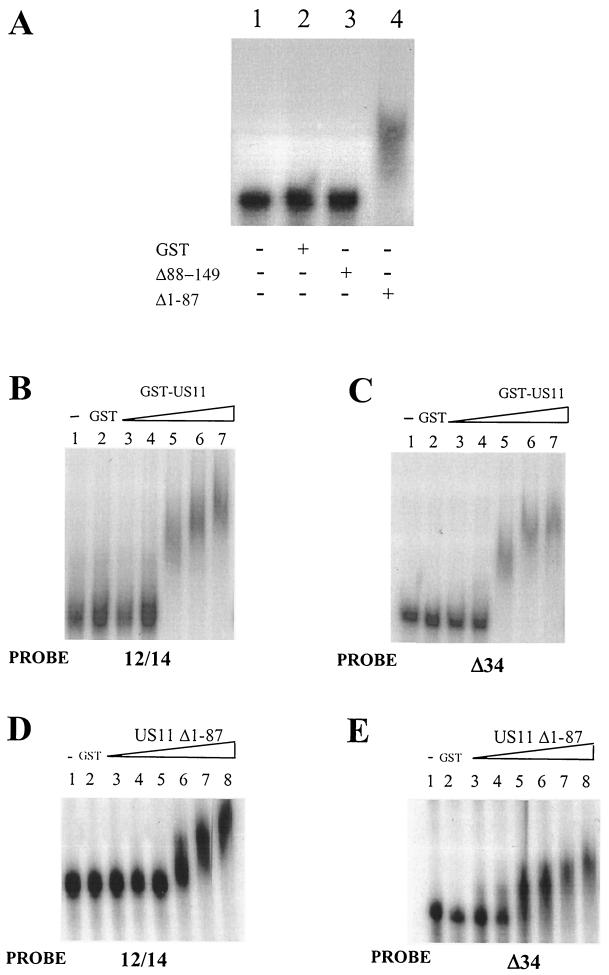
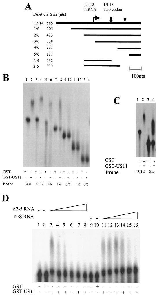
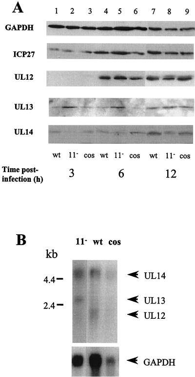
The US11 protein of herpes simplex virus type 1 (HSV-1) is a small, highly basic phosphoprotein expressed at late times during infection. US11 localizes to the nucleolus in infected cells, can associate with ribosomes, and has been shown to bind RNA. The RNA substrates of US11 identified thus far have no apparent role in the virus lytic cycle, so we set out to identify a novel, biologically relevant RNA substrate(s) for this protein in HSV-1-infected cells. We designed a reverse transcriptase PCR-based protocol that allowed specific selection of a 600-bp RNA binding partner for US11. This RNA sequence, designated 12/14, is present in the coterminal HSV-1 mRNAs UL12, UL13, and UL14. We show that the binding of US11 to 12/14 is sequence-specific and mediated by the C-terminal domain of the protein. To elucidate the role of US11 in the virus life cycle, we infected cells with wild-type virus, a cosmid-reconstructed US11 HSV-1 null mutant, and a cosmid-reconstructed wild-type virus and analyzed expression of UL12, -13, and -14 during a time course of infection. These experiments revealed that this interaction has biological activity; at early times of infection, US11 down-regulates UL13 protein kinase mRNA and protein.


Figures

FIG. 1.
FIG. 1.
Scheme for the isolation and identification of RNAs that bind US11. The thin straight lines represent RNA molecules. The thick straight lines represent single-stranded (ss) cDNA molecules. The doubly thick straight lines represent double-stranded cDNA molecules. The solid hooked lines represent single-stranded random primers. The dotted hooked lines represent 5′ RACE primers (Life Technologies). Abbreviations: AAP, abridged anchor primer; AUAP, abridged universal amplification primer; (C)n, dC tail.
FIG. 2.
FIG. 2.
Analysis of the products of the 6-mer and 9-mer RT-PCRs of the US11 RNA pull down. (A) Fractionation of the products of the 6-mer RT-PCR on an ethidium bromide-stained agarose gel. Lanes: 1, 1-kb ladder marker; 2, RT-PCR with RNA isolated using GST alone in the pull down; 3, RT-PCR with RNA isolated using GST-US11 fusion protein in the pull down (the arrowhead indicates the band that hybridizes to the Δ34 probe in the blot in panel B); 4, 100-bp marker. (B) Southern blot of the gel shown in panel A hybridized with an in vitro-transcribed 32P-labeled Δ34 probe. (C) Fractionation of the products of the 9-mer RT-PCR on an ethidium bromide-stained agarose gel. Lanes: 1, 1-kb ladder marker; 2, control RT-PCR using Δ34-specific primers and RNA isolated using GST alone in the pull down; 3, control RT-PCR using Δ34-specific primers and RNA isolated using GST-US11 fusion protein in the pull down; 4, RT-PCR with random and anchor primers and RNA isolated using GST alone in the pull down; 5, RT-PCR with random and anchor primers and RNA isolated using GST-US11 fusion protein in the pull down. (D) Diagram of the three coterminal transcripts UL12, UL13, and UL14 showing the position of the US11-pulled-out 12/14 RNA and the sequence of the 585-nt 12/14 RNA. Thick lines, coding regions; thin lines, 5′ and 3′ UTRs; (A)n, end of the message.
FIG. 3.
FIG. 3.
EMSA analysis of binding of radiolabeled 12/14 RNA to US11 protein. (A) EMSA of the binding of US11 to 32P-labeled RNAin vitro transcribed from PCR-amplified Δ34 or 12/14 DNA. Protein (0.25 μg) was added (+) in each reaction. Lanes 1 and 4, no added protein; lanes 2 and 5, GST alone; lanes 3 and 6, GST-US11 fusion protein. (B) EMSA of US11/-12/-14 binding in the presence of nonspecific competitor tRNA. Lanes: 1, 12/14 probe incubated with GST protein; 2 to 4, 12/14 probe and GST-US11 protein bound in the presence of increasing concentrations of tRNA. (C) EMSA of binding of US11 to 32P-labeled 12/14 RNA in the presence of cold specific competitor Δ34 RNA. Lanes: 1, no protein; 2, GST protein; 3 to 6, 12/14 probe (1.5 pmol) and GST-US11 protein bound in the presence of increasing concentrations (one- to eightfold molar excess) of unlabeled competitor Δ34 RNA.
FIG. 4.
FIG. 4.
EMSA of binding of wild-type and truncated US11 proteins to radiolabeled 12/14 RNA. (A) Binding of N- and C-terminal truncations of US11 to 12/14 RNA using 32P-labeled 12/14 probe. Lane 1, no protein; lane 2, GST protein; lane 3, C-terminal truncation; lane 4, N-terminal truncation. +, present; −, absent. (B) Multimerization of US11 on 12/14 RNA. Lane 1, no added protein; lane 2, GST protein; lanes 3 to 7, increasing concentrations (110 to 550 nM) of wild-type GST-US11 fusion protein. (C) Multimerization of US11 on Δ34 RNA. Lane 1, no added protein; lane 2, GST protein; lanes 3 to 7, increasing concentrations (110 to 550 nM) of wild-type GST-US11 fusion protein. (D) Multimerization of the US11 N-terminal truncation protein on 12/14 RNA. Lane 1, no added protein; lane 2, GST protein; lanes 3 to 8, increasing concentrations (150 to 1,500 nM) of GST-US11Δ1-87 fusion protein. (E) Multimerization of the US11 N-terminal truncation protein on Δ34 RNA. Lane 1, no added protein; lane 2, GST protein; lanes 3 to 8, increasing concentrations (150 to 1,500 nM) of GST-US11Δ1-64 fusion protein.
FIG. 5.
FIG. 5.
Deletion analysis of the US11 binding site on 12/14 RNA. (A) Diagram of the 12/14 RNA showing locations of the deletion probes used. The bent arrow indicates the start of the UL12 RNA. The open arrow indicates the position of the UL13 stop codon. The arrowhead represents the start codon of the UL12 RNA. (B) EMSA of GST and GST-US11 protein binding to sequential 5′-3′ deletions of the 12/14 RNA. Protein (0.25 μg) was added in each reaction. The RNA probes used are indicated beneath the panel. (C) EMSA comparing GST-US11 binding to α-32P-labeled RNA in vitro transcribed from PCR-amplified 12/14 and 2-4 deletion DNAs. Lanes 1 and 3, GST protein; lanes 2 and 4, GST-US11 protein. +, present; −, absent. (D) EMSA of binding of US11 protein to α-32P-labeled Δ2-5 RNA in the presence of specific (Δ2-5 RNA, lanes 3 to 8) and nonspecific (N/S) (HPV-31 late 3′ UTR; lanes 11 to 16) cold competitor RNA. Protein (0.5 μg) was added in each reaction. Lanes 1 and 9, no protein; lanes 2 and 10, GST protein; lanes 3 to 8 and 11 to 16, GST-US11 protein.
FIG. 6.
FIG. 6.
US11 specifically regulates expression of UL13 RNA and protein. (A) Western blot analysis of levels of ICP27; UL12, -13, and -14 proteins; and GAPDH (to assess protein loading) during a time course of infection of HeLa cells with wild-type (wt), cosmid-reconstructed wild-type (cos), and US11 null mutant (11) viruses. Only half the amount of protein was electrophoresed in each lane for the 12-h samples. Mock-infected cell extracts did not show binding to any HSV-1 protein antibody (data not shown). Each blot was repeated three times with very similar results. (B) Northern blot analysis of abundances of UL12, -13, and -14 polyadenylated transcripts in HeLa cells at 6 h postinfection with wild-type, cosmid-reconstructed wild-type, and US11 deletion mutant viruses. The probe was α-32P-radiolabeled RNA in vitro transcribed from the cloned portion of the HSV-1 genome containing the UL12/-13/-14 coterminal RNAs. The bottom blot shows the result of stripping and rehybridizing the blot with a probe for GAPDH RNA. The blot was hybridized in 50% formamide-5× SSC at 42°C for 16 h and was washed to 0.1× SSC at 65°C.

References

    1. Brand, S. R., R. Kobayashi, and M. B. Matthews. 1997. The Tat protein of human immunodeficiency virus type 1 is a substrate and inhibitor of the interferon-induced, virally activated protein kinase, PKR. J. Biol. Chem. 272:8388-8395. - PubMed
    1. Brooks, S. A., and W. F. Rigby. 2000. Characterisation of the mRNA ligands bound by the RNA binding protein hnRNP A2 utilizing a novel in vivo technique. Nucleic Acids Res. 28:E49. - PMC - PubMed
    1. Cassady, K. A., M. Gross, and B. Roizman. 1998. The herpes simplex virus Us11 protein effectively compensates for the gamma134.5 gene if present before activation of protein kinase R by precluding its phosphorylation and that of the α subunit of eukaryotic translation initiation factor 2. J. Virol. 72:8620-8626. - PMC - PubMed
    1. Cassady, K. A., M. Gross, and B. Roizman. 1998. The second-site mutation in the herpes simplex virus recombinants lacking the gamma1 34.5 genes precludes shutoff of protein synthesis by blocking the phosphorylation of eIF-2α. J. Virol. 72:7005-7011. - PMC - PubMed
    1. Chou, J., and B. Roizman. 1992. The gamma134.5 gene of herpes simplex virus 1 precludes neuroblastoma cells from triggering total shutoff of protein synthesis characteristic of programmed cell death in neuronal cells. Proc. Natl. Acad. Sci. USA 89:3266-3270. - PMC - PubMed

MeSH terms