. 2002 Aug;76(16):8101-9.
doi: 10.1128/jvi.76.16.8101-8109.2002.

Role of a highly conserved NH(2)-terminal domain of the human parainfluenza virus type 3 RNA polymerase

Affiliations

Role of a highly conserved NH(2)-terminal domain of the human parainfluenza virus type 3 RNA polymerase

Achut G Malur et al. J Virol. 2002 Aug.

Abstract

The RNA polymerase complex of human parainfluenza virus type 3 (HPIV 3), a member of the family Paramyxoviridae, is composed of two virally encoded polypeptides: a multifunctional large protein (L, 255 kDa) and a phosphoprotein (P, 90 kDa). From extensive deduced amino acid sequence analyses of the cDNA clones of a number of L proteins of nonsegmented negative-strand RNA viruses, a cluster of high-homology sequence segments have been identified within the body of the L proteins. Here, we have focused on the NH(2)-terminal domain of HPIV 3 L protein that is also highly conserved. Following mutational analyses within this domain, we examined the ability of the mutant L proteins to (i) transcribe an HPIV 3 minireplicon, (ii) transcribe the viral RNA in vitro using the HPIV 3 nucleocapsid RNA template, and (iii) interact with HPIV 3 P protein. Our results demonstrate that the first 15 amino acids of the NH(2)-terminal domain spanning a highly conserved motif is directly involved in transcription of the genome RNA and in forming a functional complex with the P protein. Substitution of eight nonconserved amino acids within this domain by the corresponding Sendai virus L protein residues yielded mutants with variable transcriptional activities. However, one mutant in which all eight amino acids were replaced with the corresponding residues of Sendai virus L protein failed to both transcribe the minireplicon and interact with HPIV 3 P and the Sendai virus P protein. The possible functional significance of the NH(2)-terminal domain of paramyxovirus L protein is discussed.


Figures

FIG. 1.
FIG. 1.
Schematic representation of paramyxovirus L proteins. The arbitrary locations of the conserved domains (I to VI) are indicated by boxes. The amino-terminal sequences of paramyxovirus L proteins were aligned by a CLUSTAL sequence program. Dashes represents gaps introduced to optimize alignment. Stretches of strictly or conservatively maintained amino acids are shaded and are shown in boxes. PIV3, human parainfluenza virus type 3; SV, Sendai virus; MV, measles virus; CDV, canine distemper virus; SV5, simian virus 5; PIV 2, human parainfluenza virus type 2; MU, mumps virus; NDV, New Castle disease virus.
FIG. 2.
FIG. 2.
(A) Activity of mutant L proteins in minigenome replication in vivo. HeLa cells were infected with MVA and transfected with pHPIV3-MG(-) and the support plasmids encoding N, P, and Lwt or Lmut as detailed in Materials and Methods. At 24 h postinfection, cell extracts were prepared and luciferase activity was determined. The data are the averages of three independent experiments ± standard deviation. (B) Primer extension analysis of positive-sense RNA products using total RNA extract. Primer extension was performed as described in Materials and Methods from total RNA extracted from HeLa cells transfected with plasmids as described for panel A. The dideoxy sequencing ladder was made using the same primer and pHPIV3-MG(-) as the template. Relevant regions of the gel with bands corresponding to the transcription (trans) and replication (rep) products are shown. Mock, vaccinia virus-infected and mock-transfected cell extract. (C) In vitro transcription of HPIV 3 L mutants. HeLa cells were infected with vTF7-3 and transfected with Lwt or Lmut along with P plasmid. Cell extracts containing the coexpressed L and P proteins were used in transcription reaction mixtures containing N-RNA template in the presence of [32P]UTP, and the mRNA products were analyzed in a 5% polyacrylamide-urea gel. Mock, vaccinia virus-infected and mock-transfected cell extract. (D) Analysis of L-P interaction by coimmunoprecipitation assay in vitro. 35S-labeled cell extracts prepared after transfection as described for panel C were immunoprecipitated with anti-FLAG antibody and analyzed by SDS-PAGE followed by fluorography as described in Materials and Methods (top). Western blot analysis of the cell extracts using anti-RNP antibody (bottom). The positions of L and P proteins are indicated. Mock, vaccinia virus-infected and mock-transfected cell extract.
FIG. 3.
FIG. 3.
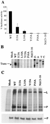
(A) Functional analysis of mutant L proteins by using luciferase assay. Cell lysates prepared 24 h posttransfection were assayed for luciferase activity as detailed in Materials and Methods and as described for Fig. 2A. (B) Analysis of positive-sense RNA products by primer extension. Total RNA extracted after infection and transfection with plasmids was used in primer extension as described in Materials and Methods and for Fig. 2B. Relevant regions of the gel with the bands corresponding to the transcription (trans) products are shown. Mock, vaccinia virus-infected and mock-transfected cell extract. (C) L-P complex formation with HPIV 3 L mutants. [35S]methionine-labeled cell extracts were immunoprecipitated with anti-FLAG antibody and analyzed by SDS-PAGE followed by fluorography (top) as described in Materials and Methods and for Fig. 2D. Shown is a Western blot analysis of the cell extracts using anti-RNP antibody (bottom). The positions of L and P proteins are indicated. Mock, vaccinia virus-infected and mock-transfected cell extract.
FIG. 4.
FIG. 4.
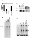
(A) Sequence alignment of the amino termini of HPIV 3 (top) and Sendai virus L (bottom) proteins. The nonconserved amino acid residues are shown in boxes. (B) Luciferase activity from cell extracts was measured for mutant L proteins as described in Materials and Methods and for Fig. 2A. (C) Primer extension was performed using total RNA extract transfected with plasmids as detailed in Materials and Methods and for Fig. 2B. Relevant regions of the gel with the bands corresponding to the transcription (trans) and replication (rep) products are shown. (D) Coimmunoprecipitation of L and P proteins in vitro. 35S-labeled cell extracts were immunoprecipitated as described for Fig. 3C (top). Shown is a Western blot analysis of the cell extracts using anti-RNP antibody (bottom). The positions of L and P proteins are indicated. Mock, vaccinia virus-infected and mock-transfected cell extract.
FIG. 5.
FIG. 5.
(A) Sequences of mutant HPIV 3 L proteins. The amino-terminal residues of HPIV 3 and Sendai virus L proteins shown. The amino acid residue(s) within the boxes represent the corresponding residue(s) from Sendai virus L protein that were substituted within the HPIV 3 L protein. (B) Analysis of mutant L proteins using luciferase assay. The luciferase activity was determined for mutant L proteins as described in Materials and Methods and for Fig. 2A. (C) L-P complex formation between HPIV 3 L mutants and P in vitro. [35S]methionine-labeled cell extracts were immunoprecipitated and analyzed by SDS-PAGE followed by fluorography as described earlier in Materials and Methods and for Fig. 2D (top). Shown is a Western blot analysis of the cell extracts using anti-RNP antibody (bottom). Mock, vaccinia virus-infected and mock-transfected cell extract.
FIG. 6.
FIG. 6.
Interaction of HPIV 3 L mutants with HPIV 3 P and Sendai virus P protein (SV-P). HeLa cells were infected with vTF7-3 and transfected with Lwt or Lmut plasmids along with HPIV 3 P plasmid or Sendai virus P plasmid as indicated. [35S]methionine-labeled cell extracts were immunoprecipitated with anti-FLAG antibody conjugated to agarose beads as described in Materials and Methods and for Fig. 2D or anti-SV-P antibody (MAb M56) previously conjugated to Sepharose A and analyzed by SDS-PAGE followed by fluorography. Mock, vaccinia virus-infected and mock-transfected cell extract.

References

    1. Banerjee, A. K. 1987. Transcription and replication of rhabdoviruses. Microbiol. Rev. 51:66-87. - PMC - PubMed
    1. Banerjee, A. K., S. Barik, and B. P. De. 1991. Gene expression of nonsegmented negative strand RNA viruses. Pharmacol. Ther. 51:47-70. - PubMed
    1. Canter, D. M., R. L, Jackson, and J. Perrault. 1993. Faithful and efficient in vitro reconstitution of vesicular stomatitis virus transcription using plasmid-encoded L and P proteins. Virology 194:518-529. - PubMed
    1. Chandrika, R., S. M. Horikami, S. Smallwood, and S. A. Moyer. 1995. Mutations in conserved domain I of the Sendai virus L polymerase protein uncouple transcription and replication. Virology 213:352-363. - PubMed
    1. Collins, P. L., R. M. Chanock, and K. McIntosh. 1996. Parainfluenza viruses, p. 1205-1241. In B. N. Fields, D. M. Knipe, and P.M. Howley (ed.), Fields virology, 3rd ed. Lippincott-Raven Publishers, Philadelphia, Pa.

Publication types

MeSH terms

LinkOut - more resources