. 2002 Aug;76(16):8169-78.
doi: 10.1128/jvi.76.16.8169-8178.2002.

Bacteriophage PM2 has a protein capsid surrounding a spherical proteinaceous lipid core

Affiliations

Bacteriophage PM2 has a protein capsid surrounding a spherical proteinaceous lipid core

Hanna M Kivelä et al. J Virol. 2002 Aug.

Abstract

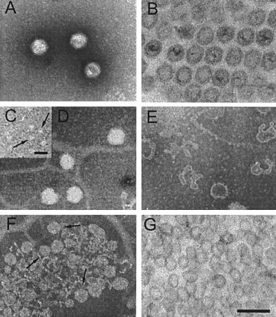
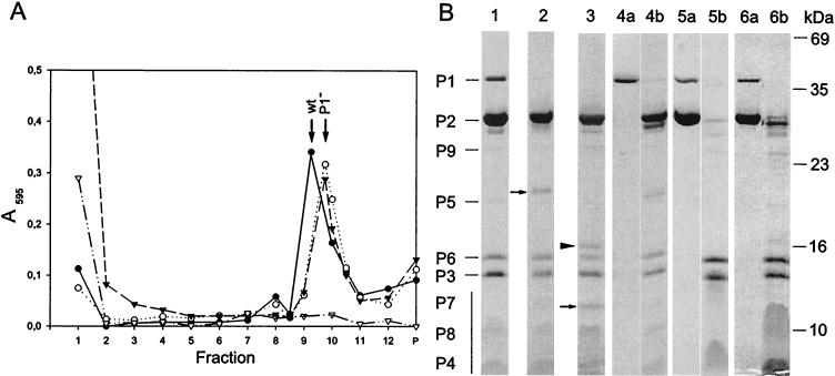
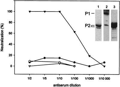
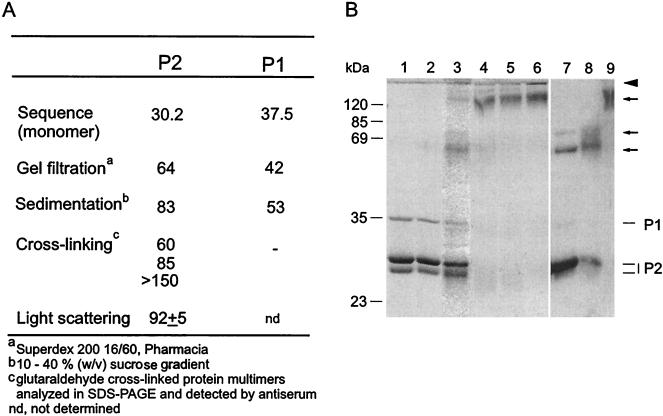
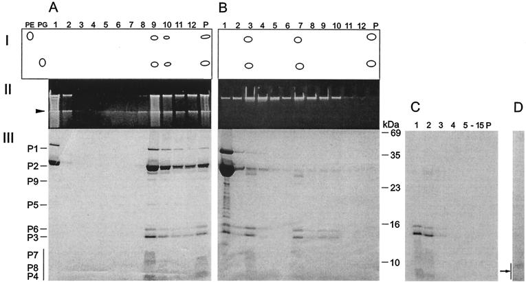
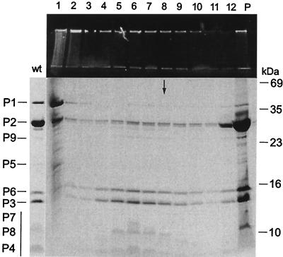
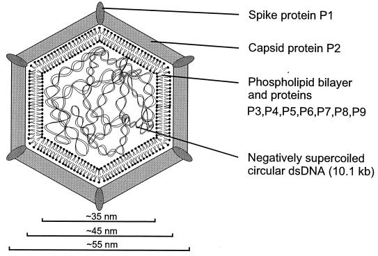
The marine double-stranded DNA (dsDNA) bacteriophage PM2, studied since 1968, is the type organism of the family Corticoviridae, infecting two gram-negative Pseudoalteromonas species. The virion contains a membrane underneath an icosahedral protein capsid composed of two structural proteins. The purified major capsid protein, P2, appears as a trimer, and the receptor binding protein, P1, appears as a monomer. The C-terminal part of P1 is distal and is responsible for receptor binding activity. The rest of the structural proteins are associated with the internal phospholipid membrane enclosing the viral genome. This internal particle is designated the lipid core. The overall structural organization of phage PM2 resembles that of dsDNA bacteriophage PRD1, the type organism of the family TECTIVIRIDAE:


Figures

FIG. 1.
FIG. 1.
Electron micrographs of PM2 virions and subviral particles. (A and B) Negative-stain electron microscopy (A) and thin-section electron microscopy (B) of untreated PM2 virions. (C to E) Fresh PM2 virions disrupted in water prior to negative staining. (C) Soluble fraction containing capsid proteins P1 and P2. Released structures were identified as multimers of protein P2 (arrows) by negative staining of purified protein P2 (see the text). (D) Spherical lipid cores. (E) Pleomorphic PM2 membrane vesicles. (F and G) PM2 lipid cores obtained after dissociation of the virion with 4.5 M urea. (F) Negatively stained lipid cores (arrows) purified by rate zonal centrifugation (see Fig. 5B, lane 7). (G) Thin section of lipid cores. Bars, 100 nm in A, B, and D to G and 20 nm in C.
FIG. 2.
FIG. 2.
(A) Sedimentation analysis of fresh PM2 virions (filled circles), protein P1-deficient particles obtained by proteinase K treatment (50 μg/ml; open circles) or bromelain treatment (5 mg/ml; filled triangles), and particles after freezing and thawing (open triangles). Protein concentrations in the fractions and in the pellet (P) were assayed by the Coomassie blue method. Sedimentation positions of untreated virions (wt) and protease-treated particles (P1) are indicated. (B) SDS-PAGE analysis of dissociated particles. Protein patterns of the peak fractions of PM2 virions (lane 1) and particles treated with either proteinase K (lane 2) or bromelain (lane 3) are shown. N-terminal amino acid sequences were determined for the new protein bands which appeared after protease treatments (arrows; see the text). The protein band marked by an arrowhead (lane 3) was derived from the bromelain enzyme. PM2 dissociation products either in 20 mM Tris-HCl (pH 7.2) or in sterilized water were separated in a sucrose gradient; the top fraction and the pellet are shown in lanes 4a and 4b, respectively. Calcium ions were removed from the virions with a 20 M excess of EGTA; the top fraction and the pellet are shown in lanes 5a and 5b, respectively. For particles after one cycle of freezing and thawing, the top fraction (lane 6a) and the pellet (lane 6b) are shown. The positions of the PM2 structural proteins are indicated on the left (the vertical bar denotes the small membrane proteins forming diffuse bands), and the molecular masses of the standard proteins are indicated on the right.
FIG. 3.
FIG. 3.
Neutralization of PM2 virions with anti-P1 serum (filled circles) and anti-P2 serum (filled triangles). P1 preimmune serum (open circles) and P2 preimmune serum (open triangles) were used as negative controls. Purified P2 protein used for the production of polyclonal anti-P2 serum is shown on a Coomassie brilliant blue-stained SDS-polyacrylamide gel (lane 1). The specificities of anti-P1 serum (1:20,000 dilution) and anti-P2 serum (1:100,000 dilution) are shown in lanes 2 and 3 (Western blots). The positions of proteins P1 and P2 determined from the corresponding Coomassie brilliant blue-stained gel are indicated on the left.
FIG. 4.
FIG. 4.
(A) Sizes of PM2 capsid proteins P1 and P2 (kilodaltons) obtained by different methods. (B) Cross-linking of proteins with glutaraldehyde. Lanes 1 to 6 show a Coomassie brilliant blue-stained polyacrylamide gel, and lanes 7 to 9 show a Western blot with anti-P2 serum. Non-cross-linked proteins P1 and P2 are shown in lane 1, and their positions are indicated on the right. Mixtures of proteins P1 and P2 (250 μg of protein/ml) were cross-linked with increasing glutaraldehyde concentrations (0.001% [vol/vol] [lane 2], 0.01% [lane 3], 0.05% [lane 4], 0.1% [lane 5], and 0.5% [lane 6]) as described in Materials and Methods. Multimeric forms detected by anti-P2 serum in samples cross-linked with 0.01% (lane 7), 0.05% (lane 8), and 0.1% (lane 9) glutaraldehyde are indicated by arrows. The boundary between the upper gel and the lower gel is indicated by an arrowhead. Numbers on the left indicate the molecular masses of the standard proteins.
FIG. 5.
FIG. 5.
Dissociation of PM2 particles with urea and separation of the products by rate zonal centrifugation (see Materials and Methods). (A) Untreated particles. (B) Particles dissociated with 4.5 M urea. (Panel I) Major lipids separated by thin-layer chromatography with PE and PG as standards. (Panel II) Viral DNA in upper SDS-polyacylamide gel stained with ethidium bromide. (Panel III) Structural proteins in Coomassie brilliant blue-stained SDS-polyacrylamide gel. P, pellet. The boundary between the upper gel and the lower gel is indicated by an arrowhead. The positions of the PM2 structural proteins are indicated on the left (the vertical bar denotes the small membrane proteins forming diffuse bands). The numbers to the right of panel B indicate the molecular masses of the standard proteins. (C) Isolated phage lipid cores in 4.5 M urea were solubilized with 0.1% SDS and analyzed with a rate zonal sucrose gradient containing 2 M urea, 0.1% SDS, and PM2 buffer. Fifteen fractions and the pellet were collected and analyzed by SDS-PAGE. The area indicated by an arrow was subjected to N-terminal amino acid sequencing. (D) Isolated phage lipid cores in 4.5 M urea were treated with trypsin (50 μg/ml) and soybean trypsin inhibitor (500 μg/ml) and analyzed with a rate zonal sucrose gradient (PM2 buffer). One visible light-scattering zone was detected and analyzed by SDS-PAGE. No other PM2 proteins were detected in the gradient fractions.
FIG. 6.
FIG. 6.
Reconstitution of bacteriophage PM2. PM2 virions were dissociated with 4.5 M urea-20 mM 2-mercaptoethanol in PM2 buffer. The separation of dissociated phage components by rate zonal centrifugation is shown in Fig. 5B. Reconstitution was carried out by dialyzing the dissociation mixture against a reduced urea concentration with 20 mM 2-mercaptoethanol in PM2 buffer overnight at 4°C and was followed by rate zonal centrifugation (see Materials and Methods). Viral DNA and protein compositions of collected gradient fractions and the pellet (P) were analyzed by SDS-PAGE with an ethidium bromide-stained upper gel and a Coomassie brilliant blue-stained separation gel. The figure shows the outcome of the experiment after dialysis against 0.5 M urea (for results obtained with other concentrations, see the text). The sedimentation position of PM2 virions is indicated by an arrow. The protein pattern of intact virions (wt) is indicated on the left (the vertical bar denotes the small membrane proteins forming diffuse bands), and the molecular masses of the standard proteins are indicated on the right.
FIG. 7.
FIG. 7.
Schematic representation of PM2 virion organization.

Similar articles

Cited by

References

    1. Akutsu, H., H. Satake, and R. M. Franklin. 1980. Phosphorus nuclear magnetic resonance studies on the lipid-containing bacteriophage PM2. Biochemistry 19:5264-5270. - PubMed
    1. Armour, G. A., and G. J. Brewer. 1990. Membrane morphogenesis from cloned fragments of bacteriophage PM2 DNA that contain the sp6.6 gene. FASEB J. 4:1488-1493. - PubMed
    1. Bamford, D. H., J. Caldentey, and J. K. H. Bamford. 1995. Bacteriophage PRD1: a broad host range dsDNA tectivirus with an internal membrane. Adv. Virus Res. 45:281-319. - PubMed
    1. Bamford, D. H., and L. Mindich. 1980. Electron microscopy of cells infected with nonsense mutants of bacteriophage φ6. Virology 107:222-228. - PubMed
    1. Bamford, D. H., and L. Mindich. 1982. Structure of the lipid-containing bacteriophage PRD1: disruption of wild-type and nonsense mutant phage particles with guanidine hydrochloride. J. Virol. 44:1031-1038. - PMC - PubMed

Publication types

LinkOut - more resources