. 2002 Aug;76(16):8189-99.
doi: 10.1128/jvi.76.16.8189-8199.2002.

Alpha interferon inhibits hepatitis C virus replication in primary human hepatocytes infected in vitro

Affiliations

Alpha interferon inhibits hepatitis C virus replication in primary human hepatocytes infected in vitro

Valérie Castet et al. J Virol. 2002 Aug.

Abstract

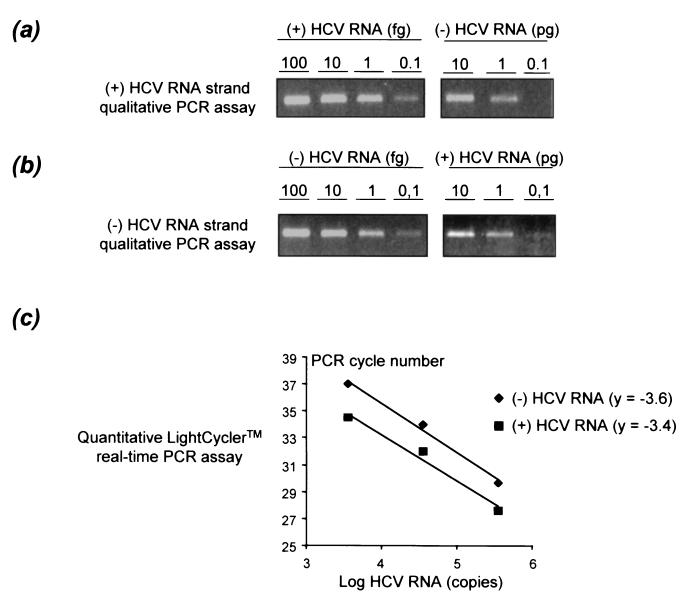
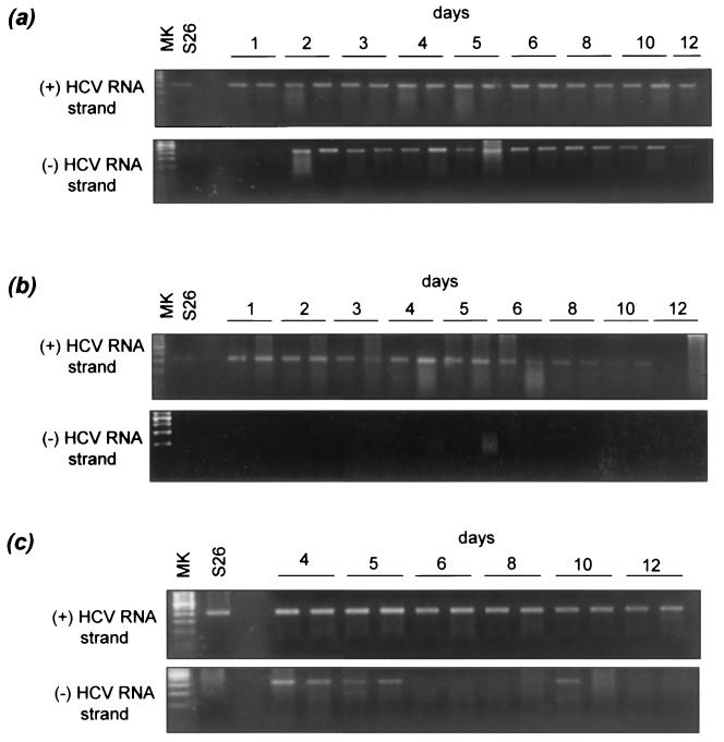
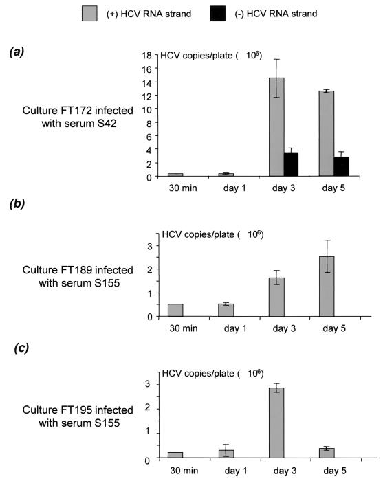
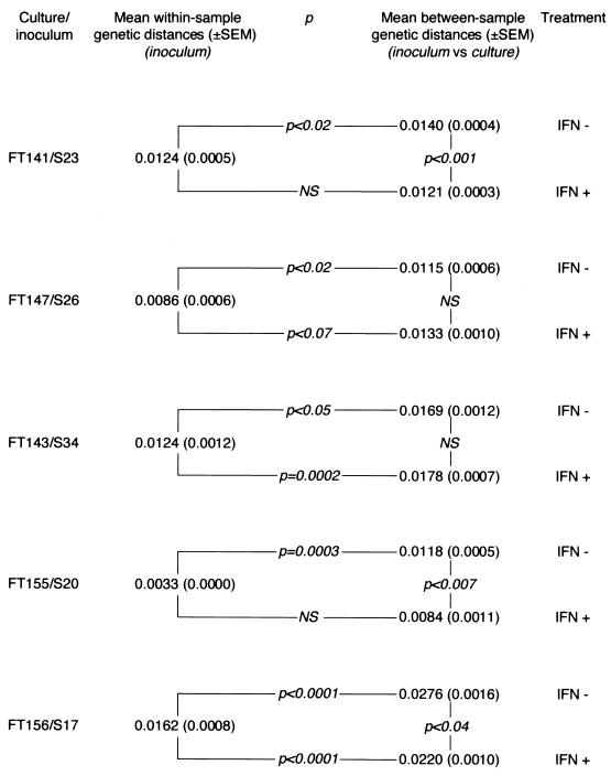
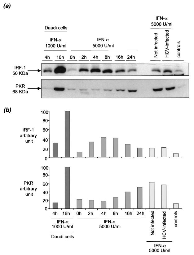
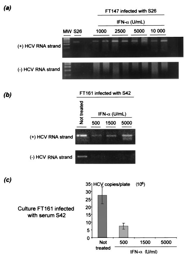
Chronic hepatitis C is a common cause of liver disease, the complications of which include cirrhosis and hepatocellular carcinoma. Treatment of chronic hepatitis C is based on the use of alpha interferon (IFN-alpha). Recently, indirect evidence based on mathematical modeling of hepatitis C virus (HCV) dynamics during human IFN-alpha therapy suggested that the major initial effect of IFN-alpha is to block HCV virion production or release. Here, we used primary cultures of healthy, uninfected human hepatocytes to show that: (i) healthy human hepatocytes can be infected in vitro and support HCV genome replication, (ii) hepatocyte treatment with IFN-alpha results in expression of IFN-alpha-induced genes, and (iii) IFN-alpha inhibits HCV replication in infected human hepatocytes. These results show that IFN-alpha acts primarily through its nonspecific antiviral effects and suggest that primary cultures of human hepatocytes may provide a good model to study intrinsic HCV resistance to IFN-alpha.


Figures

FIG. 1.
FIG. 1.
Characteristics of the strand-specific HCV RNA assays used in this study. (a) Strand specificity of the positive-strand-specific HCV RNA rTth RT-PCR assay. Decreasing amounts of positive-strand (+) HCV RNA (100, 10, 1, and 0.1 fg) and of negative-strand (-) HCV RNA (10, 1, and 0.1 pg) synthesized from an appropriate plasmid were subjected to the rTth RT-PCR assay. The products were analyzed by agarose gel electrophoresis. (b) Strand specificity of the negative-strand-specific HCV RNA rTth RT-PCR assay. Decreasing amounts of negative-strand HCV RNA (100, 10, 1, and 0.1 fg) and positive-strand HCV RNA (10, 1, and 0.1 pg) synthesized from the same plasmid as for panel a were analyzed by the same procedure. (c) Range of linear quantification of the quantitative assay based on real-time PCR using the LightCycler technology and SYBR green I dye for detection. The range of linear quantification of the assay was studied by testing 10-fold serial dilutions of synthetic positive- and negative-sense HCV RNA strands after RT at 70°C with the rTth polymerase. Each point is the mean of three experimental values for each dilution. y is the slope of the linear plots.
FIG. 2.
FIG. 2.
Qualitative assay detection of positive- and negative-strand HCV RNA in a primary culture of healthy human hepatocytes infected in vitro with an HCV-positive serum and effect of IFN-α. The hepatocyte culture FT147, infected 3 days after plating by HCV-positive serum S26, is shown. Positive-strand (+) RNA but not negative-strand (-) RNA was present in the inoculum. (a) Primary hepatocyte culture in the absence of IFN-α. Positive-strand HCV RNA was detected with the qualitative strand-specific rTth PCR assay from day 1 to the end of the culture (day 12), whereas negative-strand RNA was detected from days 2 to 10. (b) Culture in the presence of 5,000 U of IFN-α per ml. Positive-strand HCV RNA was detected from days 1 to 10, whereas negative-strand RNA was never detected. (c) Culture treated on day 3 with 5,000 U of IFN-α per ml. Positive-strand RNA was detected throughout the culture period, whereas negative-strand RNA was no longer detected after day 5. Similar patterns (not shown) were observed with the following cultures infected with the corresponding sera: FT141 and S23, FT143 and S34, FT144 and S27, FT154 and S23, FT155 and S20, and FT156 and S17. MK, molecular size standards.
FIG. 3.
FIG. 3.
Accumulation of positive- and negative-strand HCV RNA in hepatocyte cultures FT172 (a), FT189 (b), and FT195 (c), infected with sera S42, S155, and S155, respectively, as measured by the quantitative LightCycler real-time RT-PCR assay. The hepatocyte cultures were infected 3 days after plating. The cells were harvested 30 min and 1, 3, and 5 days after infection for positive-strand (gray) and negative-strand (black) HCV RNA quantification. The amounts of HCV RNA strands are shown as means ± SEMs of three determinations, expressed in numbers of HCV RNA copies per 2 × 10⁶ cells, normalized to GAPDH mRNA. Similar results (not shown) were obtained with culture FT168 infected with serum S34.
FIG. 4.
FIG. 4.
Accumulation of nucleotide substitutions on HCV genomes during replication in five primary cultures of healthy human hepatocytes in the presence and absence of IFN-α. The accumulation of mutations on HCV genomes was assessed by comparing the mean ± SEM within-sample genetic distance (calculated by pairwise comparison of NS5A quasispecies sequences in the inoculum) with the mean ± SEM between-sample genetic distance (calculated by pairwise comparison of NS5A quasispecies sequences in the culture versus the inoculum). A significantly higher between-sample than within-sample genetic distance was interpreted as a significant accumulation of genomic mutations over time as a result of HCV replication in the culture; the lack of significant difference was interpreted as a lack of genetic evolution in the culture, reflecting inhibition of HCV replication. The between-sample genetic distances were also compared for each culture in the presence (+) and absence (-) of IFN-α. A significantly smaller between-sample genetic distance in the presence of IFN-α reflected reduced accumulation of mutations in the culture and was interpreted as an inhibition of HCV replication by IFN-α. NS, not significantly different (i.e., P > 0.05).
FIG. 5.
FIG. 5.
Effects of IFN-α on IRF-1 and PKR expression in primary cultures of human hepatocytes. Immunoblot analysis was performed with anti-IRF-1 and anti-PKR antibodies after 8 days of culture in infected and noninfected primary hepatocytes treated with 5,000 U of IFN-α per ml and in Daudi cells treated with 1,000 U of IFN-α per ml used as positive controls. Cells not treated with IFN-α were used as controls. Immunoblot experimental results (a), together with their quantitative representation after National Institutes of Health image analysis (b) are shown. (b) (Left) Effect of 1,000 U of IFN-α per ml on Daudi cells harvested after 4 and 16 h of treatment. (Center) Effect of 5,000 U of IFN-α per ml on hepatocyte culture FT172 harvested after 0, 2, 4, 8, 16, and 24 h of treatment. (Right) Effect of HCV infection of the primary hepatocyte culture on the effect of IFN-α on IRF-1 and PKR expression. Similar results (not shown) were obtained in cultures FT164 and FT171.
FIG. 6.
FIG. 6.
Effect of increasing concentrations of IFN-α on the accumulation of positive- and negative-strand HCV RNA in primary hepatocyte cultures infected in vitro. Cultures FT147 (infected with serum S26) and FT161 (infected with serum S42) were treated for 5 and 8 days with IFN-α concentrations ranging from 1,000 to 10,000 U/ml and 500 to 5,000 U/ml, respectively. Qualitative detection of positive-sense (+) and negative-sense (-) HCV RNA strands is shown in cultures FT147 (a) and FT161 (b). In both instances, positive-strand HCV RNA was detected at all concentrations used, whereas the negative strand was never detected. MW, molecular size standards. (c) In culture FT161, LightCycler real-time RT-PCR quantitative analysis of the same extracts showed a reduction in the amount of positive-sense HCV RNA strand in the culture when the IFN-α concentration increased, suggesting IFN-α concentration-dependent inhibition of HCV replication in the culture. Similar results (not shown) were obtained with culture FT187 infected with serum S155.

Similar articles

Cited by

References

    1. Agnello, V., G. Abel, G. B. Knight, and E. Muchmore. 1998. Detection of widespread hepatocyte infection in chronic hepatitis C. Hepatology 28:573-584. - PubMed
    1. Alter, H. J., and L. B. Seeff. 2000. Recovery, persistence, and sequelae in hepatitis C virus infection: a perspective on long-term outcome. Semin. Liver Dis. 20:17-35. - PubMed
    1. Chieux, V., D. Hober, J. Harvey, G. Lion, D. Lucidarme, G. Forzy, M. Duhamel, J. Cousin, H. Ducoulombier, and P. Wattre. 1998. The MxA protein levels in whole blood lysates of patients with various viral infections. J. Virol. Methods 70:183-191. - PubMed
    1. Chung, R. T., W. He, A. Saquib, A. M. Contreras, R. J. Xavier, A. Chawla, T. C. Wang, and E. V. Schmidt. 2001. Hepatitis C virus replication is directly inhibited by IFN-alpha in a full-length binary expression system. Proc. Natl. Acad. Sci. USA 98:9847-9852. - PMC - PubMed
    1. Diamond, M. S., T. G. Roberts, D. Edgil, B. Lu, J. Ernst, and E. Harris. 2000. Modulation of dengue virus infection in human cells by alpha, beta, and gamma interferons. J. Virol. 74:4957-4966. - PMC - PubMed

Publication types

MeSH terms

LinkOut - more resources