

Studies of the mechanism of transactivation of the adeno-associated virus p19 promoter by Rep protein
- PMID: 12134028
- PMCID: PMC155137
- DOI: 10.1128/jvi.76.16.8225-8235.2002
Studies of the mechanism of transactivation of the adeno-associated virus p19 promoter by Rep protein
Abstract
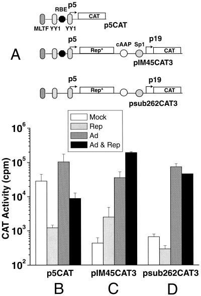
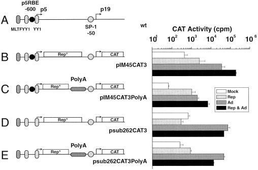
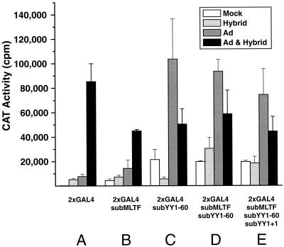
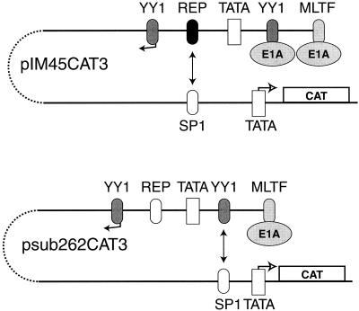
During adeno-associated virus (AAV) type 2 productive infections, the p19 promoter of AAV is activated by the AAV Rep78 and Rep68 proteins. Rep-induced activation of p19 depends on the presence of one of several redundant Rep binding elements (RBEs) within the p5 promoter or within the terminal repeats (TR). In the absence of the TR, the p5 RBE and the p19 Sp1 site at position -50 are essential for p19 transactivation. To determine how a Rep complex bound at p5 induces transcription at p19, we made a series of p19 promoter chloramphenicol acetyltransferase constructs in which the p5 RBE was inserted at different locations upstream or downstream of the p19 mRNA start site. The RBE acted like a repressor element at most positions in the presence of both Rep and adenovirus (Ad), and the level of repression increased dramatically as the RBE was inserted closer to the p19 promoter. We concluded that the RBE by itself was not a conventional upstream activation signal and instead behaved like a repressor. To understand how the Rep-RBE complex within p5 activated p19, we considered the possibility that its role was to function as an architectural protein whose purpose was to bring other p5 transcriptional elements to the p19 promoter. In order to address this possibility, we replaced both the p5 RBE and the p19 Sp1 site with GAL4 binding sites. The modified GAL4-containing constructs were cotransfected with plasmids that expressed GAL4 fusion proteins capable of interacting through p53 and T-antigen (T-ag) protein domains. In the presence of Ad and the GAL4 fusion proteins, the p19 promoter exhibited strong transcriptional activation that was dependent on both the GAL4 fusion proteins and Ad infection. This suggested that the primary role of the p5 RBE and the p19 Sp1 sites was to act as a scaffold for bringing transcription complexes in the p5 promoter into close proximity with the p19 promoter. Since Rep and Sp1 themselves were not essential for transactivation, we tested mutants within the other p5 transcriptional elements in the context of GAL4-induced looping to determine which of the other p5 elements was necessary for p19 induction. Mutation of the p5 major late-transcription factor site reduced p19 activity but did not eliminate induction in the presence of the GAL4 fusion proteins. However, mutation of the p5 YY1 site at position -60 (YY1-60) eliminated GAL4-induced transactivation. This implicated the YY1-60 protein complexes in p19 induction by Rep. In addition, both basal p19 activity and activity in the presence of Ad increased when the YY1-60 site was mutated even in the absence of Rep or GAL4 fusion proteins. Therefore, there are likely to be alternative p5-p19 interactions that are Rep independent in which the YY1-60 complex inhibits p19 transcription. We concluded that transcriptional control of the p19 promoter was dependent on the formation of complexes between the p5 and p19 promoters and that activation of the p19 promoter depends largely on the ability of Rep and Sp1 to form a scaffold that positions the p5 YY1 complex near the p19 promoter.
Figures


Similar articles
-
The adeno-associated virus (AAV) Rep protein acts as both a repressor and an activator to regulate AAV transcription during a productive infection.J Virol. 1997 Feb;71(2):1079-88. doi: 10.1128/JVI.71.2.1079-1088.1997. J Virol. 1997. PMID: 8995628 Free PMC article.
-
The adeno-associated virus type 2 p40 promoter requires a proximal Sp1 interaction and a p19 CArG-like element to facilitate Rep transactivation.J Virol. 1997 Jun;71(6):4300-9. doi: 10.1128/JVI.71.6.4300-4309.1997. J Virol. 1997. PMID: 9151818 Free PMC article.
-
Analysis of adeno-associated virus (AAV) wild-type and mutant Rep proteins for their abilities to negatively regulate AAV p5 and p19 mRNA levels.J Virol. 1994 May;68(5):2947-57. doi: 10.1128/JVI.68.5.2947-2957.1994. J Virol. 1994. PMID: 8151765 Free PMC article.
-
Negative regulation of the adeno-associated virus (AAV) P5 promoter involves both the P5 rep binding site and the consensus ATP-binding motif of the AAV Rep68 protein.J Virol. 1995 Nov;69(11):6787-96. doi: 10.1128/JVI.69.11.6787-6796.1995. J Virol. 1995. PMID: 7474090 Free PMC article.
-
The Two Sides of YY1 in Cancer: A Friend and a Foe.Front Oncol. 2019 Nov 20;9:1230. doi: 10.3389/fonc.2019.01230. eCollection 2019. Front Oncol. 2019. PMID: 31824839 Free PMC article. Review.
Cited by
-
The Expression and Function of the Small Nonstructural Proteins of Adeno-Associated Viruses (AAVs).Viruses. 2024 Jul 29;16(8):1215. doi: 10.3390/v16081215. Viruses. 2024. PMID: 39205189 Free PMC article. Review.
-
An inducible system for highly efficient production of recombinant adeno-associated virus (rAAV) vectors in insect Sf9 cells.Proc Natl Acad Sci U S A. 2009 Mar 31;106(13):5059-64. doi: 10.1073/pnas.0810614106. Epub 2009 Mar 11. Proc Natl Acad Sci U S A. 2009. PMID: 19279219 Free PMC article.
-
Two Sp1/Sp3 binding sites in the major immediate-early proximal enhancer of human cytomegalovirus have a significant role in viral replication.J Virol. 2005 Aug;79(15):9597-607. doi: 10.1128/JVI.79.15.9597-9607.2005. J Virol. 2005. PMID: 16014922 Free PMC article.
-
Advances and Challenges in Adeno-Associated Virus Gene Therapy Applications of Localized Delivery Strategies.Curr Med Sci. 2025 Aug;45(4):683-698. doi: 10.1007/s11596-025-00084-6. Epub 2025 Jul 15. Curr Med Sci. 2025. PMID: 40665138 Review.
-
Identification of an adeno-associated virus Rep protein binding site in the adenovirus E2a promoter.J Virol. 2005 Jan;79(1):28-38. doi: 10.1128/JVI.79.1.28-38.2005. J Virol. 2005. PMID: 15596798 Free PMC article.
References
-
- Batchu, R. B., and P. L. Hermonat. 1995. The trans-inhibitory Rep78 protein of adeno-associated virus binds to TAR region DNA of the human immunodeficiency virus type 1 long terminal repeat. FEBS Lett. 367:267-271. - PubMed
-
- Batchu, R. B., R. M. Kotin, and P. L. Hermonat. 1994. The regulatory rep protein of adeno-associated virus binds to sequences within the c-H-ras promoter. Cancer Lett. 86:23-31. - PubMed
Publication types
MeSH terms
Substances
Grants and funding
LinkOut - more resources
Full Text Sources
Other Literature Sources
Research Materials
Miscellaneous
