. 2002 Aug;76(16):8236-43.
doi: 10.1128/jvi.76.16.8236-8243.2002.

Inhibition of chemokine expression by adenovirus early region three (E3) genes

Affiliations

Inhibition of chemokine expression by adenovirus early region three (E3) genes

Alexander M Lesokhin et al. J Virol. 2002 Aug.

Abstract

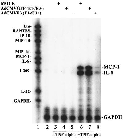
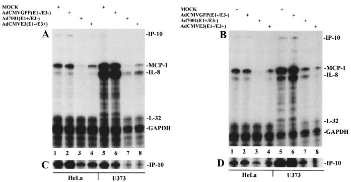
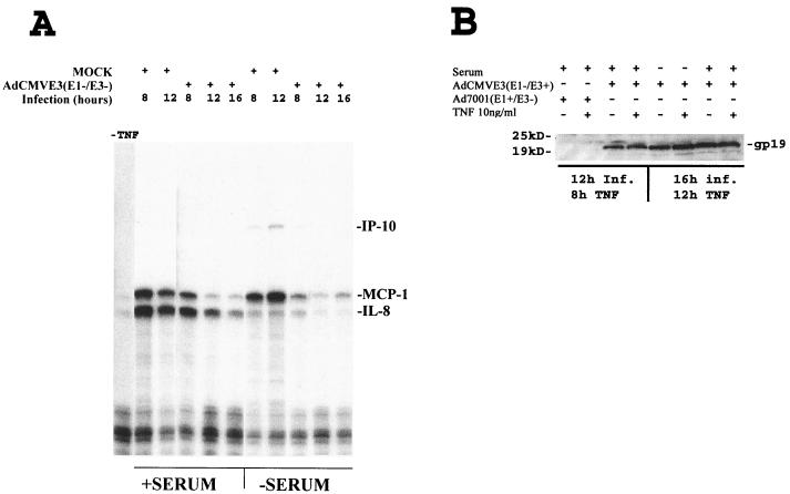
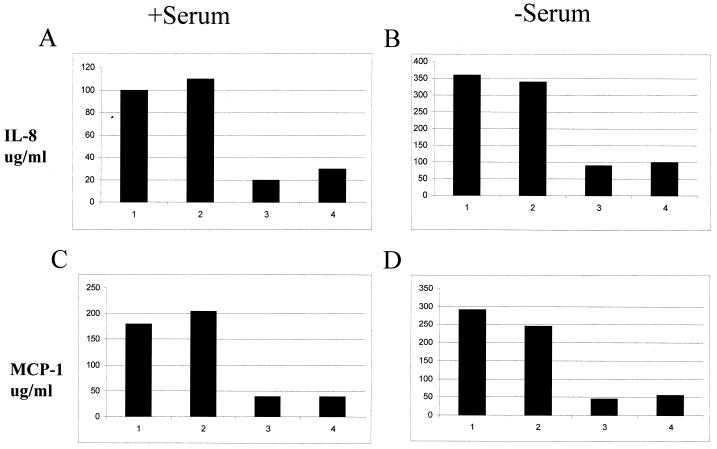
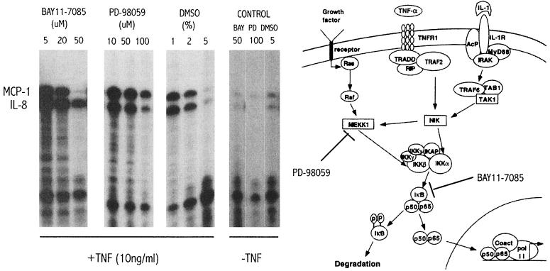
Adenoviruses (Ad) have a variety of immunoregulatory genes, many of which are clustered in a 3.5-kb segment of DNA known as early region 3 (E3). Ad E3 codes for proteins that downregulate surface expression of class I major histocompatibility antigens and also inhibit tumor necrosis factor alpha (TNF-alpha)- and Fas-induced cytolysis. We were interested in determining whether chemokine production or activity might also be inhibited by Ad E3 and we have studied this function in a human astrocytoma cell line, U373. Astrocytes constitute a part of the blood-brain barrier, and chemokines (IP-10, IL-8, MCP-1-4, and MIPs) expressed by them may contribute to leukocyte infiltration within the brain during inflammation. When U373 cells are activated by the proinflammatory molecule TNF-alpha, the increase in chemokine MCP-1, IL-8, and IP-10 transcripts is blocked by a recombinant Ad expressing the E3 genes under cytomegalovirus promoter control. Comparable Ads expressing green fluorescent protein in place of E3 have no effect on these chemokines. Ads also have been extensively studied as gene therapy vectors and most have a deletion of the E3 region to permit the insertion of larger fragments of foreign DNA. Our results suggest that construction of Ad vectors to include E3 expression cassettes will improve the efficacy and safety of such viral-based gene therapy protocols.


Figures

FIG. 1.
FIG. 1.
Ad E3 inhibition of TNF-α-induced chemokines in U373. U373 cells (106) were grown in α-MEM medium supplemented with 10% FBS and infected with 4,000 particles of virus per cell; TNF-α was added at 12 hpi and the samples were harvested as described in the text at 16 hpi. RNase protection was performed with the hck-5 probe set using 5 μg of sample RNA. Lane 1, α-32P-labeled, undigested hck-5 probe; lane 2, probes from lane 1 digested with RNase but without an RNA sample; lanes 3 to 5, U373 cells infected with mock, AdCMVGFP, and AdCMVE3 virus, respectively, in the absence of TNF-α treatment; lanes 6 to 8, U373 cells infected with mock, AdCMVGFP, and AdCMVE3 virus, respectively, and treated with 10 ng of TNF-α per ml. The protected bands, indicated by the labels on the right (MCP-1, IL-8, and GAPDH), migrate faster than undigested probes, as expected.
FIG. 2.
FIG. 2.
Ad E1 and E3 independently inhibit chemokines in U373 and HeLa cells. (A) Cells (106) grown in DMEM supplemented with 10% FBS were infected with 2,000 particles of virus per cell, treated with 10 ng of TNF-α per ml at 12 hpi, and harvested as described in the text at 16 hpi. (B) Cells (106) were first serum starved for 14 h and then treated as in panel A and harvested at 16 hpi. For panels A and B, lanes 1 to 4, HeLa cells; and lanes 5 to 8, U373 cells; lanes 1 and 5, mock-infected cells; lanes 2 and 6, AdCMVGFP-infected cells; lanes 3 and 7, Ad7001-infected cells; lanes 4 and 8, AdCMVE3-infected cells. MCP-1, IL-8, and to a lesser degree IP-10 were the predominant chemokines induced with TNF-α; L32 and GAPDH were used for normalization of data points. Each lane represents RNase protection of the hck-5 labeling probe set by 5 μg of RNA. (C) The same experiment as for panel A is shown; however, the gel was exposed for 72 h instead of 3 h to visualize the IP-10 results. (D) The gel shown in panel B was reexposed for 72 h. The amounts of extracts loaded per lane were equivalent as measured by the intensity of GAPDH; however, in panels A and C, the amounts loaded in lanes 7 and 8 were 54% of the controls in lanes 5 and 6 as analyzed by densitometry.
FIG. 3.
FIG. 3.
Kinetics of MCP-1 and IL-8 inhibition by Ad E3. U373 cells (106) were grown in α-MEM medium supplemented with 10% FBS or depleted of serum for 14 h prior to infection. Cells were subsequently infected with 4,000 particles of virus per cell; 10 ng of TNF-α per ml was added 4 h prior to harvesting each sample at the time points indicated. (A) RNase protection assay. The first lane is a negative control (no virus or TNF). The time of harvest of the mock- or AdCMVE3 (E1 and E3 positive)-infected cells and the presence or absence of serum are indicated for each lane. (B) Western blot. U373 cells were infected with Ad7001 and AdCMVE3 and some samples were treated with TNF-α 4 h prior to harvesting at 12 or 16 hpi for protein and analysis as described in Materials and Methods. The infecting adenovirus and the presence of serum or TNF stimulation is indicated for each lane. The Ad E3-gp19K band, detected by rabbit polyclonal antibodies, is labeled
FIG. 4.
FIG. 4.
E1 and E3 genes impair secretion of MCP-1 and IL-8. U373 cells (5 × 10⁵ per well) were mock infected or infected with AdCMVGFP, Ad7001, or AdCMVE3 for 12 h with 6,000 particles/cell. At that time, 10 ng of TNF-α per ml was added per well. After 12 h of stimulation with TNF-α, the supernatants were collected and analyzed by ELISA for IL-8 (A and B) and MCP-1 (C and D) as described in Materials and Methods. One group of cells was maintained with normal FBS in the medium (A and C), while the others were treated for 12 h with serum-depleted medium before infection began (B and D). The numbers at the bottom of each bar represent different treatments as follows: 1, mock; 2, AdCMVGFP (E1 and E3 negative); 3, Ad7001 (E1 positive and E3 negative); and 4, AdCMVE3 (E1 negative and E3 positive).
FIG. 5.
FIG. 5.
MCP-1 and IL-8 chemokine upregulation induced by TNF-α is not coordinately affected by inhibitors of the NF-κB pathway. Confluent wells with 5 × 10⁵ U373 cells were pretreated for 1 h with different chemical compounds before TNF-α stimulation for an additional 4 h before harvest and total RNA isolation for RNase protection assay analysis. The inhibitors were tested at different concentrations. BAY11-7085 (BIOMOL), a specific inhibitor of IκB phosphorylation, PD-98059 (BIOMOL), a specific inhibitor of MEKK1 kinase activity, and DMSO, a solvent with antioxidant properties that has been shown to inhibit NF-κB in a nonspecific manner, were tested. In the control panel, the chemicals were tested at the maximal concentration and under the same conditions but without TNF-α stimulation. A diagram of the NF-κB signal transduction pathway (reproduced with permission from reference 9) is shown together with the steps inhibited by BAY11-7085 and PD-98059.

Similar articles

Cited by

References

    1. Alessi, D. R., A. Cuenda, P. Cohen, D. T. Dudley, and A. R. Saltiel. 1995. PD 098059 is a specific inhibitor of the activation of mitogen-activated protein kinase kinase in vitro and in vivo. J. Biol. Chem. 270:27489-27494. - PubMed
    1. Amin, R., R. Wilmott, Y. Schwarz, B. Trapnell, and J. Stark. 1995. Replication-deficient adenovirus induces expression of interleukin-8 by airway epithelial cells in vitro. Hum. Gene Ther. 6:145-153. - PubMed
    1. Bennett, E. M., J. R. Bennink, J. W. Yewdell, and F. M. Brodsky. 1999. Cutting edge: adenovirus E19 has two mechanisms for affecting class I MHC expression. J. Immunol. 162:5049-5052. - PubMed
    1. Bett, A. J., W. Haddara, L. Prevec, and F. L. Graham. 1994. An efficient and flexible system for construction of adenovirus vectors with insertions or deletions in early regions 1 and 3. Proc. Natl. Acad. Sci. USA 91:8802-8806. - PMC - PubMed
    1. Bodaghi, B., T. R. Jones, D. Zipeto, C. Vita, L. Sun, L. Laurent, F. Arenzana-Seisdedos, J. L. Virelizier, and S. Michelson. 1998. Chemokine sequestration by viral chemoreceptors as a novel viral escape strategy: withdrawal of chemokines from the environment of cytomegalovirus-infected cells. J. Exp. Med. 188:855-866. - PMC - PubMed

Publication types

MeSH terms