Comparative Study
. 2002 Aug;76(16):8244-51.
doi: 10.1128/jvi.76.16.8244-8251.2002.

The long noncoding region of the human parainfluenza virus type 1 f gene contributes to the read-through transcription at the m-f gene junction

Affiliations
Comparative Study

The long noncoding region of the human parainfluenza virus type 1 f gene contributes to the read-through transcription at the m-f gene junction

Tatiana Bousse et al. J Virol. 2002 Aug.

Abstract

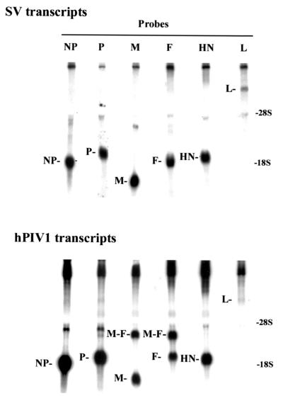
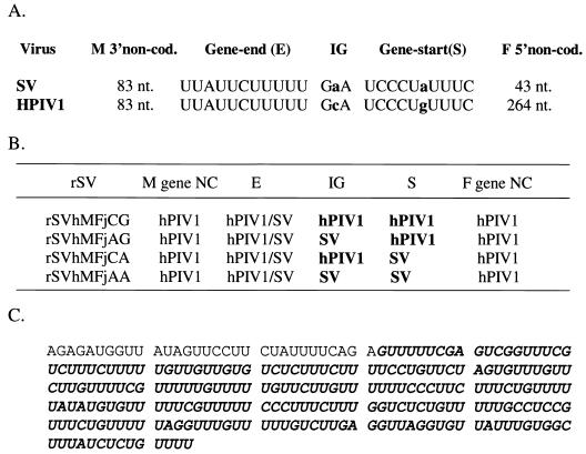
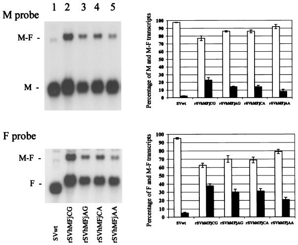
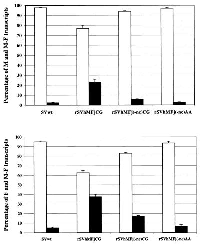
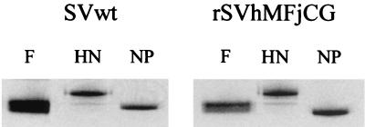
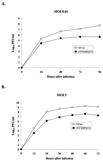
Sendai virus (SV) and human parainfluenza virus type 1 (hPIV1) have genomes consisting of nonsegmented negative-sense RNA in which the six genes are separated by well-conserved intergenic (IG) sequences and transcriptional start (S) and end signals. In hPIV1-infected cells, transcriptional termination at the M-F gene junction is ineffective; a large number of M-F read-through transcripts are produced (T. Bousse, T. Takimoto, K. G. Murti, and A. Portner, Virology 232:44-52, 1997). In contrast, few M-F read-through transcripts are detected in SV-infected cells. Sequence analysis indicated that the hPIV1 IG and S sequences in the M-F junction differ from those of SV. Furthermore, the hPIV1 F gene contains an unusually long noncoding sequence. To identify the cis-acting elements that prevent transcriptional termination at the M-F junction, we rescued recombinant SV (rSVhMFjCG) in which its M-F gene junction was replaced by that of hPIV1. Cells infected with rSVhMFjCG produced an abundance of M-F read-through transcripts; this result indicated that the hPIV1 M-F junction is responsible for inefficient termination. When one or both of the IG and S sites in rSVhMFjCG were replaced by those of SV, the efficiency of transcriptional termination increased but not to the level observed in wild-type SV-infected cells. Deletion of most of the long noncoding region of the hPIV1 F gene in rSVhMFjCG in addition to the mutations in IG and S signals resulted in efficient termination that was equivalent to the level observed in wild-type virus-infected cells. Therefore, the long noncoding sequence of the hPIV1 F gene contains cis-acting element(s) that affects transcriptional termination. Our evaluation of the effect of inefficient transcriptional termination on viral replication in culture revealed that cells infected with rSVhMFjCG produced less F protein than cells infected with wild-type SV and that assembly of the recombinant SV in culture was less efficient. These phenotypes seem to be responsible for the extended survival of mice infected with rSVhMFjCG.


Figures

FIG. 1.
FIG. 1.
Northern blot analysis of mRNA extracted from LLC-MK2 cells infected with SV or hPIV1. Polyadenylated RNAs were hybridized with 32P-labeled NP, P, M, F, HN, or L probes (as indicated at the top of each blot). The identity and position of the hybridizing mRNAs are marked. The positions to which 28S and 18S rRNA migrated are shown.
FIG. 2.
FIG. 2.
Nucleotide sequences at the junction between M and F genes (A) and at the origins of genes of recombinant SV (B). E, IG, and S sequences at the M-F gene junction of the SV and hPIV1 genomes are shown in the viral RNA sense orientation. Nucleotide differences are indicated by lowercase letters. NC, noncoding sequence. (C) Nucleotide sequence of the noncoding region of the hPIV1 F gene. The nucleotides truncated from this region in SVhMFj(-nc)CG are shown in bold italics.
FIG. 3.
FIG. 3.
Analysis of M and F transcripts extracted from cells infected with wild-type SV (SVwt) or various rSVs. Polyadenylated RNA was isolated from SV-infected cells and transferred to Northern blots, which were then hybridized with a 32P-labeled M probe (top left panel) and with a 32P-labeled F probe (bottom left panel). The blots were exposed to X-ray film, and the resulting autoradiographs are shown. The ratios of M mRNA to M-F mRNA (top right panel) and of F mRNA to M-F mRNA (bottom right panel) were quantified by using the Storm Image System. The percentages of mRNA that were monocistronic (white bars) or read-through M-F transcripts (black bars) are shown. The averages of four independent experiments (± standard deviations) are shown.
FIG. 4.
FIG. 4.
Effect of long noncoding sequence of the hPIV1 F gene on the production of M-F read-through transcripts in infected cells. Polyadenylated RNA was isolated from LLC-MK2 cells infected with wild-type SV (SVwt), rSVhMFjCG, rSVhMFj(-nc)CG, or rSVhMFj(-nc)AA (10 PFU per cell). Northern blots of the RNA were analyzed by hybridization with 32P-labeled M or F probes. The ratios of M to M-F mRNA and of F to M-F mRNA for each experiment were quantified, and the average ratios of four independent experiments (± standard deviations) are shown.
FIG. 5.
FIG. 5.
Radioimmunoprecipitation of F, NP, and HN proteins. LLC-MK2 cells were infected with wild-type SV (SVwt) or rSVhMFjCG. Twenty-four hours after infection, cells were radiolabeled for 4 h. Proteins in cell lysates were immunoprecipitated with specific anti-F, anti-HN, or anti-NP monoclonal antibodies and analyzed by electrophoresis through a sodium dodecyl sulfate-polyacrylamide gel.
FIG. 6.
FIG. 6.
Kinetics of virus growth. Multiple-step (A) or single-step (B) growth curves of wild-type SV (SVwt) and rSV in LLC-MK2 cells are shown. Cells were infected with 0.01 PFU per cell (A) or 5 PFU per cell (B). Aliquots of the medium were harvested every 24 (A) or 12 (B) h and replaced with equal volumes of fresh medium. The quantity of infectious virus in the samples was determined by plaque assays with LLC-MK2 cells.
FIG. 7.
FIG. 7.
Change in body weight of 129X1/SvJ mice infected with wild-type SV (SVwt) or rSVMFjCG. Groups of 4 mice were inoculated intranasally with various doses of virus (104 to 107 PFU per mouse). The weight gain of each mouse was measured every day for up to 16 days after inoculation. The symbols, which indicate the dose of virus given or type of infection, are as follows: ▪, 107 PFU of virus; ▵, 106 PFU of virus; ▴, 105 PFU of virus; ○, 104 PFU of virus; •, mock infection. The body weight of one mouse that survived infection is shown by a dotted line.

Similar articles

Cited by

References

    1. Barr, J. N., S. P. J. Whelan, and G. W. Wertz. 1997. Role of the intergenic dinucleotide in vesicular stomatitis virus RNA transcription. J. Virol. 71:1794-1801. - PMC - PubMed
    1. Barr, J. N., S. P. J. Whelan, and G. W. Wertz. 1997. cis-acting signals involved in termination of vesicular stomatitis virus mRNA synthesis include the conserved AUAC and U7 signal for polyadenylation. J. Virol. 71:8718-8725. - PMC - PubMed
    1. Bousse, T., T. Takimoto, W. L. Gorman, T. Takahashi, and A. Portner. 1994. Regions on the hemagglutinin-neuraminidase protein of human parainfluenza virus type 1 and Sendai virus important for membrane fusion. Virology 204:506-514. - PubMed
    1. Bousse, T., T. Takimoto, T. Matrosovich, and A. Portner. 2001. Two regions of the P protein are required to be active with L protein for human parainfluenza virus type 1 RNA polymerase activity. Virology 283:306-314. - PubMed
    1. Bousse, T., T. Takimoto, K. G. Murti, and A. Portner. 1997. Elevated expression of the human parainfluenza virus type 1 F gene downregulates HN expression. Virology 232:44-52. - PubMed

Publication types

MeSH terms

LinkOut - more resources