. 2002 Aug;76(16):8318-34.
doi: 10.1128/jvi.76.16.8318-8334.2002.

The block in assembly of modified vaccinia virus Ankara in HeLa cells reveals new insights into vaccinia virus morphogenesis

Affiliations

The block in assembly of modified vaccinia virus Ankara in HeLa cells reveals new insights into vaccinia virus morphogenesis

M Carmen Sancho et al. J Virol. 2002 Aug.

Abstract

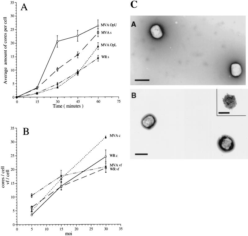
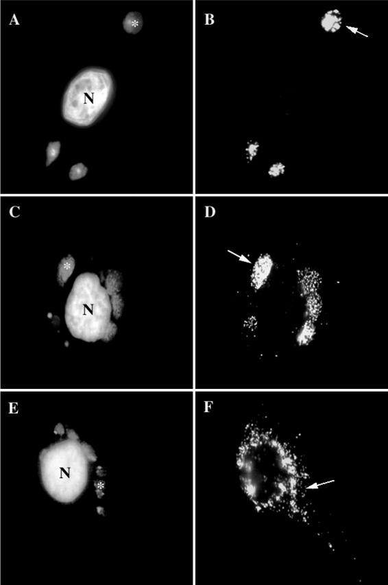
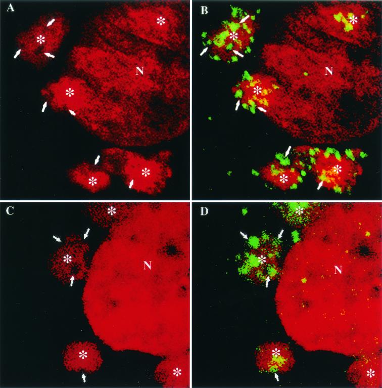
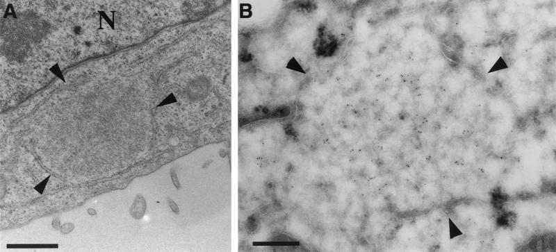
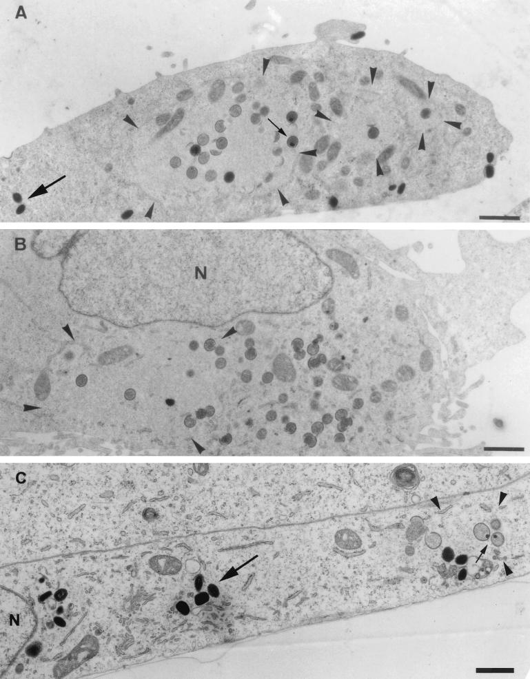
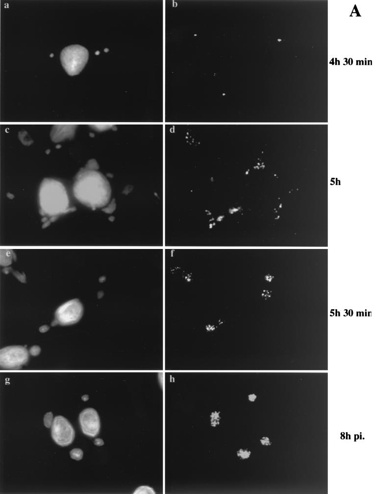
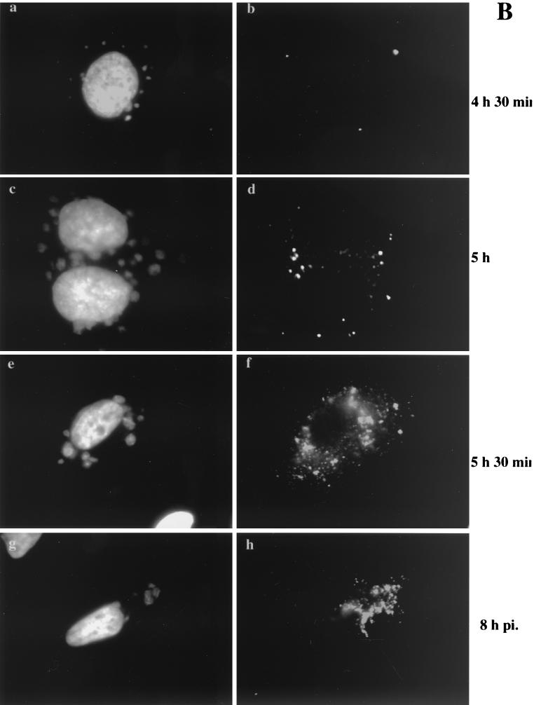
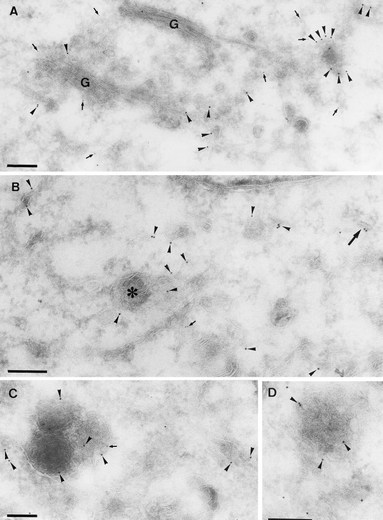
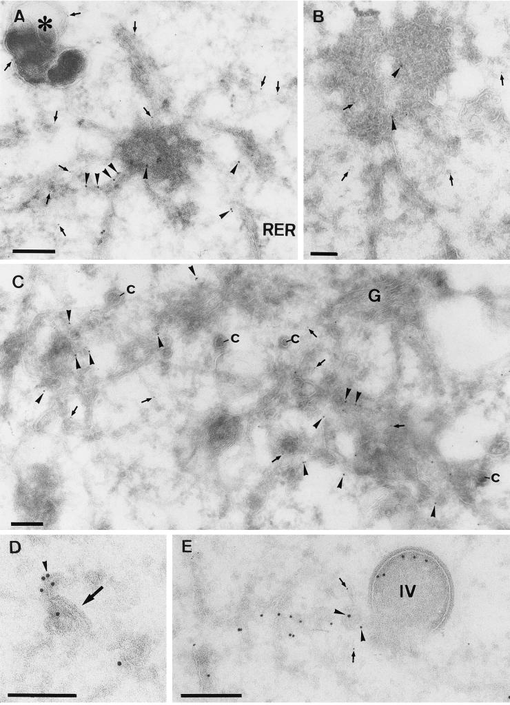
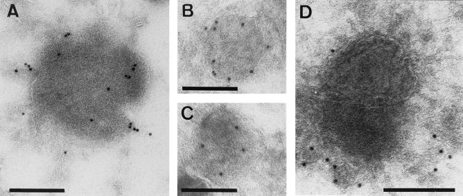
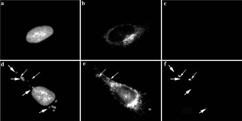
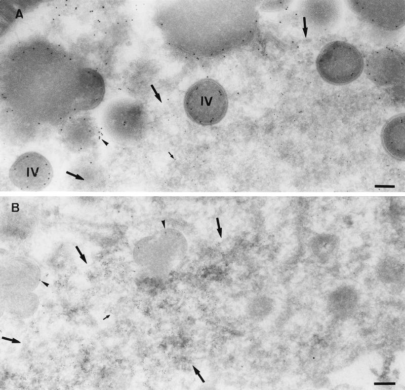
It has previously been shown that upon infection of HeLa cells with modified vaccinia virus Ankara (MVA), assembly is blocked at a late stage of infection and immature virions (IVs) accumulate (G. Sutter and B. Moss, Proc. Natl. Acad. Sci. USA 89:10847-10851, 1992). In the present study the morphogenesis of MVA in HeLa cells was studied in more detail and compared to that under two conditions that permit the production of infectious particles: infection of HeLa cells with the WR strain of vaccinia virus (VV) and infection of BHK cells with MVA. Using several quantitative and qualitative assays, we show that early in infection, MVA in HeLa cells behaves in a manner identical to that under the permissive conditions. By immunofluorescence microscopy (IF) at late times of infection, the labelings for an abundant membrane protein of the intracellular mature virus, p16/A14L, and the viral DNA colocalize under permissive conditions, whereas in HeLa cells infected with MVA these two structures do not colocalize to the same extent. In both permissive and nonpermissive infection, p16-labeled IVs first appear at 5 h postinfection. In HeLa cells infected with MVA, IVs accumulated predominantly outside the DNA regions, whereas under permissive conditions they were associated with the viral DNA. At 4 h 30 min, the earliest time at which p16 is detected, the p16 labeling was found predominantly in a small number of distinct puncta by IF, which were distinct from the sites of DNA in both permissive and nonpermissive infection. By electron microscopy, no crescents or IVs were found at this time, and the p16-labeled structures were found to consist of membrane-rich vesicles that were in continuity with the cellular endoplasmic reticulum. Over the next 30 min of infection, a large number of p16-labeled crescents and IVs appeared abruptly under both permissive and nonpermissive conditions. Under permissive conditions, these IVs were in close association with the sites of DNA, and a significant amount of these IVs engulfed the viral DNA. In contrast, under nonpermissive conditions, the IVs and DNA were mostly in separate locations and relatively few IVs acquired DNA. Our data show that in HeLa cells MVA forms normal DNA replication sites and normal viral precursor membranes but the transport between these two structures is inhibited.


Figures

FIG. 1.
FIG. 1.
Entry of WR and MVA in HeLa cells (A) and relationship between MOI, intracellular cores, and DNA replication sites (B). (A) HeLa cells grown on coverslips were infected at an MOI of 30 in the presence of 300 μg of cycloheximide per ml with sucrose-purified WR (WR s) or MVA (MVA s), the upper band obtained from MVA purified on Optiprep gradients (MVA OpU) (enriched for EEV), or the lower band of the same gradient (MVA OpL) (containing IMV only). Cells were fixed at the indicated times postinfection and labeled with an anticore antibody followed by anti-rabbit-FITC to visualize intracellular cores. The graph represents the average amounts of cores per cell in 30 cells and the standard errors of the means. (B) HeLa cells grown on coverslips were infected with sucrose-purified WR or MVA at the indicated MOI. After 60 min at 37°C, one set of cells was fixed and labeled with the anticore antibody. A parallel set of cells was washed three times to remove unpenetrated virus and incubated for an additional 3 h before fixation. Intracellular cores and replication sites were visualized by IF (see Materials and Methods) and counted. The values represent the average amounts of cores per cell and standard errors of the means at 60 min postinfection with WR (WR c) or MVA (MVA c) or the average amounts of viral factories (vf) per cell and standard errors of the means at 4 h postinfection (WR vf or MVA vf) in 30 cells. (C) Negative-staining EM and immunolabeling with anti-p42 (B5R; 10-nm-diameter gold) of sucrose-purified WR (panel A) and MVA (panel B) isolated from infected HeLa or BHK cells, respectively. Bars, 300 μm.
FIG. 2.
FIG. 2.
As determined by IF, p16 and the viral DNA do not colocalize to the same extent in MVA-infected HeLa cells as with permissive infections of MVA in BHK cells or WR in HeLa cells. BHK cells (A and B) or HeLa cells (E and F) infected with MVA or HeLa cells infected with WR (C and D), all at an MOI of 10, were fixed at 8 h postinfection. Fixed cells were double labeled with DAPI (A, C, and E) and anti-p16 (A14L) (B, D, and F). In panels A, C, and E, asterisks indicate DNA factories, while in panels B, D, and F, arrows point to the same DNA factories. In panels B and D, p16 overlaps completely with the DNA factories in panels A and C, while in panel F, the p16 labeling is scattered in the cytoplasm, not obviously colocalizing with the DNA in panel E. N, nucleus.
FIG. 3.
FIG. 3.
Confocal sections of VV-infected cells double labeled for DNA (DAPI) and p16 (A14L). BHK cells infected with MVA (A and B) and HeLa cells infected with WR (C and D) were fixed at 6 h postinfection and double labeled with DAPI (red) and anti-p16 (green). In panels A and C the DAPI labeling is shown alone, and in panels B and D the merge of DAPI and p16 labeling is shown. The arrows indicate clusters of p16 labeling in panels B and D and the corresponding area in panels A and C. Asterisks indicate a DNA factory region. N, nucleus.
FIG. 4.
FIG. 4.
Viral DNA replication site in HeLa cells infected with MVA at 4 h 30 min postinfection. HeLa cells were infected with MVA at a MOI of 10 and fixed at 4 h 30 min postinfection. (A) Epon section in which one DNA replication site that is almost entirely surrounded by membranes of the ER can be seen. (B) Cryosections were double labeled with anti-p16 (10-nm-diameter gold) and anti-DNA (5-nm-diameter gold). The image shows a replication site that is almost completely ER enwrapped. The central part of this region is labeled abundantly with anti-DNA. The ER membrane around the replication site is not significantly labeled with anti-p16. Bars, 200 nm. N, nucleus.
FIG. 5.
FIG. 5.
Epon sections of HeLa cells infected with WR (A) or MVA (B) or BHK cells infected with MVA (C) at 8 h postinfection. In all cases cells were infected at an MOI of 10. The region of viral DNA accumulation in the cytoplasm of infected cells is indicated with arrowheads. This region is covered with IVs in HeLa cells infected with WR (A) and BHK cells infected with MVA (C), while no IVs are observed outside this region. Small arrows point to IVs containing a nucleoid. In HeLa cells infected with MVA (B), the region of DNA accumulation (arrowheads) has a few IVs, but more IVs can be found in the rest of the cytoplasm, not localizing to the DNA site. IMVs in panels A and C are indicated with large arrows and do not appear to be present in the DNA region. The latter viral form cannot be detected in HeLa cells infected with MVA (B). N, nucleus. Bars, 1 μm.
FIG. 6.
FIG. 6.
Localization of p16 at different times of infection. BHK cells (A) or HeLa cells (B) were grown on coverslips and infected with MVA at an MOI of 10. The cells were fixed at the indicated times postinfection (pi.) and double labeled with DAPI (a, c, e, and g) and anti-p16 (A14L) followed by anti-rabbit antibody coupled to rhodamine (b, d, f, and h). Note that p16 perfectly overlaps with the regions labeled with DAPI in BHK cells infected with MVA from 5 h 30 min postinfection onwards. p16 does not overlap to the same extent in HeLa cells infected with MVA. At 4 h 30 min postinfection under both permissive and nonpermissive conditions, p16-positive spots that do not colocalize with the viral DNA are detected.
FIG. 6.
FIG. 6.
Localization of p16 at different times of infection. BHK cells (A) or HeLa cells (B) were grown on coverslips and infected with MVA at an MOI of 10. The cells were fixed at the indicated times postinfection (pi.) and double labeled with DAPI (a, c, e, and g) and anti-p16 (A14L) followed by anti-rabbit antibody coupled to rhodamine (b, d, f, and h). Note that p16 perfectly overlaps with the regions labeled with DAPI in BHK cells infected with MVA from 5 h 30 min postinfection onwards. p16 does not overlap to the same extent in HeLa cells infected with MVA. At 4 h 30 min postinfection under both permissive and nonpermissive conditions, p16-positive spots that do not colocalize with the viral DNA are detected.
FIG. 7.
FIG. 7.
Double labeling of p16 and gp27 at 4 h 30 min postinfection. Cryosections were labeled with anti-p16 (10-nm-diameter gold) (arrowheads) and anti-gp27 (5-nm-diameter gold) (arrows). (A, C, and D) MVA in HeLa cells; (B) MVA in BHK cells. Panel A shows scattered labeling for both antigens in the vicinity of the Golgi stack (G). Note the concentration of p16 in structures that are in continuity with the smooth ER membranes. These domains are mostly devoid of gp27. In panel B, in addition to scattered ER labeling of p16, some is found on a multivesicular body-like structure (asterisk). The large arrow in panel B indicates the first hint, at 4 h 30 min, of a small crescent domain labeled for p16. In panels C and D examples of the membrane-rich vesicles described in the text are shown. Bars, 200 nm.
FIG. 8.
FIG. 8.
Double labeling of p16 and gp27 at 5 h postinfection. (A, B, and C) HeLa cells (A and B) or BHK cells (C) infected with MVA. All sections were double labeled with anti-p16 (10-nm-diameter gold) (arrowheads) and anti-gp27 (5-nm-diameter gold) (arrows). (A and B) Variable p16 labeling of the multimembrane vesicle that is connected to the rough ER (RER). These structures contain little to no labeling for gp27, while the membranes in continuity with them are clearly labeled for the cellular protein. In panel A gp27 but not p16 also labels a distinct dense vesicular structure (asterisk). (C) Relatively large amounts of label for both antigens in membranes adjacent to the Golgi complex are seen. The significant number of cop buds-vesicles (c) in continuity with these membranes indicates that they are on the cis side of the Golgi stack (G). (D) What we believe is the beginning of crescent formation (arrow) that is labeled with anti-p16 (arrowhead) is shown. (E) Double labeling with p16 (10-nm-diameter gold) (arrowheads) and gp27 (5-nm-diameter gold) (arrows), showing an IV, the inner membranes of which are labeled with anti-p16 and that is in continuity with tubular membranes that are labeled with both p16 and gp27. Bars, 200 nm.
FIG. 9.
FIG. 9.
Examples of p16-labeled membrane-rich vesicles in HeLa cells infected with MVA. Cells were fixed at 4 h 30 min (A to C) or 8 h (D) postinfection. Bars, 200 nm.
FIG. 10.
FIG. 10.
IF localization of β-COP in HeLa cells infected with MVA at 4 h 30 min postinfection. HeLa cells grown on coverslips were infected (d to f) or mock infected (a to c) with MVA at an MOI of 10 and fixed at 4 h 30 min after infection. Fixed cells were triple labeled with DAPI (a and d), anti-β-COP (b and e), and anti-p16 (c and f). The large arrows indicate viral DNA replication sites, and the small arrows indicate p16-labeled structures. In panel e the small arrows indicate p16 structures that are adjacent to COP-labeled structures.
FIG. 11.
FIG. 11.
BHK cells infected with MVA and fixed at 5 h postinfection. (A) Sections were double labeled with anti-p16 (10-nm-diameter gold) (arrowheads) and anti-p35 (H5R) (5-nm-diameter gold) (small arrow). The image shows an extended p35-positive area, the boundary of which is indicated with large arrows, that contains several IVs. (B) Sections were double labeled with anti-DNA (5-nm-diameter gold) (small arrow) and p16 (10-nm-diameter gold) (arrowheads). Also in this example there is an abrupt boundary (large arrows) where the DNA labeling stops. p16-labeled IVs are found within this region. Bars, 200 nm.

Similar articles

Cited by

References

    1. Antoine, G., F. Scheiflinger, F. Dorner, and F. G. Falkner. 1998. The complete genomic sequence of the modified vaccinia Ankara strain: comparison with other orthopoxviruses. Virology 244:365-396. - PubMed
    1. Beaud, G., and R. Beaud. 1997. Preferential virosomal location of underphosphorylated H5R protein synthesized in vaccinia virus-infected cells. J. Gen. Virol. 78:3297-3302. - PubMed
    1. Belyakov, I. M., L. S. Wyatt, J. D. Ahlers, P. Earl, D. Pendleton, B. L. Kelsall, W. Strober, B. Moss, and J. A. Berzofsky. 1998. Induction of a mucosal cytotoxic T-lymphocyte response by intrarectal immunization with a replication-deficient recombinant vaccinia virus expressing human immunodeficiency virus 89.6 envelope protein. J. Virol. 72:8264-8272. - PMC - PubMed
    1. Carroll, M. W., and B. Moss. 1997. Host range and cytopathogenicity of the highly attenuated MVA strain of vaccinia virus: propagation and generation of recombinant viruses in a nonhuman mammalian cell line. Virology 238:198-211. - PubMed
    1. Carroll, M. W., W. W. Overwijk, R. S. Charmberlain, S. A. Rosenberg, B. Moss, and N. P. Restifo. 1997. Highly attenuated modified vaccinia virus Ankara (MVA) as an effective recombinant vector: a murine tumor model. Vaccine 15:387-394. - PMC - PubMed

Publication types

LinkOut - more resources