. 2002 Aug 6;99(16):10765-70.
doi: 10.1073/pnas.132272199. Epub 2002 Jul 19.

Cerebral energetics and spiking frequency: the neurophysiological basis of fMRI

Affiliations

Cerebral energetics and spiking frequency: the neurophysiological basis of fMRI

Arien J Smith et al. Proc Natl Acad Sci U S A. .

Abstract

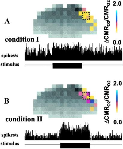
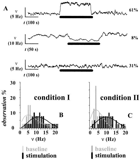
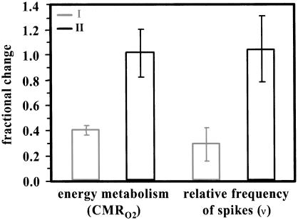
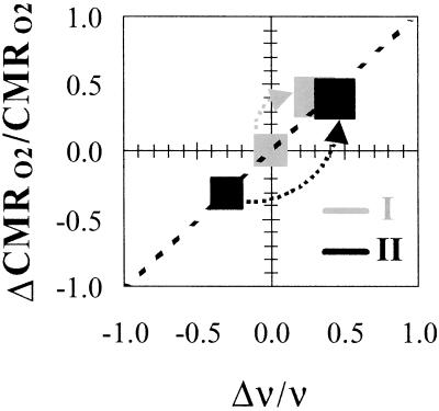
Functional MRI (fMRI) is widely assumed to measure neuronal activity, but no satisfactory mechanism for this linkage has been identified. Here we derived the changes in the energetic component from the blood oxygenation level-dependent (BOLD) fMRI signal and related it to changes in the neuronal spiking frequency in the activated voxels. Extracellular recordings were used to measure changes in cerebral spiking frequency (Deltanu/nu) of a neuronal ensemble during forepaw stimulation in the alpha-chloralose anesthetized rat. Under the same conditions localized changes in brain energy metabolism (DeltaCMR(O2)/CMR(O2)) were obtained from BOLD fMRI data in conjunction with measured changes in cerebral blood flow (DeltaCBF/CBF), cerebral blood volume (DeltaCBV/CBV), and transverse relaxation rates of tissue water (T(2)(*) and T(2)) by MRI methods at 7T. On stimulation from two different depths of anesthesia DeltaCMR(O2)/CMR(O2) approximately Deltanu/nu. Previous (13)C magnetic resonance spectroscopy studies, under similar conditions, had shown that DeltaCMR(O2)/CMR(O2) was proportional to changes in glutamatergic neurotransmitter flux (DeltaV(cyc)/V(cyc)). These combined results show that DeltaCMR(O2)/CMR(O2) approximately DeltaV(cyc)/V(cyc) approximately Deltanu/nu, thereby relating the energetic basis of brain activity to neuronal spiking frequency and neurotransmitter flux. Because DeltaCMR(O2)/CMR(O2) had the same high spatial and temporal resolutions of the fMRI signal, these results show how BOLD imaging, when converted to DeltaCMR(O2)/CMR(O2), responds to localized changes in neuronal spike frequency.


Figures

Fig 1.
Fig 1.
Calibrated BOLD maps (Upper) of ΔCMRO2/CMRO2 (Table 1) and changes in spiking activity (Lower) extracted from extracellular recordings in cortical layer 4 (Table 2) during contralateral forepaw stimulation. The resting and stimulated signals were made under high basal activity (A, condition I) followed by low basal activity (B, condition II) in the same rats. For the fMRI measurements, the colored bar shows the scale for ΔCMRO2/CMRO2 from the respective baselines. For the electrophysiology measurements, the black bar shows the stimulation period where the horizontal axis represents 900 s of data acquisition and the vertical axis (same range in A and B) represents number of spikes per second (1-s bins). The magnitudes of changes in CMRO2 and spiking activity were greater from baseline of condition II.
Fig 2.
Fig 2.
(A) Experimental variations across electrophysiological measurements (shown for condition II only) from the contralateral forepaw region. Significant increases and decreases in ν were observed in ≈60% and ≈10% of the recordings, respectively (Top and Middle), whereas in ≈30% of the recordings the stimulation did not induce any significant changes in ν (Bottom). The vertical and horizontal bars represent the scales for ν and time, respectively (see text and Table 2 for details). The thick black horizontal bar represents the stimulus duration. (B and C) The comparison in a small neuronal ensemble (72 neurons; see Table 2) between basal activity achieved with two dosages of α-chloralose shows significantly different spiking frequencies at rest (P < 0.01), whereas on stimulation the spiking frequencies became similar (P > 0.25).
Fig 3.
Fig 3.
Relative changes in CMRO2 (from black dotted regions in Fig. 1) and ν (guided by fMRI) during stimulation obtained from baseline conditions I and II are shown in gray and black, respectively. The baseline condition II was lowered by ≈30% from baseline condition I because of higher dosage of α-chloralose; however, the incremental change on stimulation from condition II was larger (see Tables 1 and 2). In each modality, on stimulation approximately the same levels of activation are reached from both starting baseline levels. The larger incremental change from condition II reflects the lowered starting baseline value, but the same stimulated final value.
Fig 4.
Fig 4.
Relationship between ΔCMRO2/CMRO2 and Δν/ν in α-chloralose-anesthetized rats. The sizes of boxes represent the mean ± the largest standard deviation from all rats studied. The data from conditions I and II are shown in gray and black, respectively, where the arrows show the increment on stimulation from the respective baseline levels. The final level reached on stimulation from both baseline conditions are similar within experimental errors. The dotted line represents the best linear regression (R2 = 0.98) of the pooled data. The overall changes in CMRO2 and ν from all rats studied are plotted to show a relationship of ≈1:1 between ΔCMRO2/CMRO2 and Δν/ν.

Comment in

  • Appraising the brain's energy budget.
    Raichle ME, Gusnard DA. Raichle ME, et al. Proc Natl Acad Sci U S A. 2002 Aug 6;99(16):10237-9. doi: 10.1073/pnas.172399499. Epub 2002 Jul 29. Proc Natl Acad Sci U S A. 2002. PMID: 12149485 Free PMC article. Review. No abstract available.

Similar articles

Cited by

References

    1. Roy C. S. & Sherrington, C. S. (1890) J. Physiol. (London) 11, 85-108. - PMC - PubMed
    1. Sokoloff L. (1993) Dev. Neurosci. 15, 194-206. - PubMed
    1. Siesjo B. K., (1978) Brain Energy Metabolism (Wiley, New York).
    1. Rothman D. L., Sibson, N. R., Hyder, F., Shen, J., Behar, K. L. & Shulman, R. G. (1999) Philos. Trans. R. Soc. London B 354, 1165-1177. - PMC - PubMed
    1. Sibson N. R., Dhankhar, A., Mason, G. F., Rothman, D. L., Behar, K. L. & Shulman, R. G. (1998) Proc. Natl. Acad. Sci. USA 95, 316-321. - PMC - PubMed

Publication types