

Tissue-specific silencing of a transgene in rice
- PMID: 12134059
- PMCID: PMC125067
- DOI: 10.1073/pnas.152330299
Tissue-specific silencing of a transgene in rice
Abstract
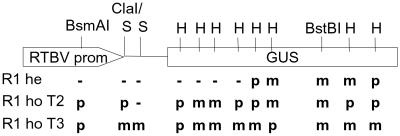
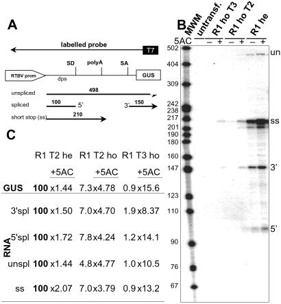
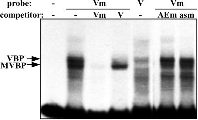
In a transgenic rice line, a beta-glucuronidase reporter gene under the control of the rice tungro bacilliform virus promoter became gradually methylated, and gene activity was lost concomitantly. Methylation was observed only in the homozygous offspring and was initially restricted to the promoter region and accompanied by loss of expression in the vascular bundle tissue only. This expression pattern was similar to that of a promoter with a deletion of a vascular bundle expression element. The gene activity could be reestablished by treatment with 5-azacytidine. Methylation per se did not inhibit the binding to the promoter region of protein factors which also bound to the unmethylated sequence. Instead, promoter methylation enabled the alternative binding of a protein with specificity for sequence and methylation. In further generations of homozygous offspring the methylation spread into the transcribed region and gene activity was completely repressed also in nonvascular cells. The results indicate that different stages are involved in DNA methylation-correlated gene inactivation, and that at least one of them may involve the attraction of a sequence and methylation-specific DNA-binding protein.
Figures
Similar articles
-
Sequence-specific and methylation-dependent and -independent binding of rice nuclear proteins to a rice tungro bacilliform virus vascular bundle expression element.J Biol Chem. 2001 Jan 26;276(4):2644-51. doi: 10.1074/jbc.M006653200. Epub 2000 Oct 17. J Biol Chem. 2001. PMID: 11036074
-
Upstream and downstream sequence elements determine the specificity of the rice tungro bacilliform virus promoter and influence RNA production after transcription initiation.Plant Mol Biol. 1999 May;40(2):249-66. doi: 10.1023/a:1006119517262. Plant Mol Biol. 1999. PMID: 10412904
-
Specificity of a promoter from the rice tungro bacilliform virus for expression in phloem tissues.Plant J. 1993 Jul;4(1):71-9. doi: 10.1046/j.1365-313x.1993.04010071.x. Plant J. 1993. PMID: 8220476
-
Transcription factor RF2a alters expression of the rice tungro bacilliform virus promoter in transgenic tobacco plants.Proc Natl Acad Sci U S A. 2001 Jun 19;98(13):7635-40. doi: 10.1073/pnas.121186398. Epub 2001 Jun 5. Proc Natl Acad Sci U S A. 2001. PMID: 11390974 Free PMC article.
-
Contribution of downstream promoter elements to transcriptional regulation of the rice tungro bacilliform virus promoter.Nucleic Acids Res. 2002 Jan 15;30(2):497-506. doi: 10.1093/nar/30.2.497. Nucleic Acids Res. 2002. PMID: 11788712 Free PMC article.
Cited by
-
Relationship between allelic state of T-DNA and DNA methylation of chromosomal integration region in transformed Arabidopsis thaliana plants.Plant Mol Biol. 2005 Jun;58(3):295-303. doi: 10.1007/s11103-005-4808-0. Plant Mol Biol. 2005. PMID: 16021396
-
Promoter diversity in multigene transformation.Plant Mol Biol. 2010 Jul;73(4-5):363-78. doi: 10.1007/s11103-010-9628-1. Epub 2010 Mar 31. Plant Mol Biol. 2010. PMID: 20354894 Review.
-
Agrobacterium tumefaciens-mediated creeping bentgrass (Agrostis stolonifera L.) transformation using phosphinothricin selection results in a high frequency of single-copy transgene integration.Plant Cell Rep. 2004 Apr;22(9):645-52. doi: 10.1007/s00299-003-0734-2. Epub 2003 Nov 13. Plant Cell Rep. 2004. PMID: 14615907
-
Rice transposable elements are characterized by various methylation environments in the genome.BMC Genomics. 2007 Dec 20;8:469. doi: 10.1186/1471-2164-8-469. BMC Genomics. 2007. PMID: 18093338 Free PMC article.
-
Epigenetic switch from posttranscriptional to transcriptional silencing is correlated with promoter hypermethylation.Plant Physiol. 2003 Nov;133(3):1240-50. doi: 10.1104/pp.103.023796. Epub 2003 Oct 9. Plant Physiol. 2003. PMID: 14551338 Free PMC article.
References
-
- Narlikar G. J., Fan, H.-Y. & Kingston, R. E. (2002) Cell 108, 475-487. - PubMed
-
- Richards E. J. & Elgin, S. C. R. (2002) Cell 108, 489-500. - PubMed
-
- Bird A. (2002) Genes Dev. 16, 6-21. - PubMed
-
- Jones P. A. & Takai, D. (2001) Science 293, 1068-1070. - PubMed
-
- Martienssen R. A. & Colot, V. (2001) Science 293, 1070-1074. - PubMed
Publication types
MeSH terms
Substances
LinkOut - more resources
Full Text Sources
Other Literature Sources
