. 2002 Jul;13(7):2207-22.
doi: 10.1091/mbc.e02-03-0175.

Ordered nucleation and spreading of silenced chromatin in Saccharomyces cerevisiae

Affiliations

Ordered nucleation and spreading of silenced chromatin in Saccharomyces cerevisiae

Laura N Rusché et al. Mol Biol Cell. 2002 Jul.

Abstract

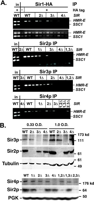
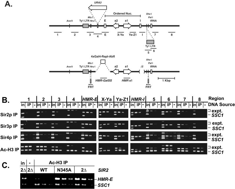
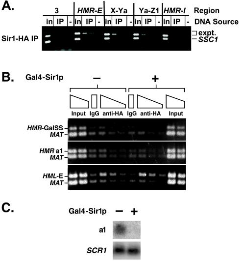
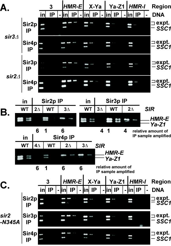
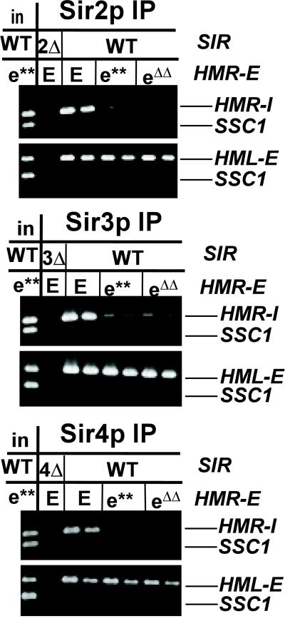
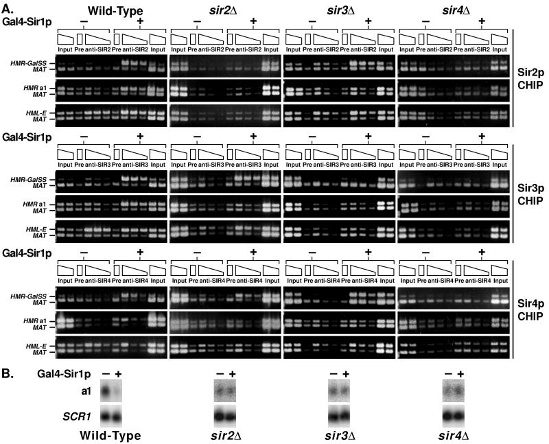
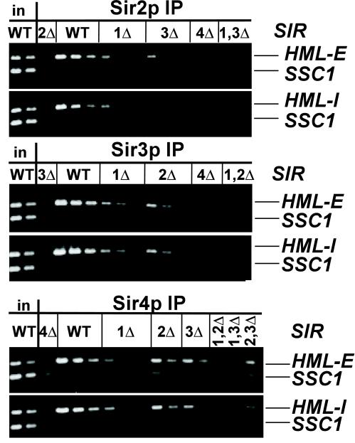
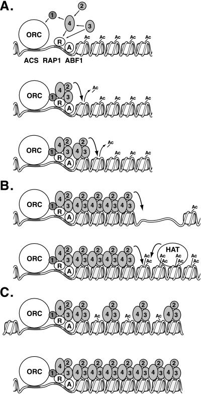
In Saccharomyces cerevisiae, silencing at the HM loci depends on Sir proteins, which are structural components of silenced chromatin. To explore the structure and assembly of silenced chromatin, the associations of Sir proteins with sequences across the HMR locus were examined by chromatin immunoprecipitation. In wild-type cells, Sir2p, Sir3p, and Sir4p were spread throughout and coincident with the silenced region at HMR. Sir1p, in contrast, associated only with the HMR-E silencer, consistent with its role in establishment but not maintenance of silencing. Sir4p was required for the association of other Sir proteins with silencers. In contrast, in the absence of Sir2p or Sir3p, partial assemblies of Sir proteins could form at silencers, where Sir protein assembly began. Spreading across HMR required Sir2p and Sir3p, as well as the deacetylase activity of Sir2p. These data support a model for the spreading of silenced chromatin involving cycles of nucleosome deacetylation by Sir2p followed by recruitment of additional Sir2p, Sir3p, and Sir4p to the newly deacetylated nucleosome. This model suggests mechanisms for boundary formation, and for maintenance and inheritance of silenced chromatin. The principles are generalizable to other types of heritable chromatin states.


Figures

Figure 1
Figure 1
Association of Sir proteins with the HMR-E silencer. (A) Chromatin immunoprecipitations from MATα SIR (JRY4013), sir2Δ (JRY4563), sir3Δ (JRY4605), sir4Δ (JRY4580), sir1Δ sir2Δ (JRY7296), sir1Δ sir3Δ (JRY7297), or sir2Δ sir3Δ (JRY7298) cells. Strains used in panels 1 and 2 also expressed SIR1–3xHA and were SIR (JRY7299), sir2Δ (JRY7300), sir3Δ (JRY7301), or sir4Δ (JRY7302). DNA immunoprecipitated with antibodies against HA tag (to monitor Sir1-HAp; panels 1 and 2), Sir2p (panel 3), Sir3p (panel 4), or Sir4p (panel 5) was analyzed by simultaneous amplification of HMR-E (top bands) and SSC1 (bottom bands). Panel 2 is a longer exposure of panel 1. For each sample, serial twofold dilutions of the template DNA are shown, starting with 1/19,000 of the input DNA or 2/75 (panels 1 and 2), 1/250 (panels 3 and 4), or 1/125 (panel 5) of the immunoprecipitated DNA. For the negative controls (lanes 3 in each panel), a single lane is shown, amplified from the largest fraction of the immunoprecipitated DNA used in that panel. These negative controls contained samples immunoprecipitated from strains lacking Sir2p (panel 3), Sir3p (panel 4), or Sir4p (panel 5), or bearing an untagged version of Sir1p (panels 1 and 2). (B) Expression of Sir proteins. Whole cell lysates were analyzed by immunoblots with antibodies against Sir2p (panels 2 and 5), Sir3p (panel 1), Sir4p, (panel 4) tubulin (panel 3), or phosphoglycerate kinase (panel 6). For panels 1–3, whole cell lysates were from SIR (JRY7131), sir2Δ (JRY7303), sir3Δ (JRY7304), or sir4Δ (JRY7305) cells. For panels 4–6, whole cell lysates were from strains used in Figure 1A and represent 0.75 OD equivalents. Sir2p was consistently reduced up to about twofold in sir3Δ and sir4Δ strains. Sir3p levels did not vary significantly in multiple cell lines tested.
Figure 2
Figure 2
Distribution of Sir proteins across HMR in a SIR strain. (A) Scale diagram of the HMR locus. Wild-type (top) and synthetic silencer-bearing (bottom) alleles are shown. The regions amplified are shown below each allele. The positions of the silencers, open reading frames, tRNA gene (boundary), and Ty1 LTRs (5′ LTR is a boundary) are shown on the line. The 3′ LTR is particular to these strains and is inserted at the known XhoI site. The Mlu I site just 3′ of the 5′ Ty1 LTR is less accessible to cleavage in SIR vs. sir strains, whereas the AvaII site 5′ of the Ty1 LTR is cut equally well in either strain (Loo and Rine, 1994). The region of ordered nucleosomes, the position at which URA3 was inserted (Donze et al., 1999), and the positions at which FRT sites were inserted are indicated. (B) Chromatin immunoprecipitations from MATα SIR cells (JRY4013). DNA immunoprecipitated with antibodies against Sir2p (panel 1), Sir3p (panel 2), Sir4p (panel 3), or di-acetylated histone H3 (panel 4) was analyzed by simultaneous amplification of the indicated region of HMR (top bands) and the SSC1 promoter (bottom bands). Negative controls (lanes −) had samples immunoprecipitated from strains lacking Sir2p (panel 1), Sir3p (panel 2), Sir4p (panel 3), or represent a mock precipitation with no antibody from SIR cells (panel 4). 1/19,000 of the input DNA, 1/250 or 1/500 (panels 1 and 2), 1/125 or 1/250 (panel 3), and 1/500 or 1/1000 (panel 4) of immunoprecipitated DNA was analyzed. The primers used to amplify region 6 generated a product of the predicted size (middle band) as well as a larger product of unknown identity, presumably involving another Ty1 LTR. (C) Chromatin immunoprecipitations from MATa SIR2, sir2-N345A, or sir2Δ cells (all W303-AR background). DNA was immunoprecipitated with antibodies against di-acetylated histone H3. Negative control (lane −) represents a mock precipitation with no antibody. The template DNA was serially diluted twofold, starting with 1/19,000 of the input DNA or 1/375 of the immunoprecipitated DNA.
Figure 3
Figure 3
Distribution of Sir1p across HMR. (A) Chromatin immunoprecipitation from MATα SIR cells bearing an HA-tagged Sir1p (JRY7299). DNA immunoprecipitated with antibodies against HA tag was analyzed by simultaneous amplification of the indicated region of HMR (top bands) and the SSC1 promoter (bottom bands). Negative controls (lanes −) had samples immunoprecipitated from a strain (JRY4013) bearing an untagged Sir1p. 1/23,000 of the input DNA or 1/50 or 1/100 of immunoprecipitated DNA was analyzed. (B) Chromatin immunoprecipitation from cells bearing a synthetic silencer in place of HMR-E. Cells constitutively expressed Sir1-HAp in the absence (lanes 1–6) or presence (lanes 7–12) of Gal4-Sir1p. DNA immunoprecipitated with antibodies against HA tag was analyzed by simultaneous amplification of the synthetic silencer (panel 1), a1 open reading frame (panel 2), or HML-E (panel 3) and the MAT locus (lower bands). Negative controls (lanes IgG) had samples immunoprecipitated with rabbit sera from an unimmunized rabbit. 1/7500 and 1/15,000 of the input DNA or 1/100, 1/200, or 1/400 of the immunoprecipitated DNA was analyzed. In this experiment, the ratio of immunoprecipitated HMR to MAT DNA was smaller than the ratio of immunoprecipitated HMR to SSC1 DNA in previous experiments. Consequently, these images reflect longer exposures than those in other figures, and the negative control is more visible. (C) RNA was isolated from the same samples used in B in the absence (lane 1) or presence (lane 2) of Gal4-Sir1p and analyzed for a1 or SCR1 mRNA.
Figure 4
Figure 4
Distribution of Sir proteins across HMR in sir strains. (A) Chromatin immunoprecipitations from MATα sir3Δ (JRY4605; panels 1 and 2) or sir2Δ (JRY4563; panels 3 and 4) cells. DNA immunoprecipitated with antibodies against Sir2p (panel 1), Sir3p (panel 3), or Sir4p (panels 2 and 4) was analyzed by simultaneous amplification of the indicated region of HMR (top bands) and the SSC1 promoter (bottom bands). Negative controls (lanes −) had samples immunoprecipitated from strains lacking Sir2p (panel 1), Sir3p (panel 3), or Sir4p (panels 2,4). 1/19,000 of the input DNA, 1/125 or 1/250 (panels 1 and 3), 1/50 or 1/100 (panel 2), or 2/125 or 1/125 (panel 4) of immunoprecipitated DNA was analyzed. (B) Chromatin immunoprecipitations from MATα SIR (JRY4013), sir2Δ (JRY4563), sir3Δ (JRY4605), or sir4Δ (JRY4580) cells. Immunoprecipitated DNA was analyzed by simultaneous amplification of HMR-E (top bands) and Ya-Z1 (bottom bands). The template DNA was serially diluted twofold, starting with 1/28,000 of the input DNA or 1/750 of the DNA immunoprecipitated from SIR cells. The relative amount of each immunoprecipitated sample amplified is indicated below the gel. (C) Chromatin immunoprecipitations from MATa sir2-N345A cells. Immunoprecipitated DNA was analyzed as in Figure 4A. Negative controls (lanes −) had samples immunoprecipitated from strains lacking Sir2p (panel 1), or represent a mock precipitation with no antibody (panels 2 and 3). 1/23,000 of the input DNA, 1/250 or 1/500 (panels 1 and 2), 2/125 or 1/125 (panel 3) of immunoprecipitated DNA was analyzed.
Figure 5
Figure 5
Association of Sir proteins with HMR-I silencer. Chromatin immunoprecipitations from MATα SIR cells bearing wild-type (JRY4013) or mutated HMR-E silencers (e** = JRY5472, eΔΔ = YGM1). Immunoprecipitated DNA was analyzed by simultaneous amplification of HMR-I (panels 1, 3, and 5) or HML-E (panels 2, 4, and 6) and the SSC1 promoter (bottom bands). 1/19,000 of the input DNA or 1/250 or 1/500 (panels 1–4) or 1/125 or 1/250 (panels 5 and 6) of the immunoprecipitated DNA was analyzed.
Figure 6
Figure 6
Loading and spreading of Sir proteins at a synthetic silencer. (A) Chromatin immunoprecipitations from SIR, sir2Δ, sir3Δ, or sir4Δ strains (left-to-right columns; JRY7131, JRY7303, JRY7304, and JRY7305, respectively). Cells contained 4xGal4-RAP1-ABF1 HMRa ΔI at HMR, and Gal4-Sir1p was repressed (−, first six lanes) or expressed (+, second six lanes). Wild-type Sir1p was constitutively expressed and acted at HML but not the synthetic silencer at HMR. DNA immunoprecipitated with antibodies against Sir2p (top), Sir3p (middle), or Sir4p (bottom) was analyzed by simultaneous amplification of the synthetic silencer (top band, top gels), the a1 open reading frame (top band, middle gels), or HML-E (top band, bottom gels) and sequences adjacent to MAT (bottom band, all gels). Negative controls (lanes Pre) had samples immunoprecipitated with rabbit sera from unimmunized rabbits. 1/7500, 1/15,000, or 1/30,000 of the input DNA or 1/100, 1/200, or 1/400 of the immunoprecipitated DNA was analyzed. In this experiment, the ratio of immunoprecipitated HMR to MAT DNA was smaller than the ratio of immunoprecipitated HMR to SSC1 DNA in previous experiments. Consequently, these images reflect longer exposures than those in other figures, and the negative control is more visible. (B) RNA was isolated from the same samples used in part A in the absence (lane 1) or presence (lane 2) of Gal4-Sir1p and analyzed for a1 or SCR1 mRNA.
Figure 7
Figure 7
Association of Sir proteins with HML silencers. Chromatin immunoprecipitations from MATα strains described in Figure 1. Immunoprecipitated DNA was analyzed by simultaneous amplification of HML-E (panels 1, 3, and 5) or HML-I (panels 2, 4, and 6) and the SSC1 promoter (bottom bands). The template DNA was serially diluted twofold, starting with 1/19,000 of the input DNA or 1/250 (panels 1 and 2) or 1/125 (panel 3) of immunoprecipitated DNA.
Figure 8
Figure 8
Models for Sir protein action. (A) Formation of silenced chromatin. Sir proteins (1–4) associate with ORC, Rap1p (R), and Abf1p (A) DNA binding proteins through protein–protein interactions (straight arrows). Sir2p then deacetylates the H3 and H4 tails of the neighboring nucleosome (line 2), creating binding sites for Sir3p and Sir4p (line 3). Sir2p deacetylates the next nucleosome, and the process repeats itself. (B) Boundaries of silenced chromatin. Sir protein spreading is interrupted by a gap in the nucleosome array (line 1) or a highly acetylated nucleosome (line 2). (C) Inheritance of silenced chromatin. The newly replicated silenced locus contains a mixture of hypoacetylated nucleosomes associated with Sir proteins and newly synthesized acetylated nucleosomes (line 1). Sir2p deacetylates neighboring nucleosomes, creating binding sites for Sir3p and Sir4p and reforming silenced chromatin.

Similar articles

Cited by

References

    1. Abraham J, Nasmyth KA, Strathern JN, Klar AJ, Hicks JB. Regulation of mating-type information in yeast. Negative control requiring sequences both 5′ and 3′ to the regulated region. J Mol Biol. 1984;176:307–331. - PubMed
    1. Axelrod AR. Role of a Cell-Cycle Gene in Transcriptional Silencing. Ph.D. Thesis. Berkeley, CA: University of California, Berkeley; 1991.
    1. Axelrod A, Rine J. A role for CDC7 in repression of transcription at the silent mating-type locus HMR in Saccharomyces cerevisiae. Mol Cell Biol. 1991;11:1080–1091. - PMC - PubMed
    1. Bi X, Braunstein M, Shei G, Broach J. The yeast HML I silencer defines a heterochromatin domain boundary by directional establishment of silencing. Proc Natl Acad Sci USA. 1999;96:11934–11939. - PMC - PubMed
    1. Bi X, Broach JR. UASrpg can function as a heterochromatin boundary element in yeast. Genes Dev. 1999;13:1089–1101. - PMC - PubMed

Publication types

MeSH terms