. 2002 Jul;13(7):2289-300.
doi: 10.1091/mbc.01-11-0535.

Disruption of centrosome structure, chromosome segregation, and cytokinesis by misexpression of human Cdc14A phosphatase

Affiliations

Disruption of centrosome structure, chromosome segregation, and cytokinesis by misexpression of human Cdc14A phosphatase

Brett K Kaiser et al. Mol Biol Cell. 2002 Jul.

Abstract

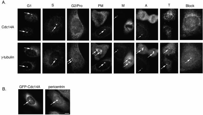
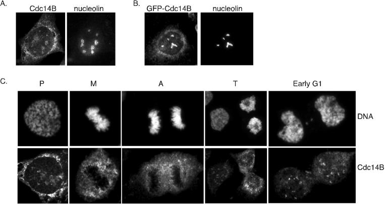
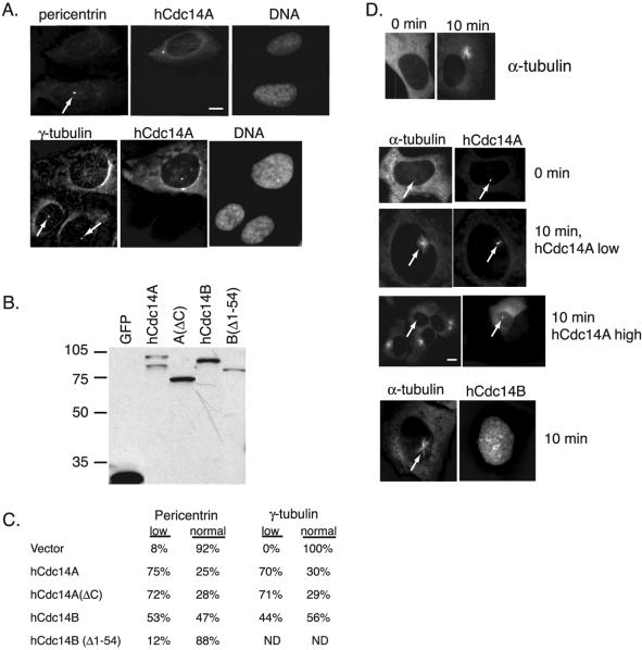
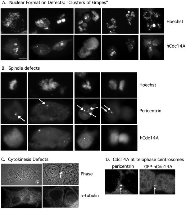
In budding yeast, the Cdc14p phosphatase activates mitotic exit by dephosphorylation of specific cyclin-dependent kinase (Cdk) substrates and seems to be regulated by sequestration in the nucleolus until its release in mitosis. Herein, we have analyzed the two human homologs of Cdc14p, hCdc14A and hCdc14B. We demonstrate that the human Cdc14A phosphatase is selective for Cdk substrates in vitro and that although the protein abundance and intrinsic phosphatase activity of hCdc14A and B vary modestly during the cell cycle, their localization is cell cycle regulated. hCdc14A dynamically localizes to interphase but not mitotic centrosomes, and hCdc14B localizes to the interphase nucleolus. These distinct patterns of localization suggest that each isoform of human Cdc14 likely regulates separate cell cycle events. In addition, hCdc14A overexpression induces the loss of the pericentriolar markers pericentrin and gamma-tubulin from centrosomes. Overproduction of hCdc14A also causes mitotic spindle and chromosome segregation defects, defective karyokinesis, and a failure to complete cytokinesis. Thus, the hCdc14A phosphatase appears to play a role in the regulation of the centrosome cycle, mitosis, and cytokinesis, thereby influencing chromosome partitioning and genomic stability in human cells.


Figures

Figure 1
Figure 1
Human Cdc14 phosphatase is specific for Cdk substrates. (A, left) Schematic of the hCdc14A and B phosphatases. hCdc14A and B represent highly related (61% identity within the catalytic domain), but independent genes with a 246 amino acid C-terminal domain specific to hCdc14A, and a 54 amino acid N-terminal peptide specific to hCdc14B. Bars indicate peptides used for immunizing rabbits. Right, bacterially expressed hCdc14A was purified as a GST fusion and used to dephosphorylate the chromogenic phosphatase substrate pNPP. Mutations in conserved residues within the active site aspartic acid (D251A), cysteine (C278S), and arginine (R284A) residues abrogated activity against pNPP. Mutation of another aspartic acid (D246A in hCdc14A), conserved in Cdc14 homologs, but not other dual specificity phosphatases, or deletion of the C-terminal domain, did not affect pNPP dephosphorylation activity. The C-terminal domain of hCdc14A had no phosphatase activity. (B) hCdc14A phosphatase dephosphorylates Cdk substrates. Validated Cdk substrates Xenopus Cdc6, human p27Kip1, and human Cdh1 were phosphorylated in vitro with cyclin E/Cdk2 (Cdc6 and p27) or cyclin B/Cdc2 (Cdh1), repurified, and mixed with 40 nM hCdc14A in phosphatase buffer for the indicated times. (C) Selectivity of the hCdc14A phosphatase. As in B, various proteins or peptides were phosphorylated by the indicated kinases, repurified, and assayed as substrates for hCdc14A. +, indicates >90% dephosphorylation; −, indicates <5% dephosphorylation; and ±, indicates <50% dephosphorylation.
Figure 2
Figure 2
hCdc14A and B protein levels and phosphatase activity fluctuate modestly through the cell cycle. (A) Antibodies specific to hCdc14A or B recognize endogenous protein. Antibodies raised against the hCdc14A-specific C terminus (residues 344–623) (left) and hCdc14A N terminus (residues 1–380) (middle) and to the specific N-terminal 54 amino acid peptide of hCdc14B (right) were affinity-purified and used to probe immunoblots of asynchronous HeLa cell lysates with (+) or without (−) prebinding to their respective antigens (see MATERIALS AND METHODS). The gradient gel system used only in the left panel was necessary to resolve the hCdc14A doublet. (B) In vitro phosphatase activities of hCdc14A and B vary modestly during the cell cycle. HeLa cells were synchronized in late G1 phase by a double thymidine block and then released from the block into fresh medium. Extracts were prepared at the indicated times (right) and DNA content analyzed by flow cytometry (major cell cycle phase shown at left). hCdc14A and B were immunoprecipitated with specific antibodies coupled to protein A-Sepharose beads, and the immune complexes were used to dephosphorylate the phosphorylated form of the APC activator human Cdh1 as a substrate (see MATERIALS AND METHODS). (C) hCdc14A protein levels are constant, whereas hCdc14B protein levels oscillate modestly during the cell cycle. HeLa cells were synchronized by double thymidine block-and-release (top) and nocodazole block-and-release (bottom) procedures and analyzed by Western blot for the abundance of endogenous hCdc14A and B protein.
Figure 3
Figure 3
hCdc14A associates with the interphase, but not mitotic, centrosome. (A) Endogenous hCdc14A staining. U2OS cells were synchronized by double thymidine block-and-release and stained with affinity-purified antibodies specific to hCdc14A and mouse antibodies to γ-tubulin. Representative images of cells in various stages of the cell cycle are shown. Arrows indicate centrosomes. Note the absence of hCdc14A staining in mitotic stages and in interphase cells where the hCdc14A antibody was preblocked with antigen (“block”). Pro, prophase; PM, prometaphase; M, metaphase; A, anaphase; T, telophase. Also note that in PM, M, and A, hCdc14A stains the centrosome of an adjacent interphase cell (indicated by an arrow). (B) GFP-hCdc14A targets to the centrosome. Full-length hCdc14A was fused to the enhanced variant of green fluorescent protein (eGFP) and transiently transfected into human U2OS cells, fixed with methanol, and costained with antibodies to GFP and pericentrin. Bar, 5 μm.
Figure 4
Figure 4
hCdc14B localizes to the nucleolus in interphase cells. (A) Endogenous staining of interphase U2OS cell with hCdc14B antibodies. Asynchronously growing U2OS cells were fixed in methanol and costained with antibodies raised against hCdc14B and nucleolin. (B) GFP-hCdc14B localizes to the nucleolus. U2OS cells were transiently transfected with a fusion between eGFP and hCdc14B, fixed in methanol and costained with antibodies against GFP and nucleolin. (C) Nucleolar staining of endogenous hCdc14B is absent during mitosis. Representative images of U2OS cells during various stages of mitosis were fixed in methanol and stained with hCdc14B antibodies. Cells were costained with Hoechst dye to visualize chromosomes. Pro, prophase; M, metaphase; A, anaphase; T, telophase.
Figure 5
Figure 5
Mapping localization determinants of hCdc14A and B. (A) U2OS cells were transfected for 24 h with the indicated GFP fusion constructs and immunostained with antibodies against nucleolin (top) or pericentrin (bottom). Arrows in the lower panels indicate centrosomes. (B) Summary of the localization of various hCdc14A or B constructs fused to GFP. Cells were fixed in methanol and stained with antibodies against GFP, and costained with either pericentrin or nucleolin as in A. NES in hCdc14A (aa 352–367) and in hCdc14B (aa 390–405) are depicted.
Figure 6
Figure 6
Overexpressed hCdc14A induces loss of pericentriolar material and inhibits microtubule regrowth from centrosomes. (A) hCdc14A overexpression causes loss of pericentriolar material from centrosomes. U2OS cells transiently transfected with pEGFP-hCdc14A were fixed and stained with the pericentriolar markers pericentrin (top) and γ-tubulin (bottom). Arrows indicate centrosomes in untransfected cells. Note that cells overexpressing hCdc14A have low levels of both pericentrin and γ-tubulin compared with untransfected cells. (B) Western blot of GFP-hCdc14 variants expressed in U2OS cells. Extracts from cells transfected with GFP (lane 1), GFP-hCdc14A (lane 2), GFP-hCdc14A(ΔC) (lane 3), GFP-hCdc14B (lane 4), and GFP-hCdc14B(Δ1–54) were resolved by SDS-PAGE and analyzed by Western blotting with antibodies to GFP. (C) Quantitation of pericentrin and γ-tubulin levels in cells transfected with variants of hCdc14A and B. GFP fusions to the indicated hCdc14A or B constructs were transfected in U2OS cells and positive cells were scored for either “low” or “normal” staining of pericentrin and γ-tubulin levels as in A. In each case, at least 150 GFP-positive cells were counted. (D) High levels of GFP-hCdc14A overexpression inhibit microtubule regrowth from centrosomes. U2OS cells transfected with GFP fusions to hCdc14A or B were treated with 10 μg/ml nocodazole at 24 h posttransfection for 2 h to depolymerize microtubules then grown in fresh media for 10 min. Coverslips were then fixed in methanol and processed for immunofluorescence with antibodies against α-tubulin to monitor regrowth of microtubules. Top, cells after treatment for 2 h with nocodazole (0 min) and after 10 min in media without nocodazole (10 min). Middle, GFP-hCdc14A-positive cells at 0 min, and at 10 min with either low or high levels of hCdc14A. Arrows indicate location of centrosomes. Bottom, a GFP-hCdc14B-positive cell after 10 min. Note that hCdc14B, even at high levels, did not inhibit microtubule regrowth. Bars 5 μm.
Figure 7
Figure 7
Transient overexpression of hCdc14A induces abnormalities in chromosome segregation, karyokinesis, and cytokinesis. U2OS cells were transiently transfected with pEGFP-hCdc14A, fixed in methanol 24 h after transfection, and stained with hCdc14A and Hoechst (A, B, and D), pericentrin (B and D), or α-tubulin (C). (A) Overexpression of hCdc14A induces karyokinesis (“micronuclei”) defects. Bar, 5 μm. (B) Overexpression of hCdc14A induces chromosome abnormalities and missegregation. Arrows indicate centrosomes. (C) Overexpression of hCdc14A induces a block to cytokinesis. Arrow indicates the position of the cytoplasmic bridge. Both cells overexpress GFP-hCdc14A. (D) GFP-hCdc14A colocalizes with centrosomes at the cleavage furrow in telophase cells. Arrows indicate the centrosomes.

Similar articles

Cited by

References

    1. Balasubramanian MK, McCollum D, Surana U. Tying the knot: linking cytokinesis to the nuclear cycle. J Cell Sci. 2000;113:1503–1513. - PubMed
    1. Brachmann CB, Sherman JM, Devine SE, Cameron EE, Pillus L, Boeke JD. The SIR2 gene family, conserved from bacteria to humans, functions in silencing, cell cycle progression, and chromosome stability. Genes Dev. 1995;9:2888–2902. - PubMed
    1. Carmena M, Riparbelli MG, Minestrini G, Tavares AM, Adams R, Callaini G, Glover DM. Drosophila Polo kinase is required for cytokinesis. J Cell Biol. 1998;143:659–671. - PMC - PubMed
    1. Carrano AC, Eytan E, Hershko A, Pagano M. SKP2 is required for ubiquitin-mediated degradation of the CDK inhibitor p27. Nat Cell Biol. 1999;1:193–199. - PubMed
    1. Chang P, Stearns T. δ-Tubulin and ε-tubulin: two new human centrosomal tubulins reveal new aspects of centrosome structure and function. Nat Cell Biol. 2000;2:30–35. - PubMed

Publication types

MeSH terms

LinkOut - more resources