. 2002 Jul;13(7):2311-22.
doi: 10.1091/mbc.e01-12-0148.

PC phosphorylation increases the ability of AFAP-110 to cross-link actin filaments

Affiliations

PC phosphorylation increases the ability of AFAP-110 to cross-link actin filaments

Yong Qian et al. Mol Biol Cell. 2002 Jul.

Abstract

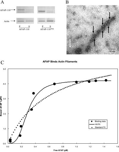
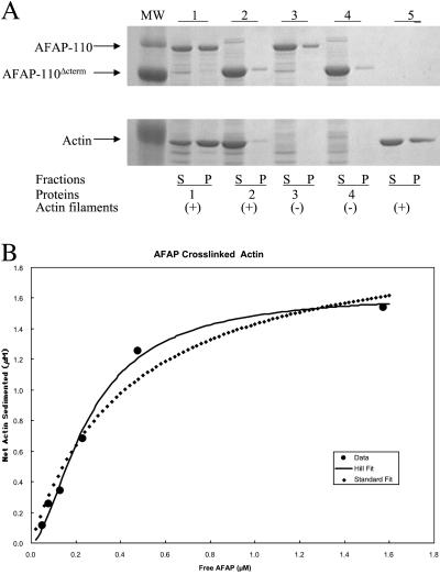
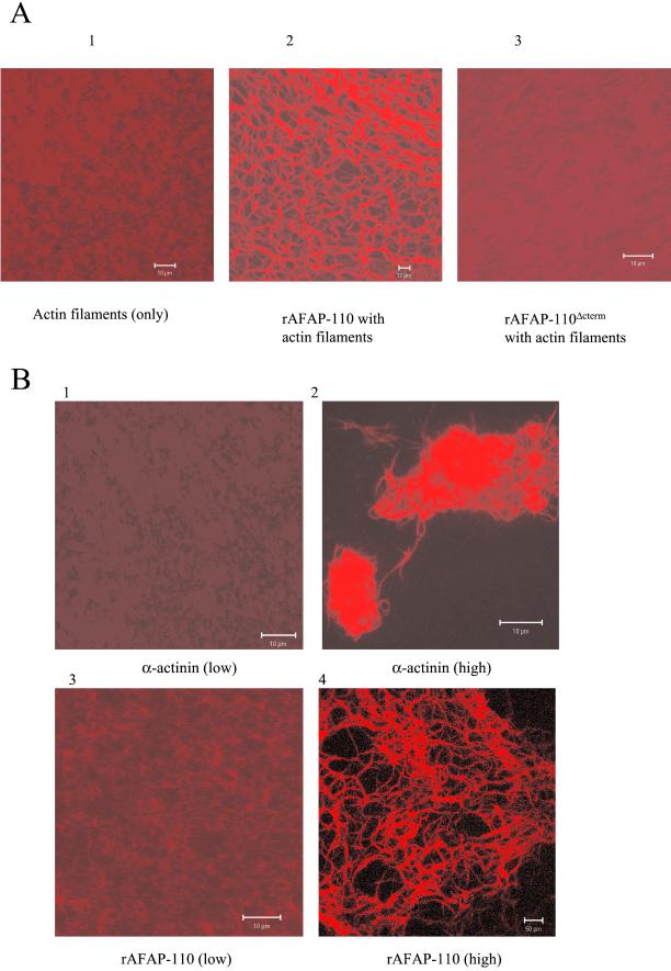
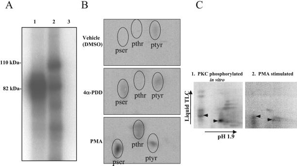
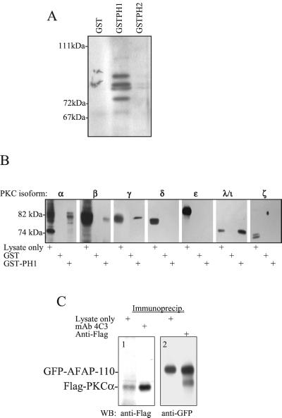
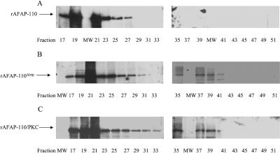
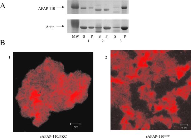
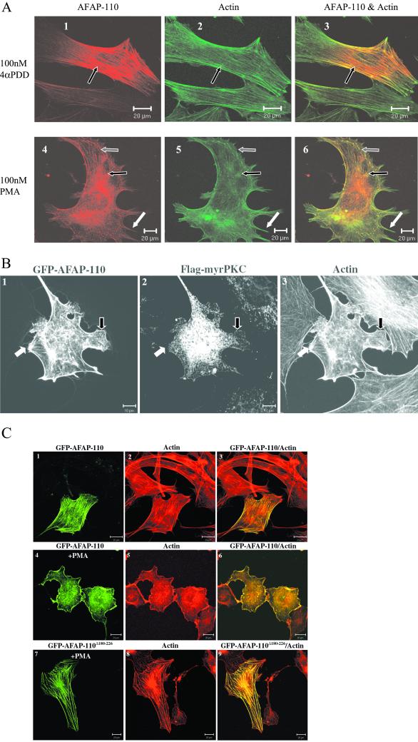
The actin filament-associated protein and Src-binding partner, AFAP-110, is an adaptor protein that links signaling molecules to actin filaments. AFAP-110 binds actin filaments directly and multimerizes through a leucine zipper motif. Cellular signals downstream of Src(527F) can regulate multimerization. Here, we determined recombinant AFAP-110 (rAFAP-110)-bound actin filaments cooperatively, through a lateral association. We demonstrate rAFAP-110 has the capability to cross-link actin filaments, and this ability is dependent on the integrity of the carboxy terminal actin binding domain. Deletion of the leucine zipper motif or PKC phosphorylation affected AFAP-110's conformation, which correlated with changes in multimerization and increased the capability of rAFAP-110 to cross-link actin filaments. AFAP-110 is both a substrate and binding partner of PKC. On PKC activation, stress filament organization is lost, motility structures form, and AFAP-110 colocalizes strongly with motility structures. Expression of a deletion mutant of AFAP-110 that is unable to bind PKC blocked the effect of PMA on actin filaments. We hypothesize that upon PKC activation, AFAP-110 can be cooperatively recruited to newly forming actin filaments, like those that exist in cell motility structures, and that PKC phosphorylation effects a conformational change that may enable AFAP-110 to promote actin filament cross-linking at the cell membrane.


Figures

Figure 1
Figure 1
rAFAP-110 cooperatively binds to actin filaments. (A) Both rAFAP-110 and rAFAP-110Δlzip bind to actin filaments directly. Either rAFAP-110 or rAFAP-110Δlzip were incubated with actin filaments at 30°C for 30 min and was centrifuged at 150,000 × g for 1 h. Both supernatant (S) and pellet (P) were applied to 10% SDS-PAGE gel, followed by Coomassie blue stain. (B) EM negative staining. The purified rAFAP-110 was incubated with actin filaments. The negative staining image was taken at 16,000 magnification. Black arrows, rAFAP-110 aggregates; white arrow, actin filaments. (C) Graph of bound rAFAP-110 versus free rAFAP-110. rAFAP-110 at 0.18, 0.29, 0.70, 1.34, 2.63, 3.73, 4.67, 5.11, 5.34, 5.48, and 5.66 μM was incubated with 2 μM concentration of G-actin polymerized into actin filaments and were then centrifuged at 150,000 × g. Both supernatants (S) and pellets (P) were applied to SDS-PAGE gel, followed by Coomassie staining. Density of AFAP-110 determined by scanning densitometry was fit as described in MATERIALS AND METHODS.
Figure 2
Figure 2
rAFAP-110 cross-links actin filaments through the carboxy terminal region. (A) Low-speed cosedimentation assay. The purified recombinant proteins were incubated with actin filaments and then were centrifuged at 20,800 × g. Both supernatants (S) and pellets (P) were applied to SDS-PAGE gel, followed by Coomassie staining. 1, rAFAP-110 with actin filaments; 2, rAFAP-110Δcterm with actin filaments; 3, rAFAP-110 only; 4, rAFAP-110Δcterm only; 5, actin filaments only. (B) Graph of cross-linked actin filaments versus free rAFAP-110. rAFAP-110 at 0.05, 0.11, 0.21, 0.43, 0.86, and 2.14 μM was incubated with the 2 μM concentration of G-actin polymerized into actin filaments. After the incubation, the reactions were centrifuged at 20,800 × g. Both supernatants and pellets were applied to SDS-PAGE gel, followed by the Western blot analysis to detect rAFAP-110 and Coomassie blue staining to detect actin, respectively. The data of cross-linked actin filaments and free rAFA-110 were gathered by scanning densitometry of SDS-PAGE gel analysis. The least-squares fit of the data to the Hill equation gave 0.26 μM for Kd, 1.63 μM for Bmax, and 1.7 for n.
Figure 3
Figure 3
rAFAP-110 cross-links actin filaments. (A) Purified rAFAP-110 or rAFAP-110Δcterm were incubated with rhodamine-phalloidin–labeled actin filaments. After the incubation, the reactions were observed with a Zeiss confocal microscope. 1, actin filaments only; 2, rAFAP-110 with actin filaments; 3, rAFAP-110Δcterm with actin filaments. (B) Confocal microscopy images. Different concentrations of α-actinin (top two panels, low = 0.0625 μM and high = 1.25 μM) and rAFAP-110 (bottom two panels, low = 0.106 μM and high = 2.1 μM) were incubated with rhodamine-phalloidin–labeled actin filaments. After the incubation, the reactions were observed with a Zeiss confocal microscope.
Figure 4
Figure 4
AFAP-110 is a substrate of PKCα. (A) PKCα phosphorylates rAFAP-110 in vitro. rAFAP-110 was incubated with or without recombinant PKCα in the presence of radiolabeled ATP. 1, PKCα (0.5 μg); 2, PKCα (0.5 μg)+rAFAP-110 (10 μg); 3, rAFAP-110 (10 μg). Data are representative of three experiments. (B) AFAP-110 is phosphorylated in vivo in response to PMA treatment. C3H10T1/2 cells were serum starved in phosphate-free media supplemented with radiolabeled 32P-orthophosphate overnight and treated with 100 nM PMA, 100 nM 4α-PDD, or DMSO, the vehicle used. Cells were lysed after 15 min and AFAP-110 was immunoprecipitated using the antibody F1. Radiolabeled AFAP-110 was isolated by SDS-PAGE and subjected to phosphoamino acid analysis. (C) Phosphotryptic analysis reveals common radioactive spots from AFAP-110 phosphorylated by PKC in vitro or phosphorylated in vivo in response to PMA. (C1) A representative tryptic map of rAFAP-110 phosphorylated in vitro by PKCα; (C2) a tryptic map of radio-labeled AFAP-110 purified from Cos-1 cells that were treated with PMA (100 nM, 1 h.). Black arrowheads, radioactive spots common between maps 1 and 2.
Figure 5
Figure 5
AFAP-110 is a binding partner of PKCα. (A) GST-PH1 affinity absorbs α pan-PKC–immunoreactive proteins. Absorptions using fusion proteins as listed were analyzed by SDS-PAGE and Western blotted with a pan-PKC antibody. This blot is representative of three experiments. (B) GST-PH1 affinity-abosorbs the classical PKC isoforms as well as PKCλ. Absorbates from affinity-absorptions using fusion proteins as listed were analyzed by SDS-PAGE and Western blotted with isoform specific PKC antibodies, as listed. Each blot is representative of two experiments. (C) Coimmunoprecipitation of AFAP-110 and PKCα. Immunoprecipitation of GFP-AFAP-110 with mAb 4C3 results in the presence of an anti-Flag immunoreactive protein from cells expressing GFP-AFAP-110 and Flag-PKCα. Likewise, an anti-GFP immunoreactive protein is seen in anti-Flag immunoprecipitation from the same lysate, shown in C2.
Figure 6
Figure 6
Either PKC phosphorylation or leucine zipper deletion destabilizes AFAP-110 multmerization. rAFAP-110(A), rAFAP-110Δlzip (B), and rAFAP-110/PKC (C) were separated by gel filtration. Fractions 17–51 were resolved by 8% SDS-PAGE gel. Western blot assays were applied using anti-AFAP-110 F1 antibody. The molecular weight markers were separated by gel filtration using FPLC and eluted as follows: thyglobulin (669 kDa) in fraction 25, ferritin (440 kDa) in fraction 30, catalase (232 kDa) in fraction 37, aldolase (158 kDa) in fraction 39, and albume (67 kDa) in fraction 44.
Figure 7
Figure 7
Either PKC phosphorylation or leucine zipper deletion increases AFAP-110's ability to cross-link actin filaments. (A) 0.5 μM purified rAFAP-110Δlzip or 0.5 μM PKC phosphorylated rAFAP-110 was incubated with 2 μM actin filaments. After the incubation, the reactions were centrifuged at 20,800 × g. Both supernatants (S) and pellets (P) were applied to SDS-PAGE gel, followed by Coomassie stain. The data are representative of two different experiments. (A1) rAFAP-110; (A2) rAFAP-110Δlzip; (A3) PKC phosphorylated rAFAP-110. (B) Confocal microscopy images. 0.5 μM purified rAFAP-110Δlzip or 0.5 μM PKC phosphorylated rAFAP-110 was incubated with 10% rhodamine-phalloidin–labeled actin filaments (2 μM). After the incubation, the reactions were observed with a Zeiss confocal microscope.
Figure 8
Figure 8
AFAP-110 mediates the effects of PKC on actin filaments. (A) AFAP-110 localizes to dynamic actin structures seen upon PMA stimulation. Immunofluorescence labeling and imaging was carried out as described in MATERIALS AND METHODS. C3H10T1/2 cells expressing AFAP-110 were treated with either 100 nM PMA (A1–A3) or 100 nM 4α-PDD (A4–A6) for 15 min after overnight serum starvation. AFAP-110 is represented in red (A1 and A4), whereas actin is represented in green (A2 and A5). (A3 and A6) Overlap images of AFAP-110 and actin. Arrows indicate colocalization of AFAP-110 with dynamic actin structures: white arrows, filopodia; gray arrows, lamellipodia; black arrows, actin filaments in the top panel and disrupted actin filaments/rosettes in the bottom panel. (B) AFAP-110 localizes to lamellipodia/filopodia seen upon expression of active PKC. Immunofluorescence labeling and imaging were carried out as described in Experimental Procedures. C3H10T1/2 cells coexpressing GFP-AFAP-110 (B1) and myristoylated, Flag-tagged PKC were labeled to show Flag-tagged PKC (B2) and actin (B3). Arrows indicate colocalization of AFAP-110 with dynamic actin structures: white arrows label filopodia and black arrows label lamellipodia. (C) AFAP-110 mediates the effects of PKC on the changes of cell structures. Immunofluorescence labeling and imaging were carried out as described in MATERIALS AND METHODS. C3H10T1/2 cells were transfected with pCMV-GFP-AFAP-110 (C1–C6) and pCMV-GFP-AFAPΔ180–226 (C7–C9). PMA at 100 nM was used to stimulate the cells for 1 h. C1, C4, and C7; green GFP fusion protein images; C2, C5, and C8; X actin images; C3, C6, and C9, overlap images of GFP fusion protein and actin images.

Similar articles

Cited by

References

    1. Baisden JM, Qian Y, Zot HG, Flynn DC. The actin filament associated protein, AFAP-110, is an adaptor protein that modulates changes in actin filament integrity. Oncogene. 2001a;20:6435–6447. - PubMed
    1. Baisden JM, Gatesman AS, Cherezova L, Jiang BH, Flynn DC. The intrinsic ability of AFAP-110 to alter actin filament integrity is linked with its ability to also activate cellular tyrosine kinases. Oncogene. 2001b;20:6607–6616. - PubMed
    1. Blikstad I, Markey F, Carlsson L, Persson T, Lindberg U. Selective assay of monomeric and filamentous actin in cell extracts using inhibition of deoxyribonuclease I. Cell. 1978;55:935–943. - PubMed
    1. Bourguignon LY, Zhu H, Shao L, Chen YW. CD44 interaction with c-Src kinase promotes cortactin-mediated cytoskeleton function and hyaluronic acid (HA)-dependent ovarian tumor cell migration. J Biol Chem. 2001;276:7327–7336. - PubMed
    1. Boyle WJ, van der Geer P, Hunter T. Phosphopeptide mapping and phosphoamino acid analysis by two-dimensional separation on thin-layer cellulose plates. Methods Enzymol. 1991;201:110–149. - PubMed

Publication types

MeSH terms

LinkOut - more resources