. 2002 Jul;13(7):2323-33.
doi: 10.1091/mbc.e02-01-0019.

Structural determinants of Ras-Raf interaction analyzed in live cells

Affiliations

Structural determinants of Ras-Raf interaction analyzed in live cells

Tzvetanka Bondeva et al. Mol Biol Cell. 2002 Jul.

Abstract

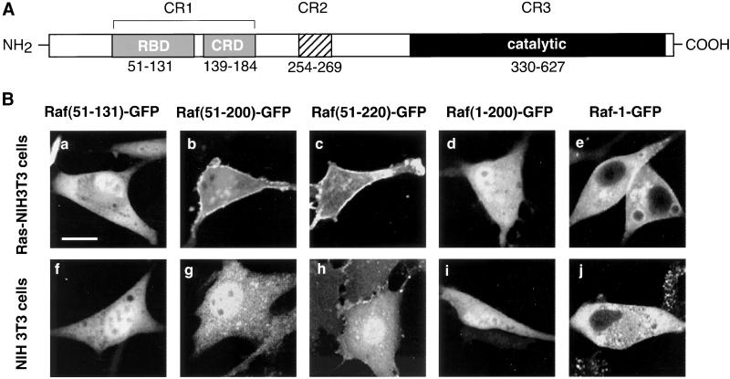
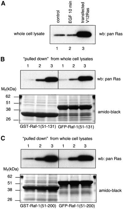
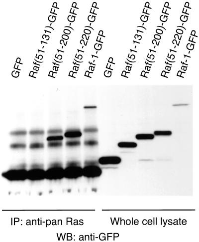
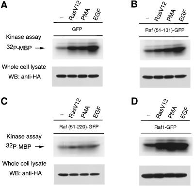
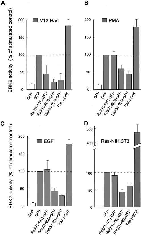
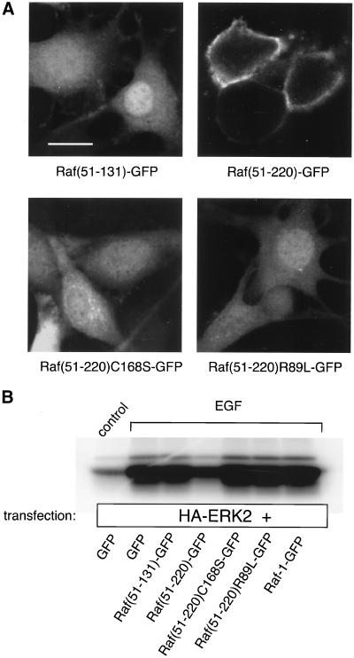
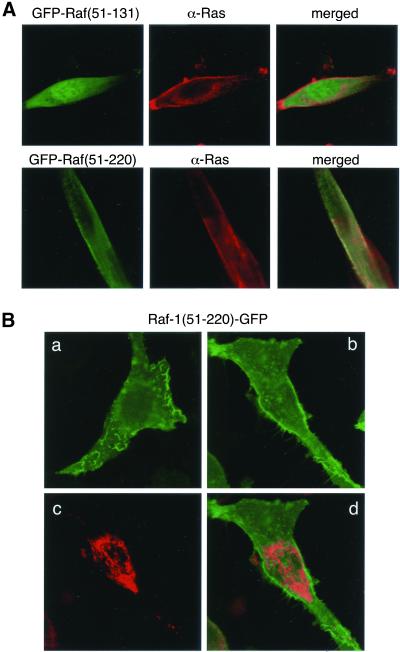
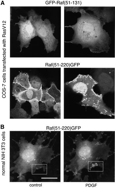
The minimum structure of the Raf-1 serine/threonine kinase that recognizes active Ras was used to create a green fluorescent fusion protein (GFP) for monitoring Ras activation in live cells. In spite of its ability to bind activated Ras in vitro, the Ras binding domain (RBD) of Raf-1 (Raf-1[51-131]GFP) failed to detect Ras in Ras-transformed NIH 3T3 fibroblasts and required the addition of the cysteine-rich domain (CRD) (Raf-1[51-220]GFP) to show clear localization to plasma membrane ruffles. In normal NIH 3T3 cells, (Raf-1[51-220]GFP) showed minimal membrane localization that was enhanced after stimulation with platelet-derived growth factor or phorbol-12-myristate-13-acetate. Mutations within either the RBD (R89L) or CRD (C168S) disrupted the membrane localization of (Raf-1[51-220]GFP), suggesting that both domains contribute to the recruitment of the fusion protein to Ras at the plasma membrane. The abilities of the various constructs to localize to the plasma membrane closely correlated with their inhibitory effects on mitogen-activated protein kinase kinase1 and mitogen-activated protein kinase activation. Membrane localization of full-length Raf-1-GFP was less prominent than that of (Raf-1[51-220]GFP) in spite of its strong binding to RasV12 and potent activation of mitogen-activated protein kinase. These finding indicate that both RBD and CRD are necessary to recruit Raf-1 to active Ras at the plasma membrane, and that these domains are not fully exposed in the Raf-1 molecule. Visualization of activated Ras in live cells will help to better understand the dynamics of Ras activation under various physiological and pathological conditions.


Figures

Figure 1
Figure 1
Domain-structure of the Raf-1 serine/threonine kinase (A) and cellular distribution of various Raf-1-GFP fusion proteins in Ras-transformed and normal NIH 3T3 fibroblasts (B). (A) The N-terminal conserved region (CR1) contains two domains, the RBD and the CRD, both of which have been implicated in the interaction and activation of Raf-1 by the small GTP binding protein, Ras. CR2 is a serine/threonine-rich region that contains several (but not all) regulatory phosphorylation sites. CR3 is the catalytic domain of the kinase. (B) Confocal images show the distribution of various Raf-1 constructs containing the Ras-interaction domains of Raf-1 fused to GFP as expressed in Ras-transformed (a-e) or normal (f-j) NIH 3T3 cells.
Figure 2
Figure 2
Interaction of recombinant GFP- and GST-fused RBDs of Raf-1 with activated Ras in vitro. Bacterially expressed and purified GFP- and GST-fused Raf(51-131) and Raf(51-200) domains were incubated with cell lysates prepared from COS-7 cells that were rendered quiescent, stimulated with EGF (100 ng/ml 10 min), or were transfected with RasV12. Ras associated with the recombinant domains was analyzed with Western blotting using an anti-pan-Ras antibody (see “Materials and Methods” for details). (A) The amount of Ras in the cell lysates. (B and C) Ras associated with the Raf(51-131) and Raf(51-200) constructs, respectively. In each case, the amounts of recombinant proteins present on the blots are shown after amido-black staining.
Figure 3
Figure 3
Interaction of various Raf-1-GFP fusion proteins with RasV12 coexpressed in COS-7 cells. COS-7 cells were transfected with plasmid DNAs encoding RasV12 and the respective Raf-1-GFP fusion constructs or GFP for 24 h. After 6 h of serum deprivation, cells were lysed and Ras was immunoprecipitated with an anti-pan-Ras antibody, followed by Western blotting for detection of the presence of GFP. The right panel shows the presence of the expressed proteins in the whole-cell lysates, and the left panel shows proteins that were associated with Ras. Representative data are shown from three similar observations.
Figure 4
Figure 4
Stimulation of ERK2 activity by RasV12, PMA, or EGF in COS-7 cells expressing various Raf-1-GFP fusion proteins. COS-7 cells were transfected with plasmid DNAs encoding HA-epitope-tagged ERK2 and GFP only (A), Raf(51-131)GFP (B), Raf(51-220)GFP (C), or Raf-1-GFP (D) with or without RasV12. One day post-transfection and after 6 h of serum deprivation, cells were stimulated with PMA (200 nM) for 15 min or with EGF (100 ng/ml) for 5 min. HA-ERK2 was then immunoprecipitated from the cell lysates and its activity was measured using [γ-32P]ATP and MBP as substrate. Similar observations were obtained in two additional experiments.
Figure 5
Figure 5
Differential ability of Raf-1-GFP fusion proteins to inhibit ERK2 activation in response to different stimuli in COS-7 cells and Ras-transformed NIH 3T3 cells. In this series of experiments, the ERK2 responses to the various stimuli (expressed as 100%) were compared between cells transfected with GFP or the various Raf-1-GFP protein constructs. For experimental details see the legend to Figure 3. Means ± SEM of four to six experiments are shown.
Figure 6
Figure 6
Mutations either within the RBD or CRD eliminate binding of Raf(51-220)GFP to Ras in the plasma membrane. Key residues within the RBD and CRD of Raf-1 known to affect its interaction with Ras were mutated within Raf(51-220)GFP, and the constructs were expressed in COS-7 or Ras-transformed NIH 3T3 cells. Both mutations (R89L and C168S) eliminate plasma membrane localization of the fusion protein in Ras-transformed NIH 3T3 cells (A) as well as its inhibitory effect on ERK2 activation in COS-7 cells (B).
Figure 7
Figure 7
Cellular distribution of Raf(51-131)GFP and Raf(51-220)GFP in Ras-transformed NIH 3T3 cells. (A) The distribution of Raf(51-131)GFP (upper) and Raf(51-220)GFP (lower) in fixed Ras-transformed NIH 3T3 cells immunostained with anti-Ras (red) antibody. (B) Panel a shows a cell in which the surface of the cell was imaged to demonstrate the high enrichment of the membrane associated fluorescence in membrane ruffles. This distribution was especially prominent in cells stimulated with PDGF. Panel b shows that fluorescence is also associated with small vesicular structures in the cytoplasm. These structures do not correspond to mitochondria as assessed by simultaneous imaging of the MitoTracker dye (c) and merging the two images (d).
Figure 8
Figure 8
Cellular distribution of GFP-Raf(51-131) and Raf(51-220)GFP in COS-7 cells expressing RasV12 (A) and the effect of PDGF stimulation on the distribution of Raf(51-220)GFP in normal NIH 3T3 fibroblasts (B). COS-7 cells were cotransfected with RasV12 and the respective GFP fusion construct, and normal NIH cells were transfected with Raf(51-220)GFP for 24 h. Live cells were examined under a confocal microscope. NIH 3T3 cells were serum-deprived (control) for 6 h before stimulation with PDGF (50 ng/ml, 2.5 min). Note the decrease in cytoplasmic intensity and the increased localization of the protein in membrane ruffles in B.

References

    1. Avruch J, Zhang X, Kyriakis JM. Raf meets Ras: completing the framework of a signal transduction pathway. Trends Biochem Sci. 1994;19:279–283. - PubMed
    1. Bondeva T, Pirola L, Bulgarelli-Leva G, Rubio I, Wetzker R, Wymann MP. Bifurcation of lipid and protein kinase signals of PI3Kγ to the protein kinases PKB and MAPK. Science. 1998;282:293–296. - PubMed
    1. Brtva TR, Drugan JK, Ghosh S, Terrell RS, Campbell-Burk S, Bell RM, Der CJ. Two distinct Raf domains mediate interaction with Ras. J Biol Chem. 1995;270:9809–9812. - PubMed
    1. Cai H, Erhardt P, Troppmair J, Diaz-Meco MT, Sithanandam G, Rapp UR, Moscat J, Cooper GM. Hydrolysis of phosphatidylcholine couples Ras activation of Raf protein kinase during mitogenic signal transduction. Mol Cell Biol. 1993;13:7645–7651. - PMC - PubMed
    1. Choy E, Chiu VK, Siletti J, Feoktistov M, Morimoto T, Michaelson D, Ivanov IE, Philips MR. Endomembrane trafficking of Ras: the CAAX motif targets proteins to the ER and Golgi. Cell. 1999;98:69–80. - PubMed

MeSH terms

Substances