. 2002 Jul;13(7):2374-82.
doi: 10.1091/mbc.e01-10-0096.

Differential recognition of tyrosine-based basolateral signals by AP-1B subunit mu1B in polarized epithelial cells

Affiliations

Differential recognition of tyrosine-based basolateral signals by AP-1B subunit mu1B in polarized epithelial cells

Hisashi Sugimoto et al. Mol Biol Cell. 2002 Jul.

Abstract

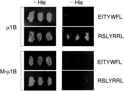
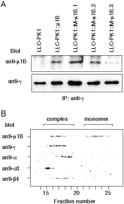
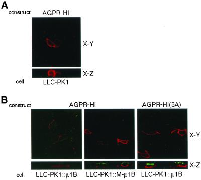
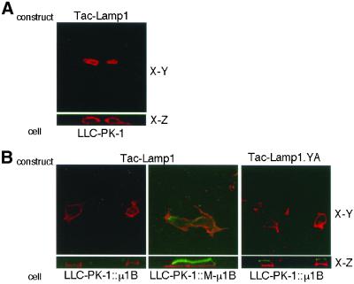
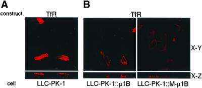
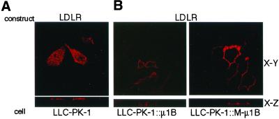
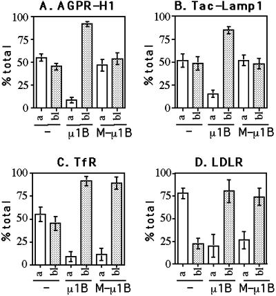
To investigate the importance of tyrosine recognition by the AP-1B clathrin adaptor subunit mu1B for basolateral sorting of integral membrane proteins in polarized epithelial cells, we have produced and characterized a mutant form of mu1B. The mutant (M-mu1B) contains alanine substitutions of each of the four conserved residues, which in the AP-2 adaptor subunit micro2 are critical for interacting with tyrosine-based endocytosis signals. We show M-mu1B is defective for tyrosine binding in vitro, but is nevertheless incorporated into AP-1 complexes in transfected cells. Using LLC-PK1 cells expressing either wild type or M-mu1B, we find that there is inefficient basolateral expression of membrane proteins whose basolateral targeting signals share critical tyrosines with signals for endocytosis. In contrast, membrane proteins whose basolateral targeting signals are distinct from their endocytosis signals (transferrin and low-density lipoprotein receptors) accumulate at the basolateral domain normally, although in a manner that is strictly dependent on mu1B or M-mu1B expression. Our results suggest that mu1B interacts with different classes of basolateral targeting signals in distinct ways.


Figures

Figure 1
Figure 1
M-μ1B does not interact with tyrosine-based sorting signals. HF7c yeast cells were cotransformed with a plasmid encoding GAL4ad fused to human μ1B or a mutant μ1B (M-μ1B) and a plasmid encoding GAL4bd fused to EITYWFL or RSLYRRL. Cotransformants were tested for their ability to grow in the presence (+His) or absence (−His) of histidine. Growth on the −His plate indicates that the products of GAL4bd and GAL4ad constructs can interact.
Figure 2
Figure 2
M-μ1B is specifically incorporated into AP-1B complexes. (A) AP-1 complexes were immunoprecipitated with an anti-γ-adaptin antibody 100/3 from lysates of LLC-PK1 cells stably expressing μ1B (LLC-PK1::μ1B) or a mutant μ1B (LLC-PK1::M-μ1B). Immunoprecipitants were subjected to SDS-PAGE, transferred onto Hybond-ECL membranes, immunoblotted with the anti-μ1B antibody or the anti-γ-adaptin antibody, and detected using the enhanced chemiluminescence system as described in MATERIALS AND METHODS. (B) Cytosol from LLC-PK1 cells stably expressing M-μ1B (LLC-PK1::μ1B) was fractionated by gel filtration chromatography on a Superose 6 column, and fractions (0.5 ml) were collected and analyzed by SDS-PAGE and Western blotting by using antibodies to various AP subunits as described in MATERIALS AND METHODS.
Figure 3
Figure 3
Basolateral sorting of AGPR-H1 depends on the tyrosine-binding ability of μ1B. (A) LLC-PK1 cells grown on the Transwell filters were transiently transfected with the AGPR-H1 expression plasmid and incubated with an anti-H1 antiserum. Subsequently, cells were fixed and stained with the Alexa Fluor 546 anti-rabbit IgG as described in MATERIALS AND METHODS. (B) LLC-PK1 cells stably expressing μ1B (LLC-PK1::μ1B) or a mutant μ1B (LLC-PK1::M-μ1B) grown on the Transwell filters were transiently transfected with AGPR-H1 or AGPR-H1(5A) expression plasmids, and incubated with an anti-H1 antiserum. Subsequently, cells were fixed and stained with the secondary antibodies, the Alexa Fluor 488 anti-rabbit IgG from the apical side, and Alexa Fluor 546 anti-rabbit IgG from the basolateral side as described in MATERIALS AND METHODS. Samples were analyzed using an LSM 510 laser scanning confocal microscope (Carl Zeiss, Thornwood, NY), and representative X-Y and X-Z sections are shown.
Figure 4
Figure 4
Basolateral sorting of Tac-Lamp1 depends on the tyrosine-binding ability of μ1B. (A) LLC-PK1 cells grown on the Transwell filters were transiently transfected with the Tac-Lamp1 or Tac-Lamp1.YA expression plasmid, and incubated with an anti-Tac mAb 7G7.B6. Subsequently, cells were fixed and stained with the Alexa Fluor 546 anti-mouse IgG as described in MATERIALS AND METHODS. (B) LLC-PK1 cells stably expressing μ1B (LLC-PK1::μ1B) or a mutant μ1B (LLC-PK1::M-μ1B) grown on the Transwell filters were transiently transfected with the Tac-Lamp1 expression plasmid and incubated with an anti-Tac mAb 7G7.B6. Subsequently, cells were fixed and stained with the secondary antibodies Alexa Fluor 488 anti-mouse IgG from the apical side and Alexa Fluor 546 anti-mouse IgG from the basolateral side as described in MATERIALS AND METHODS. Samples were analyzed using an LSM 510 laser scanning confocal microscope (Carl Zeiss), and representative X-Y and X-Z sections are shown.
Figure 5
Figure 5
Basolateral sorting of TfR does not depend on the tyrosine-binding ability of μ1B. (A) LLC-PK1 cells grown on the Transwell filters were transiently transfected with the TfR expression plasmid and incubated with an anti-TfR mAb L5.1. Subsequently, cells were fixed and stained with the Alexa Fluor 546 anti-mouse IgG as described in MATERIALS AND METHODS. (B) LLC-PK1 cells stably expressing μ1B (LLC-PK1::μ1B) or a mutant μ1B (LLC-PK1::M-μ1B) grown in the Transwell filters were transiently transfected with the TfR expression plasmid, and incubated with an anti-TfR mAb L5.1. Subsequently, cells were fixed and stained with the secondary antibodies Alexa Fluor 488 anti-mouse IgG from the apical side and Alexa Fluor 546 anti-mouse IgG from the basolateral side as described in MATERIALS AND METHODS. Samples were analyzed using an LSM 510 laser scanning confocal microscope (Carl Zeiss), and representative X-Y and X-Z sections are shown.
Figure 6
Figure 6
Basolateral sorting of LDLR does not depend on the tyrosine-binding ability of μ1B. (A) LLC-PK1 cells grown on the Transwell filters were transiently transfected with the LDLR expression plasmid and incubated with an anti-LDLR mAb C7. Subsequently, cells were fixed and stained with the Alexa Fluor 546 anti-mouse IgG as described in MATERIALS AND METHODS. (B) LLC-PK1 cells stably expressing μ1B (LLC-PK1::μ1B) or a mutant μ1B (LLC-PK1::M-μ1B) grown in the Transwell filters were transiently transfected with the LDLR expression plasmid and incubated with an anti-LDLR mAb C7. Subsequently, cells were fixed and stained with the secondary antibodies Alexa Fluor 488 anti-mouse IgG from the apical side and Alexa Fluor 546 anti-mouse IgG from the basolateral side as described in MATERIALS AND METHODS. Samples were analyzed using an LSM 510 laser scanning confocal microscope (Carl Zeiss), and representative X-Y and X-Z sections are shown.
Figure 7
Figure 7
Quantitative determination of the steady-state distribution on the plasma membrane of AGPR-H1, Tac-Lamp1, TfR, and LDLR. Parental LLC-PK1 cells, LLC-PK1 cells stably expressing μ1B (LLC-PK1::μ1B), or a mutant μ1B (LLC-PK1::M-μ1B) grown in the Transwell filters were transiently transfected with AGPR-H1 (A), Tac-Lamp1 (B), TfR (C), or LDLR (D) expression plasmid and incubated with the corresponding primary antibodies. Subsequently, cells were fixed and incubated with 125I-labeled anti-mouse or anti-rabbit IgG, and the cell-associated radioactivity was measured as described in MATERIALS AND METHODS. Values are given as the percentage of total cell surface radioactivity and represent mean ± SD of three independent experiments performed in duplicate.
Figure 8
Figure 8
M-μ1B supports the growth of LLC-PK1 cells in monolayer. Parental LLC-PK1 cells, LLC-PK1 cells stably expressing μ1B (LLC-PK1::μ1B), or a mutant μ1B (LLC-PK1::M-μ1B) were grown on the Transwell filers, fixed, permeabilized, and stained with Alexa Fluor 488 phalloidin as described in MATERIALS AND METHODS. Samples were analyzed using an LSM 510 laser scanning confocal microscope (Carl Zeiss), and representative X-Y and X-Z sections are shown.

Similar articles

Cited by

References

    1. Aguilar RC, Boehm M, Gorshkova I, Crouch RJ, Tomita K, Saito T, Ohno H, Bonifacino JS. Signal-binding specificity of the {micro}4 subunit of the adaptor protein complex AP-4. J Biol Chem. 2001;276:13145–13152. - PubMed
    1. Aroeti B, Okhrimenko H, Reich V, Orzech E. Polarized trafficking of plasma membrane proteins: emerging roles for coats, SNAREs, GTPases and their link to the cytoskeleton. Biochim Biophys Acta. 1998;1376:57–90. - PubMed
    1. Boll W, Ohno H, Songyang Z, Rapoport I, Cantley LC, Bonifacino JS, Kirchhausen T. Sequence requirements for the recognition of tyrosine-based endocytic signals by clathrin AP-2 complexes. EMBO J. 1996;15:5789–5795. - PMC - PubMed
    1. Bonifacino JS, Dell'Angelica EC. Molecular bases for the recognition of tyrosine-based sorting signals. J Cell Biol. 1999;145:923–926. - PMC - PubMed
    1. Dargemont C, Le Bivic A, Rothenberger S, Iacopetta B, Kuhn LC. The internalization signal and the phosphorylation site of transferrin receptor are distinct from the main basolateral sorting information. EMBO J. 1993;12:1713–1721. - PMC - PubMed

Publication types

MeSH terms