. 2002 Jul;13(7):2397-409.
doi: 10.1091/mbc.e02-01-0021.

Biogenesis of nanotubular network in Toxoplasma parasitophorous vacuole induced by parasite proteins

Affiliations

Biogenesis of nanotubular network in Toxoplasma parasitophorous vacuole induced by parasite proteins

Corinne Mercier et al. Mol Biol Cell. 2002 Jul.

Abstract

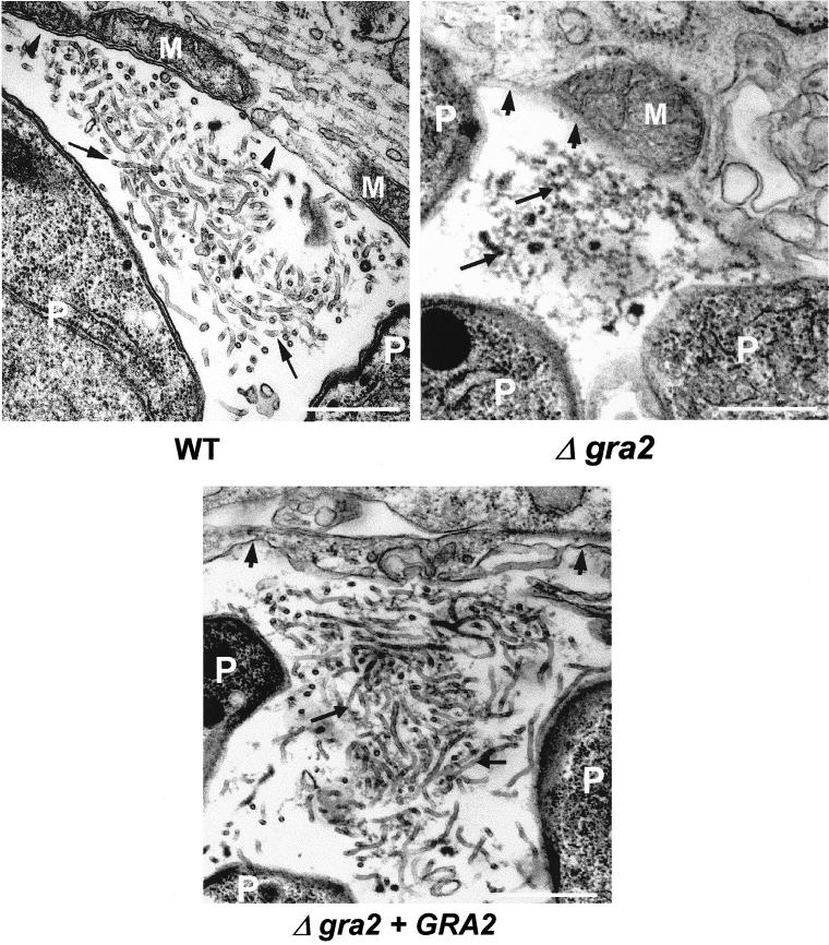
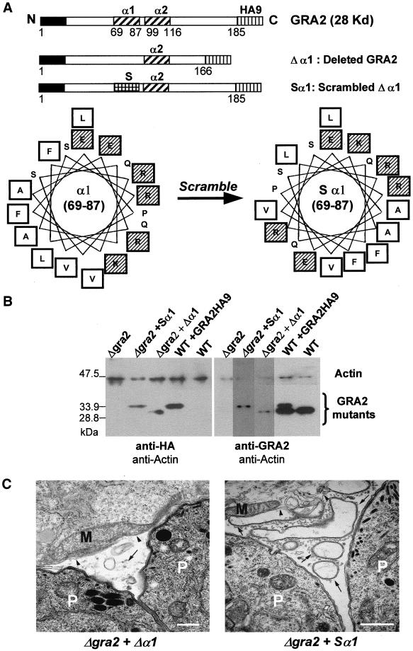
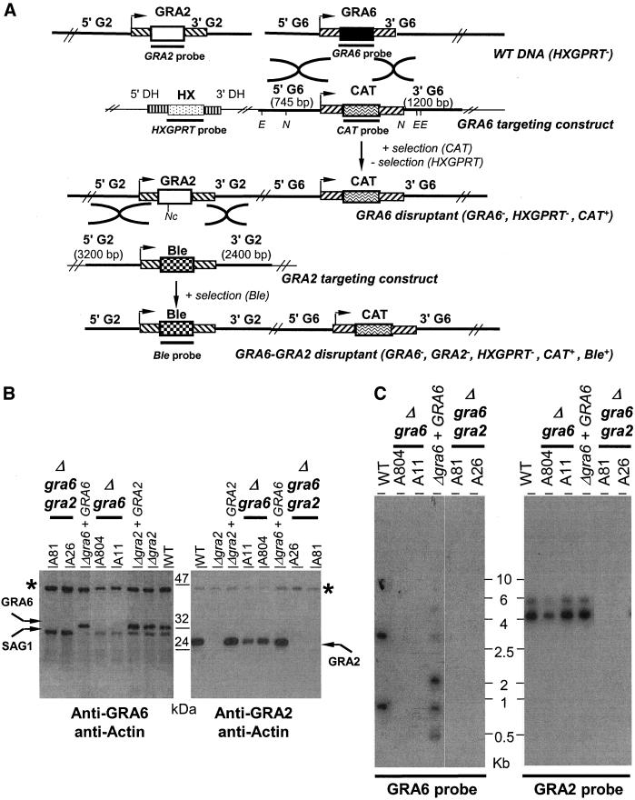
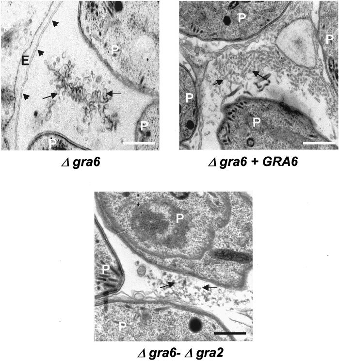
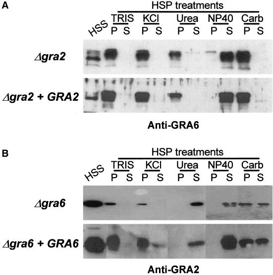
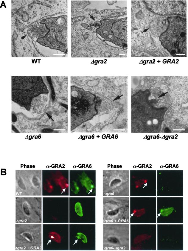
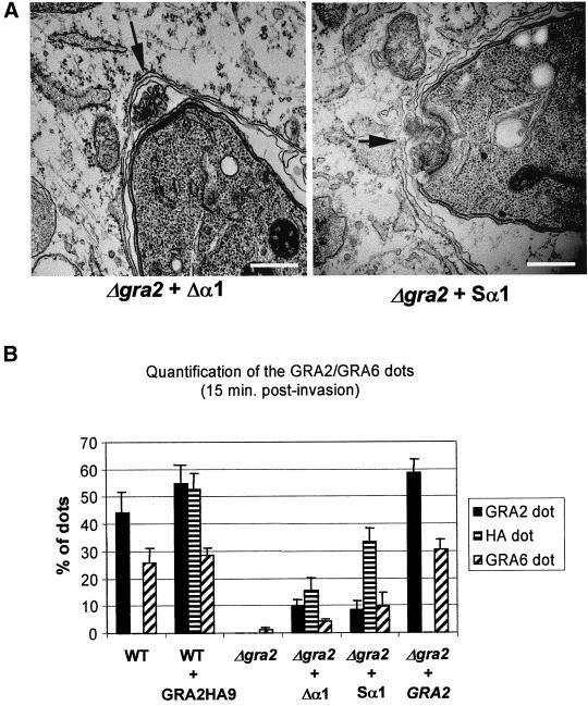
The intracellular parasite Toxoplasma gondii develops within a nonfusogenic vacuole containing a network of elongated nanotubules that form connections with the vacuolar membrane. Parasite secretory proteins discharged from dense granules (known as GRA proteins) decorate this intravacuolar network after invasion. Herein, we show using specific gene knockout mutants, that the unique nanotubule conformation of the network is induced by the parasite secretory protein GRA2 and further stabilized by GRA6. The vacuolar compartment generated by GRA2 knockout parasites was dramatically disorganized, and the normally tubular network was replaced by small aggregated material. The defect observed in Deltagra2 parasites was evident from the initial stages of network formation when a prominent cluster of multilamellar vesicles forms at a posterior invagination of the parasite. The secretory protein GRA6 failed to localize properly to this posterior organizing center in Deltagra2 cells, indicating that this early conformation is essential to proper assembly of the network. Construction of a Deltagra6 mutant also led to an altered mature network characterized by small vesicles instead of elongated nanotubules; however, the initial formation of the posterior organizing center was normal. Complementation of the Deltagra2 knockout with mutated forms of GRA2 showed that the integrity of both amphipathic alpha-helices of the protein is required for correct formation of the network. The induction of nanotubues by the parasite protein GRA2 may be a conserved feature of amphipathic alpha-helical regions, which have also been implicated in the organization of Golgi nanotubules and endocytic vesicles in mammalian cells.


Figures

Figure 1
Figure 1
Ultrastructural alteration of the intravacuolar tubular network after deletion of the GRA2 gene. Transmission electron micrographs depicting the parasitophorous vacuole of wild-type RH hxgprt (WT), Δgra2 mutant (Δgra2), or complemented Δgra2 (Δgra2 + GRA2) parasites grown in HFF cells. Cells were fixed at 20–24 h postinfection. The PV membrane is indicated by the arrowheads, and the network is indicated by the arrows. P, parasite; M, host cell mitochondrion. Scale, 500 nm.
Figure 2
Figure 2
Complementation of the Δgra2 deletion mutant (Δgra2) with altered forms of GRA2. (A) Schematic representation of the GRA2-HA9 cassettes used to complement the Δgra2 knockout mutant. The open boxes indicate the GRA2 coding sequence, with the black boxes, the N-terminal signal peptide; the vertically shaded boxes, the nine amino acid-HA9 epitope tag fused to the C-terminal end of the coding sequence; the diagonally shaded boxes, the amphipathic alpha-helices of GRA2; and the squared box, the rearranged amino acids at the 69–87 region, in the Sα1 scrambled construct. The total number of GRA2 amino acids is indicated below each cassette. Amino acids 69–87 of the first amphipathic alpha-helix are displayed as an Edmunson helical wheel below. The amino acids were rearranged to destroy the amphipathicity of the helix. (referred to as Sα1). Hydrophobic amino acids are in open squares and charged amino acids in shaded squares. (B) Western blot analysis of the Δgra2 mutant complemented with either the Δα1 form of GRA2-HA9, or the scrambled form (Sα1) of GRA2-HA9. For comparison, the Δgra2 mutant, the wild-type parasite (WT) and the transgenic parasite expressing GRA2-HA9 (WT+ GRA2HA9) are shown. The left panel was probed with the rabbit polyclonal antibody against HA (anti-HA) and the right panel, with the anti-GRA2 mAb TG17-179 (anti-GRA2). The rabbit polyclonal antibody against actin was used as an internal control of the quantity of proteins loaded in each lane. (C) Ultrastructural modifications of the vacuole after complementation of the Δgra2 mutant with either the deleted form of GRA2 HA9 (Δgra2 + Δα1) or the scrambled form of GRA2-HA9 (Δgra2 + Sα1). The network is present as small vesicles (Δgra2 + Δα1) or as large vesicles and membrane sheets (Δgra2 + Sα1) (arrows). Arrowheads indicate the PVM. P, parasite; M, host cell mitochondrion. Scale, 500 nm.
Figure 3
Figure 3
Targeted disruption of the GRA6 gene and construction of the double knockout mutant, Δgra6-Δgra2. (A) Schematic genomic representation of both the GRA2 and the GRA6 loci and of the plasmid constructs used to target both the GRA6 and the GRA2 genes. The 5′- and 3′-untranslated regions (UTRs) of both GRA2 and GRA6 are represented by diagonally shaded boxes (inclined to the right in the case of GRA6 and, to the left in the case of GRA2); stripped boxes represent the DHFR 5′- and 3′-UTR. Arrowheads indicate the transcriptional start site of either the GRA6 or the GRA2 gene. Restriction sites used for the genetic analyses are indicated: E (EcoRI), N (NsiI), and Nc (NcoI). Probes used to hybridize the southern blots are represented by the thick lines underneath the coding sequences. (B) Western blot analysis of sample clones showing the lack of GRA6 expression in the Δgra6 mutants (clone A11 and clone A804) and the lack of expression of both GRA2 and GRA6 in the Δgra6-Δgra2 mutants (clone A26 and clone A81). For comparison, the WT parasite, the Δgra2 mutant and the respective complemented mutants (Δgra2 + GRA2; Δgra6 + GRA6) are shown. Western blots were incubated with either the rabbit polyclonal antibody to GRA6 (left) or the anti-GRA2 mAb TG17-179 (right). Asterisk (*) indicates Toxoplasma actin used as an internal control of the quantity of proteins loaded in each lane and revealed by a rabbit anti-T. gondii actin. (C) Southern blot analysis of both the Δgra6 and Δgra6-Δgra2 mutants in comparison with the WT parasite and the complemented Δgra6 (Δgra6 + GRA6). The left panel shows that the Δgra6 mutants, clones A804 and A11, lack the GRA6 open-reading-frame, in contrast to the parental RH HXGPRT (WT). The complemented Δgra6 (Δgra6 + GRA6) has multiple integrations of the GRA6 gene in the genome, including one copy at the GRA6 locus. DNA was digested with XhoI and hybridized with the GRA6 probe (690 bp) shown in A. The right panel showed that the Δgra6-Δgra2 mutants, clones A81 and A26, lack the GRA2 open-reading-frame. In contrast, the GRA2 locus was unaltered by the targeted deletion of the GRA6 gene (clones A804 and A11). DNA was digested with NcoI and hybridized with the GRA2 probe (550 bp) shown in A.
Figure 4
Figure 4
Disruption of GRA6 causes loss of organization of the mature network. Ultrastructural alteration of the intravacuolar tubular network in the Δgra6 mutant, a complemented mutant (Δgra6 + GRA6) and the Δgra6-Δgra2 double mutant examined at 20–24 h postinfection. The PV membrane is indicated by the arrowheads, and the network is indicated by arrows. P, parasite; E, endoplasmic reticulum of the host cell. Scale, 500 nm.
Figure 5
Figure 5
Despite the lack of network organization, GRA6 and GRA2 still behave as integral membrane proteins in the Δgra2 and in the Δgra6 mutants, respectively. Western blot analysis of the partitioning of GRA6 and GRA2 in single knock mutants. (A) In Δgra2 mutants, GRA6 was present as both a soluble (HSS) and a membranous form (HSP) within the vacuole, with the soluble GRA6 migrating faster than the membranous form. The GRA6 membranous form was resistant to high salt concentration (KCl), high pH (Carb), 6 M urea but was fully solubilized by NP-40. TRIS refers to a control treatment with buffer alone. The behavior of GRA6 in the complemented mutant (Δgra2 + GRA2) was similar. (B) Two forms of GRA2 were detected in the Δgra6 mutant (Δgra6), a soluble (HSS) and a membrane-associated form (HSP). The membrane-associated form of GRA2 was displaced from the HSP by NP40 and by 6 M urea. The behavior of GRA2 in a complemented mutant (Δgra6 + GRA6) was similar. Fractions were analyzed by SDS-PAGE followed by Western blotting by using either the rabbit polyclonal serum against GRA6 and the mAb TG 17-179 anti-GRA2, respectively.
Figure 6
Figure 6
Disruption of GRA2 causes loss of the posterior organizing center and results in failure to target GRA6 to the posterior end of the parasite shortly after invasion. (A) Transmission electron micrographs depicting the invaginated posterior end of the parasite, 20 min postinvasion. Wild-type parasites (WT), single deletion mutants (Δgra2 and Δgra6), complemented mutants (Δgra2 + GRA2 and Δgra6 + GRA6) and double deletion mutant (Δgra6-Δgra2). Arrows indicate the posterior invagination. Scale, 500 nm. (B) IF localization of GRA proteins in the parasitophorous vacuole at 15 min postinvasion. In both the WT and complemented mutants, both GRA2 (red channel) and GRA6 (green channel) were found throughout the vacuole and concentrated in a prominent dot of fluorescence at the posterior end of the parasite (arrows). In contrast, in the Δgra2 mutant, GRA6 was secreted into the vacuolar compartment but failed to accumulate at the posterior end. Remarkably, no alteration of the typical GRA2 posterior accumulation was observed in the Δgra6 mutant. GRA2 was detected using the mAb TG17-179 followed by Texas-Red–conjugated anti-mouse IgG. GRA6 was detected using the rabbit polyclonal antibody followed by BODIPY-conjugated goat anti-rabbit IgG.
Figure 7
Figure 7
Complementation of the Δgra2 mutant with partial forms of the protein does not restore posterior organization of the network or targeting of GRA6. (A) Complementation of the Δgra2 mutant with GRA2 containing either the deleted first helix (Δgra2 + Δα1) or the scrambled first helix (Δgra2 + Sα1). Transmission electron micrographs of cells processed 20 min postinfection. Arrows indicate the posterior invagination. Scale, 500 nm. (B) Quantification of the posterior localization of GRA2 and GRA6 by IF as described in Figure 6B. Wild-type parasites (WT), wild-type parasites expressing GRA2 tagged with HA9 (WT + GRA2HA9). GRA2 deletion mutant (Δgra2), deletion mutant complemented with GRA2 lacking the first alpha-helix (Δgra2 + Δα1), deletion mutant complemented with the scrambled α1 form of GRA2 (Δgra2 + Sα1), deletion mutant complemented with the full-length gene (Δgra2 + GRA2). Values represent the mean + SD from three coverslips of a representative experiment.

Similar articles

Cited by

References

    1. Carruthers VB, Sibley LD. Sequential protein secretion from three distinct organelles of Toxoplasma gondii accompanies invasion of human fibroblasts. Eur J Cell Biol. 1997;73:114–123. - PubMed
    1. Cesbron-Delauw MF. Dense granule proteins in Toxoplasma gondii: their role in the host-parasite relationship. Parasitol Today. 1994;10:293–296. - PubMed
    1. Charif H, Darcy F, Torpier G, Cesbron-Delauw MF, Capron A. Toxoplasma gondii: characterization and localization of antigens secreted from tachyzoites. Exp Parasitol. 1990;71:114–124. - PubMed
    1. Coppens I, Sinai AP, Joiner KA. Toxoplasma gondii exploits host low-density lipoprotein receptor-mediated endocytosis for cholesterol acquisition. J Cell Biol. 2000;149:167–180. - PMC - PubMed
    1. Dobrowolski JM, INiesman IR, Sibley LD. Actin in the parasite Toxoplasma gondii is encoded by a single copy gene, ACT1 and exists primarily in a globular form. Cell Motil Cytoskeleton. 1997;37:253–262. - PubMed

Publication types

Substances

LinkOut - more resources