. 2002 Jul;13(7):2421-35.
doi: 10.1091/mbc.e02-02-0071.

GLUT4 retention in adipocytes requires two intracellular insulin-regulated transport steps

Affiliations

GLUT4 retention in adipocytes requires two intracellular insulin-regulated transport steps

Anja Zeigerer et al. Mol Biol Cell. 2002 Jul.

Abstract

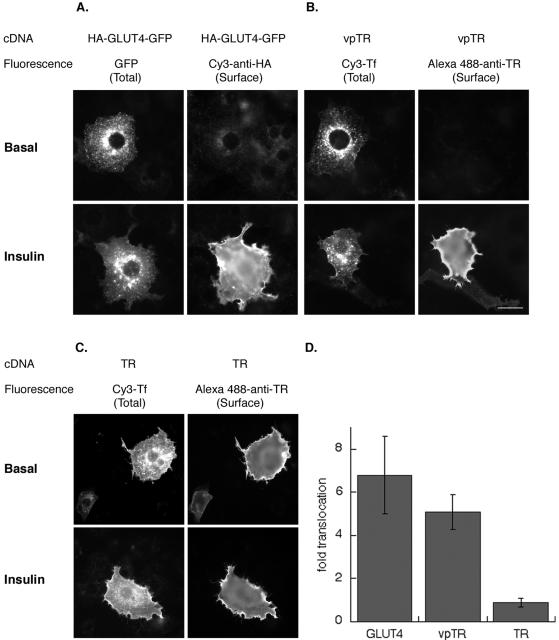
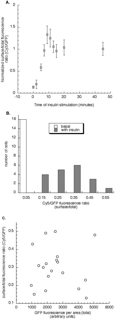
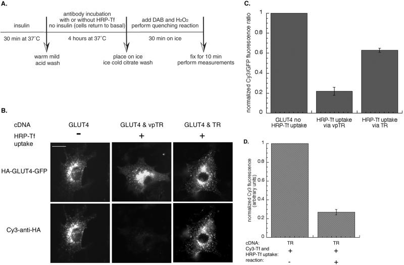
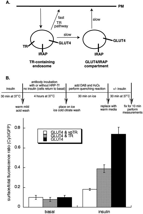
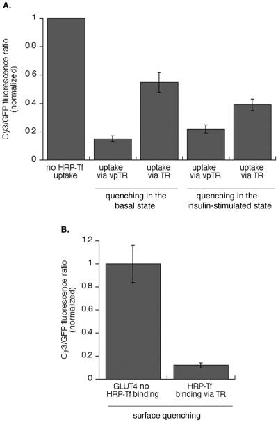
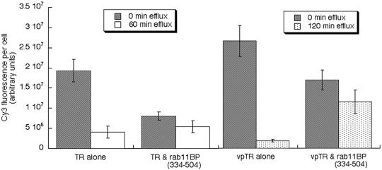
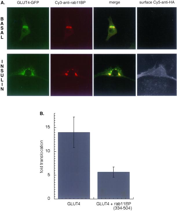
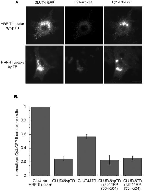
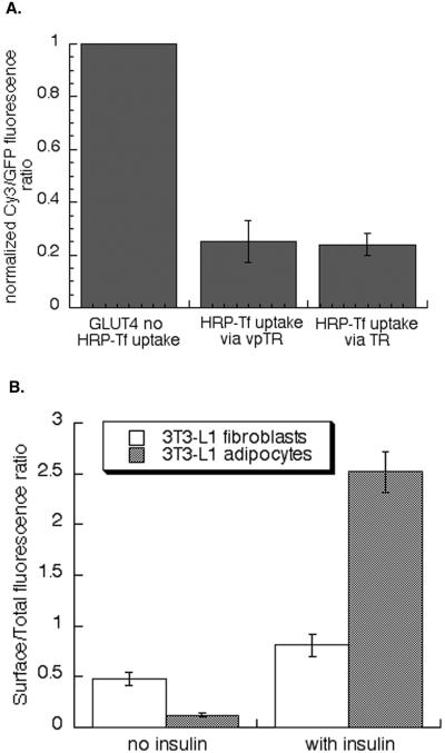
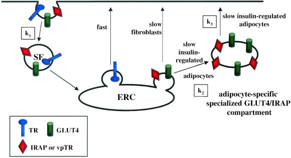
Insulin regulates glucose uptake into fat and muscle by modulating the distribution of the GLUT4 glucose transporter between the surface and interior of cells. The GLUT4 trafficking pathway overlaps with the general endocytic recycling pathway, but the degree and functional significance of the overlap are not known. In this study of intact adipocytes, we demonstrate, by using a compartment-specific fluorescence-quenching assay, that GLUT4 is equally distributed between two intracellular pools: the transferrin receptor-containing endosomes and a specialized compartment that excludes the transferrin receptor. These pools of GLUT4 are in dynamic communication with one another and with the cell surface. Insulin-induced redistribution of GLUT4 to the surface requires mobilization of both pools. These data establish a role for the general endosomal system in the specialized, insulin-regulated trafficking of GLUT4. Trafficking through the general endosomal system is regulated by rab11. Herein, we show that rab11 is required for the transport of GLUT4 from endosomes to the specialized compartment and for the insulin-induced translocation to the cell surface, emphasizing the importance of the general endosomal pathway in the specialized trafficking of GLUT4. Based on these findings we propose a two-step model for GLUT4 trafficking in which the general endosomal recycling compartment plays a specialized role in the insulin-regulated traffic of GLUT4. This compartment-based model provides the framework for understanding insulin-regulated trafficking at a molecular level.


Figures

Figure 1
Figure 1
Insulin stimulated translocation of HA-GLUT4-GFP, vpTR, and the TR in 3T3-L1 adipocytes. Cells were transiently transfected by electroporation with the cDNAs of these reporters, and the effect of insulin (170 nM for 15 min) on their distribution was determined using a quantitative single-cell fluorescence assay. (A) Example of cells expressing HA-GLUT4-GFP. GFP fluorescence is a measure of total expression. The amount of HA-GLUT4-GFP on the surface is determined with a fluorescent antibody against the HA-epitope in fixed unpermeabilized cells. (B) Example of cells expressing vpTR or the TR (C). The total amount of the constructs expressed was determined by incubating cells with Cy3-Tf for 4 h at 37°C. Where noted, insulin was added to half of the samples for the last 15 min. The amount of the constructs on the surface was detected with an antibody against the extracellular domain of the human TR. Bar, 30 μm. (D) Summary of the translocation measured in five independent experiments (mean ± SEM). In each experiment the data from ∼20 cells per condition were quantified. The insulin-induced translocation was quantified by comparing surface-to-total ratio in the presence and absence of insulin. The surface-to-total ratio normalizes the data for the level of each construct expressed.
Figure 2
Figure 2
Analysis of single cell data. (A) Analysis of the extent of translocation of HA-GLUT4-GFP to the cell surface of day 5 differentiated 3T3-L1 cells stimulated with insulin for various lengths of time. 3T3-L1 cells were electroporated with HA-GLUT4-GFP cDNA and were stimulated with 170 nM insulin for 0 (basal), 2, 5, 7, 9, 11, 13, 15, 20, and 45 min. The proportion of surface HA-GLUT4-GFP was determined after each time point as described in MATERIALS AND METHODS. The data from 20 individual cells is averaged for each time point and normalized to the steady-state level. Data from a representativeexperiment are shown. The same results were seen in two additional experiments. (B) Distribution of HA-GLUT4-GFP surface-to-total ratios among cells in the basal and insulin stimulated state. Data from a representative experiment are shown. The data from individual cells are grouped according to the surface-to-total ratio. The bin size was chosen arbitrarily. The surface and total fluorescence values are in arbitrary fluorescence units and therefore the ratio value is proportional to, but not an absolute measure of, the fraction of the construct on the surface. An increase in this ratio indicates an increase of the construct on the surface. The ratios in the insulin-stimulated conditions are normally distributed. The same is true for the basal ratios. C) Distribution of HA-GLUT4-GFP surface-to-total ratio as a function of HA-GLUT4-GFP expression per cell in the insulin stimulated state. The data are from the same experiment as shown in B. The variability in the surface-to-total distribution of individual cells does not correlate with the amount of HA-GLUT4-GFP expressed.
Figure 3
Figure 3
Compartment-specific fluorescence quenching in adipocytes. (A) Scheme of designed fluorescence-quenching experiment in 3T3-L1 adipocytes. Cells were stimulated with insulin for 30 min to increase recycling of HA-GLUT4-GFP. Insulin was removed by a mild-acid wash, and the cells were incubated with Cy3-anti-HA antibody, with or without HRP-Tf (20 μg/ml) for 4 h. During this incubation all of the HA-GLUT4-GFP is labeled with Cy3 anti-HA antibody, thereby placing a fluorescent probe within the lumen of these compartments. The compartments containing vpTR or the TR contain HRP-Tf. The cells were chilled to 4°C and the HRP catalyzed DAB polymerization initiated by adding DAB and H2O2 for 30 min on ice. The samples are fixed and then examined. (B) Images from a representative experiment. Cells were transfected with HA-GLUT4-GFP and the TR, or HA-GLUT4-GFP and vpTR. The top panels are the GFP fluorescence and the bottom panels are the Cy3 fluorescence from the same cells. There is complete overlap of the two probes in control cells (no HRP-Tf uptake), indicating all the intracellular compartments of HA-GLUT4-GFP are in dynamic communication with the cell surface. Bar, 20 μm. (C) Summary of the fluorescence quenching measured in 15 independent experiments (means ± SEM). The GFP fluorescence controls for expression level of HA-GLUT4-GFP. To compare the results of the independent experiments, the data from the individual experiments were normalized to the Cy3/GFP ratio in control cells (no HRP-Tf uptake) for that experiment. (D) Summary of data from five independent experiments measuring the Cy3-Tf fluorescence quenched by HRP-Tf in cells expressing the TR (means ± SEM). The data from the individual experiments were normalized to the Cy3 fluorescence in control cells (no reaction) for that experiment. These data establish that the maximum quenching for this assay as 80% of control.
Figure 4
Figure 4
Effect of HRP-mediated compartment ablation on insulin-stimulated translocation of HA-GLUT4-GFP. (A) Schematic illustrating that the two nearly equal pools of intracellular GLUT4 in the basal state indicate that the rate of transport from endosomes must be similar to the rate of transport from the specialized compartment. (B) Schema of the designed fluorescence quenching experiment after insulin stimulation. Insulin recruits GLUT4 from both pools. Cells were incubated with HRP-Tf (20 μg/ml) for 3.5 h, incubated with DAB and H2O2 on ice for 30 min, treated with or without insulin for 30 min at 37°C, and then fixed. The control cells were not incubated with HRP-Tf but were treated with DAB and H2O2 on ice for 30 min. The HA-GLUT4-GFP translocation was measured using quantitative fluorescence microscopy. The data are from a representative experiment (means ± SEM).
Figure 5
Figure 5
Effect of insulin on the codistribution of GLUT4, the TR, and vpTR. (A) Cells were incubated with Cy3-labeled anti-HA antibody and HRP-Tf for 4 h at 37°C. Insulin was added for the last 30 min of incubation. The cells were incubated with DAB and H2O2 for 30 min on ice. The degree of quenching when HRP-Tf is taken up by the TR is increased compared with basal quenching, but is still not as efficient than quenching via vpTR. The data for the quenching in the presence of insulin are a summary of six independent experiments, and the data for the quenching in the basal state, shown for the sake of comparison, are from a representative experiment. (B) Surface quenching of the Cy3–anti-HA antibody. Cells were stimulated with insulin for 15 min, and the Cy3-anti-HA antibody as well as HRP-Tf were bound on ice for 1 h. The cells were incubated with DAB and H2O2 as described previously. The data are from a representative experiment (means ± SEM) and were normalized to the Cy3/GFP ratio in control cells (no HRP-Tf uptake).
Figure 6
Figure 6
Effect of rab11BP(334–504) on the recycling of the TR and vpTR. Cells were incubated for 3.5 h with Cy3-Tf, washed, and incubated for an additional 60 min (for the TR) or 120 min (for vpTR) in medium without Cy3-Tf. At the end of the incubation the cells were fixed and the amount of Cy3-Tf remaining was determined. The different efflux times were chosen because the vpTR is recycled more slowly than the TR (Subtil et al., 2000). Data are from a representative experiment (means ± SEM).
Figure 7
Figure 7
Effect of rab11BP(334–504) on the translocation of HA-GLUT4-GFP in 3T3-L1 adipocytes. (A) Rab11BP(334–504), a GST-fusion construct, is stained with a Cy3-anti-GST, and the surface expression of HA-GLUT4-GFP is detected with a Cy5-anti-HA. Images are from a representative experiment. (B) Summary of data from four independent experiments measuring the insulin-induced translocation of HA-GLUT4-GFP in control cells and cells expressing rab11BP(334–504) (means ± SEM).
Figure 8
Figure 8
Effect of rab11BP on the codistribution of GLUT4, the TR, and vpTR. (A) Experiment has been performed as described in Figure 3. Rab11BP(334–504) is stained with a Cy5-anti-GST. Images are from a representative experiment. Bar, 20 μm. (B) Summary of data from five independent fluorescence-quenching experiments determining the codistribution of the TR and GLUT4, or vpTR and GLUT4 (means ± SEM). The data from the individual experiments were normalized to the Cy3/GFP ratio in control cells (no HRP-Tf uptake) for that experiment.
Figure 9
Figure 9
Fluorescence quenching and translocation in 3T3-L1 fibroblasts. (A) Summary of the data from four independent experiments measuring the Cy3-Tf fluorescence quenched by HRP-Tf in cells expressing HA-GLUT4-GFP and the TR, or HA-GLUT4-GFP and vpTR. The data are means ± SEM. The data from the individual experiments were normalized to the Cy3/GFP ratio in control cells (no HRP-Tf uptake) for that experiment. Fibroblasts are preconfluent 3T3-L1 cells. (B) HA-GLUT4-GFP translocation in adipocytes and fibroblasts. Data are from a representative experiment (means ± SEM).
Figure 10
Figure 10
Two-step model for insulin-regulated traffic in adipocytes. The first retention step occurs at the TR-containing endosomes. GLUT4 and IRAP are sorted from the rapid TR recycling pathway because they are sequestered in specialized transport vesicles that bud slowly from endosomes. We propose that this step is common to differentiated and undifferentiated cells, and therefore not adipocyte specific. In adipocytes these vesicles do not fuse directly with the plasma membrane but there is an additional insulin-regulated retention step. GLUT4 and IRAP in this postendosomal pool constitute the adipocyte-specific compartment. This specialized post endosomal compartment may be a stable fusion/fission competent compartment or it may be a collection of tethered vesicles. The available data do not distinguish between these two possibilities. Proper insulin-induced redistribution of GLUT4 and IRAP to the cell surface involves the recruitment of GLUT4/IRAP from both endosomes and the adipocyte-specific compartment.

Similar articles

Cited by

References

    1. Becker C, Sevilla L, Tomas E, Palacin M, Zorzano A, Fischer Y. The endosomal compartment is an insulin-sensitive recruitment site for GLUT4 and GLUT1 glucose transporters in cardiac myocytes. Endocrinology. 2001;142:5267–5276. - PubMed
    1. Bogan J, McKee A, Lodish H. Insulin-responsive compartments containing GLUT4 in 3T3–L1 and CHO cells: regulation by amino acid concentrations. Mol Cell Biol. 2001;21:4785–4806. - PMC - PubMed
    1. Cain CC, Trimble WS, Lienhard GE. Members of the VAMP family of synaptic vesicle proteins are components of glucose transporter-containing vesicles from rat adipocytes. J Biol Chem. 1992;267:11681–11684. - PubMed
    1. Chen W, Feng Y, Chen D, Wandinger-Ness A. Rab11 is required for trans-golgi network-to-plasma membrane transport and a preferential target for GDP dissociation inhibitor. Mol Biol Cell. 1998;9:3241–3257. - PMC - PubMed
    1. Cushman SW, Wardzala LJ. Potential mechanism of insulin action on glucose transport in the isolated rat adipose cell. Apparent translocation of intracellular transport systems to the plasma membrane. J Biol Chem. 1980;255:4758–4762. - PubMed

Publication types

MeSH terms