. 2002 Jul;13(7):2436-47.
doi: 10.1091/mbc.e02-02-0102.

A conserved Drosophila transportin-serine/arginine-rich (SR) protein permits nuclear import of Drosophila SR protein splicing factors and their antagonist repressor splicing factor 1

Affiliations

A conserved Drosophila transportin-serine/arginine-rich (SR) protein permits nuclear import of Drosophila SR protein splicing factors and their antagonist repressor splicing factor 1

Eric Allemand et al. Mol Biol Cell. 2002 Jul.

Abstract

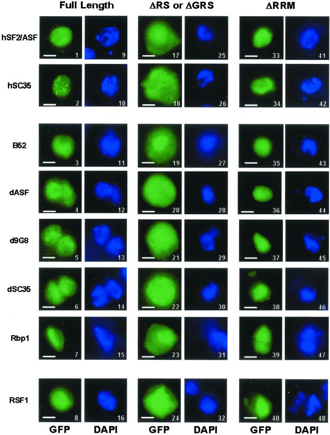
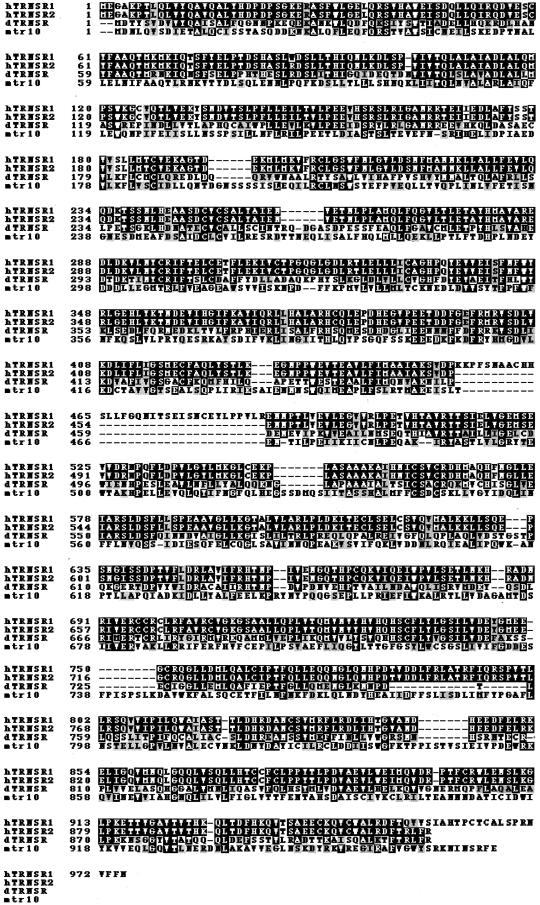
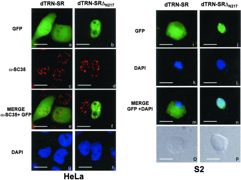
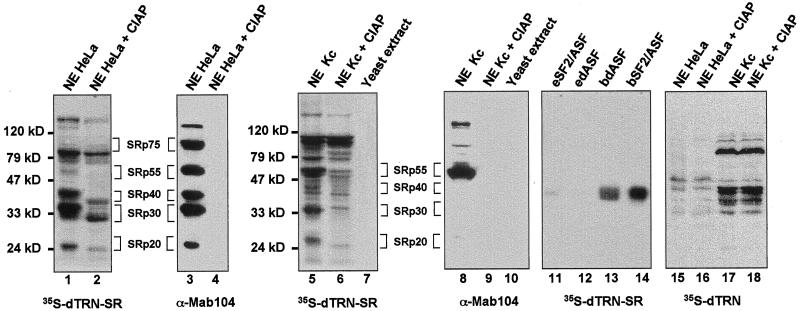
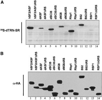
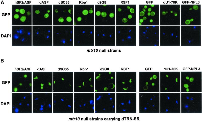
Members of the highly conserved serine/arginine-rich (SR) protein family are nuclear factors involved in splicing of metazoan mRNA precursors. In mammals, two nuclear import receptors, transportin (TRN)-SR1 and TRN-SR2, are responsible for targeting SR proteins to the nucleus. Distinctive features in the nuclear localization signal between Drosophila and mammalian SR proteins prompted us to examine the mechanism by which Drosophila SR proteins and their antagonist repressor splicing factor 1 (RSF1) are imported into nucleus. Herein, we report the identification and characterization of a Drosophila importin beta-family protein (dTRN-SR), homologous to TRN-SR2, that specifically interacts with both SR proteins and RSF1. dTRN-SR has a broad localization in the cytoplasm and the nucleus, whereas an N-terminal deletion mutant colocalizes with SR proteins in nuclear speckles. Far Western experiments established that the RS domain of SR proteins and the GRS domain of RSF1 are required for the direct interaction with dTRN-SR, an interaction that can be modulated by phosphorylation. Using the yeast model system in which nuclear import of Drosophila SR proteins and RSF1 is impaired, we demonstrate that complementation with dTRN-SR is sufficient to target these proteins to the nucleus. Together, the results imply that the mechanism by which SR proteins are imported to the nucleus is conserved between Drosophila and humans.


Figures

Figure 1
Figure 1
Cellular localization of GFP fusion proteins in Drosophila S2 Schneider cells. Direct fluorescence of GFP-hSF2ASF (1), GFP-hSC35 (2), GFP-B52 (3), GFP-dASF (4), GFP-d9G8 (5), GFP-dSC35 (6), GFP-Rpb1 (7), and GFP-RSF1 (8); GFP-hSF2ASFΔRS (17), GFP-hSC35ΔRS (18), GFP-B52ΔRS (19), GFP-dASFΔRS (20), GFP-d9G8ΔRS (21), GFP-dSC35ΔRS (22), GFP-Rbp1ΔRS (23), and GFP-RSF1ΔGRS (24) deleted of the RS domain or GRS domain; GFP-hSF2ASFΔRRM (33), GFP-hSC35ΔRRM (34), GFP-B52ΔRRM (35), GFP-dASFΔRRM (36), GFP-d9G8ΔRRM (37), GFP-dSC35ΔRRM (38), GFP-Rbp1ΔRRM (39), and GFP-RSF1ΔRRM (40) deleted of the RRM domain. Fusion proteins were analyzed 20 h after transfection. Expression of fusion proteins was confirmed by immunoblot analysis by using an anti-GFP antibody (our unpublished data). The position of nuclei was confirmed by DAPI staining of the same cells transfected with GFP fusion proteins shown in 1–8 (9–16, respectively), 17–24 (25–32, respectively), and 33–40 (41–48, respectively). Bar, 10 μm.
Figure 2
Figure 2
Amino acid sequence alignments of dTRN-SR with TRN-SR1, TRN-SR2, and Mtr10p as obtained by the ClustalW program. Identical amino acid residues are marked in dark and conservative substitutions such as RKH, IVLM, ED, FY, and ST are marked in gray. Gaps are introduced to optimize amino acid sequence alignments.
Figure 3
Figure 3
Cellular localization of dTRN-SR GFP fusion proteins in HeLa cells (a–h) and in Drosophila S2 Schneider cells (i–p). Direct fluorescence of GFP-dTRN-SR (a and i) and GFP-dTRN-SRΔN217 (b and j) fusion proteins were analyzed 20 h posttransfection. Expression of fusion proteins was confirmed by immunoblot analysis by using an anti-GFP antibody (our unpublished data). The position of nuclei was confirmed by DAPI staining of the same transfected HeLa (g and h) and S2 (k and l) cells with GFP-dTRN-SR (g and k) or GFP-dTRN-SRΔN217 (h and l). Indirect immunofluorescence staining of HeLa cells in a and b with the αSC35 mAb (c and d, respectively) showed the cellular localization of endogenous SR protein SC35. A merge between a and c (e) and between b and d (f) shows that dTRN-SRΔN217 and SC-35 colocalize in the speckles. The Normarski interference-contrast of the same S2 cells transfected with GFP-dTRN-SR (o) and GFP-dTRN-SRΔN217 (p) are shown. Bar, 10 μm
Figure 4
Figure 4
Far Western analysis showing a physical interaction between dTRN-SR and SR proteins from both HeLa and Drosophila Kc cells. Proteins from HeLa (lanes 1–4, 15, and 16), Drosophila Kc (lanes 5, 6, 8, 9, 17, and 18) nuclear extracts or yeast S. cerevisae total extracts (lanes 7 and 10) either untreated (lanes 1, 3, 5, 7, 8, 10, 15, and 17) or treated (lanes 2, 4, 6, 9, 16, and 18) with calf intestinal alkaline phosphatase (CIAP) as well as purified recombinant dASF and SF2/ASF 100 ng each, expressed either in a baculovirus system (bdASF, lane 13, and bSF2/ASF, lane 14, receptively) or in E. coli (edASF, lane 12, and eSF2/ASF, lane 11, respectively) were separated on a SDS-PAGE, transferred to nitrocellulose, renatured, and probed either with 35-S-labeled dTRN-SR (lanes 1, 2, 5–7, and 11–14) or 35-S-labeled dTRN (lanes 15–18). Proteins (lanes 3, 4, 8, and 9) were subjected to Western blot analysis by using mAb 104. Molecular weight markers are indicated on the left of the Far Western panels, and SR protein species are indicated on the right.
Figure 5
Figure 5
Binding of dTRN-SR to SR proteins, RSF1 and deletion mutants expressed in wild-type yeast strains. Total extracts prepared from yeast expressing indicated recombinant proteins (lanes 1–14) were assayed by either Far Western analysis by using 35-S-labeled dTRN-SR (A) or Western blot analysis by using 3F10 anti-HA antibody (B). Both panels represent two independent migrations.
Figure 6
Figure 6
Nuclear import of SR proteins and RSF1 requires dTRN-SR in yeast cells. Direct fluorescence of GFP-hSF2ASF, GFP-dASF, GFP-dSC35, GFP-Rpb1, GFP-d9G8, GFP-RSF1, GFP, GFP-dU1–70K, and GFP-NPL3 was analyzed after transformation in either mtr 10::HIS3 cells (A) or mtr 10::HIS3 cells complemented with c-myc-dTRN-SR (B). The position of nuclei was confirmed by DAPI staining of the transformed cells. Bar, 5 μm.

References

    1. Aitchison JD, Blobel G, Rout MP. Kap104p: a karyopherin involved in the nuclear transport of messenger RNA binding proteins. Science. 1996;274:624–627. - PubMed
    1. Allemand E, Gattoni R, Bourbon HM, Stevenin J, Caceres JF, Soret J, Tazi J. Distinctive features of Drosophila alternative splicing factor RS domain: implication for specific phosphorylation, shuttling, and splicing activation. Mol Cell Biol. 2001;21:1345–1359. - PMC - PubMed
    1. Amrein H, Hedley ML, Maniatis T. The role of specific protein-RNA and protein-protein interactions in positive and negative control of pre-mRNA splicing by Transformer 2. Cell. 1994;76:735–746. - PubMed
    1. Barnard DC, Patton JG. Identification and characterization of a novel serine-arginine-rich splicing regulatory protein. Mol Cell Biol. 2000;20:3049–3057. - PMC - PubMed
    1. Blencowe BJ. Exonic splicing enhancers: mechanism of action, diversity and role in human genetic diseases. Trends Biochem. 2000;25:106–110. - PubMed

Publication types

MeSH terms