. 2002 Jul;13(7):2448-60.
doi: 10.1091/mbc.01-12-0589.

Molecular dissection of cytokinesis by RNA interference in Drosophila cultured cells

Affiliations

Molecular dissection of cytokinesis by RNA interference in Drosophila cultured cells

Maria Patrizia Somma et al. Mol Biol Cell. 2002 Jul.

Abstract

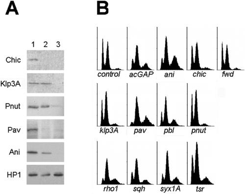
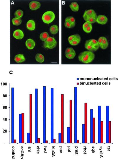
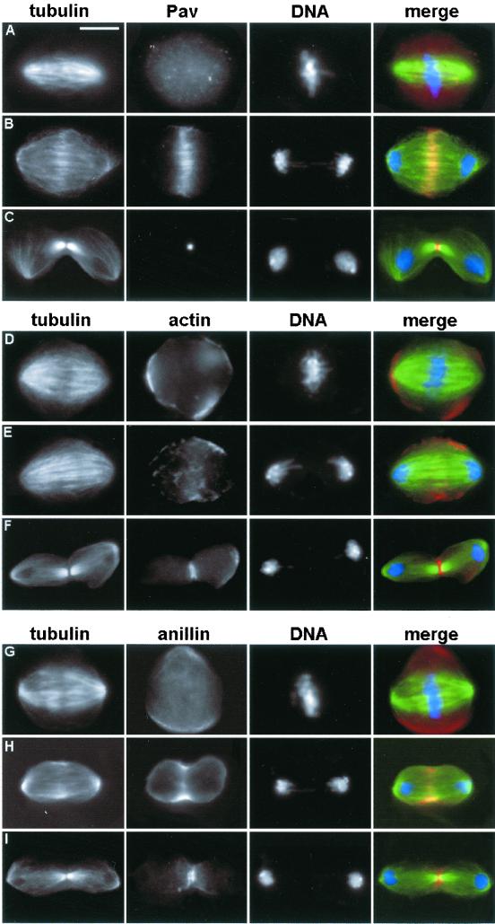
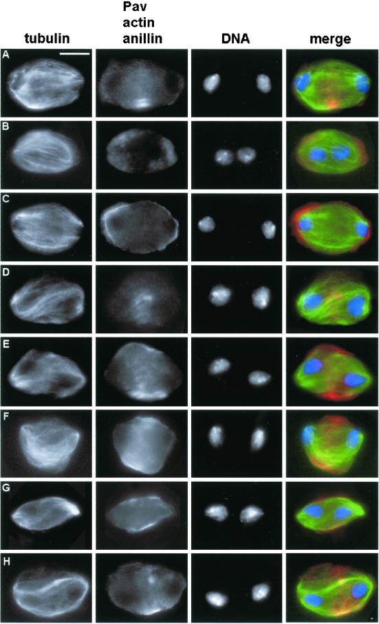
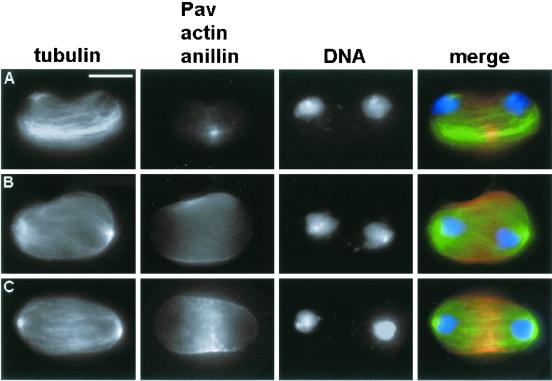
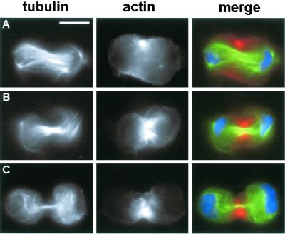
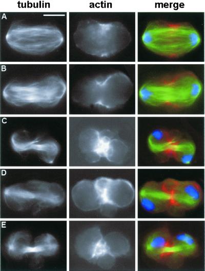
We have used double-stranded RNA-mediated interference (RNAi) to study Drosophila cytokinesis. We show that double-stranded RNAs for anillin, acGAP, pavarotti, rho1, pebble, spaghetti squash, syntaxin1A, and twinstar all disrupt cytokinesis in S2 tissue culture cells, causing gene-specific phenotypes. Our phenotypic analyses identify genes required for different aspects of cytokinesis, such as central spindle formation, actin accumulation at the cell equator, contractile ring assembly or disassembly, and membrane behavior. Moreover, the cytological phenotypes elicited by RNAi reveal simultaneous disruption of multiple aspects of cytokinesis. These phenotypes suggest interactions between central spindle microtubules, the actin-based contractile ring, and the plasma membrane, and lead us to propose that the central spindle and the contractile ring are interdependent structures. Finally, our results indicate that RNAi in S2 cells is a highly efficient method to detect cytokinetic genes, and predict that genome-wide studies using this method will permit identification of the majority of genes involved in Drosophila mitotic cytokinesis.


Figures

Figure 1
Figure 1
Effects of RNAi with Drosophila genes involved in cytokinesis. (A) Western blots from cells grown for 72 h in the presence of mock (plasmid) RNA (lane 1) or incubated for either 24 h (lane 2) or 72 h (lane 3) with the dsRNA for the protein indicated. The HP1 protein was used as a loading control in all cases. Note that 72-h treatments cause a dramatic depletion of all gene products. (B) FACS profiles of cells treated for 72 h with dsRNA; abscissa, DNA content; ordinate, cell number. Note that the control profile exhibits two main peaks of 2C and 4C cells and a very small peak of 8C cells. FACS profiles of chic (profilin), fwd (phospholipid kinase), klp3A (kinesin-like), and pnut (septin) (RNAi) cells do not differ from control, whereas the profiles of acGAP (rhoGAP), ani (anillin), pav (kinesin-like), pbl (rhoGEF), rho1, sqh (myosin II regulatory light chain), syx1A (t-SNARE), and tsr (cofilin) (RNAi) cells display a decrease of the 2C peak (G1 diploid cells) and a relative increase of both the 4C (G2-M diploid cells and G1 tetraploid cells) and the 8C peak (G2-M tetraploid cells and G1 octoploid cells).
Figure 2
Figure 2
Binucleated cells in cultures treated with dsRNA of genes involved in cytokinesis. (A and B) Control cells (A) and cells treated for 72 h with pav dsRNA (B) stained for tubulin (green) and DNA (with 4,6-diamidino-2-phenylindole, red). (C) Frequencies of binucleated cells after 72-h treatments with dsRNAs. Bar, 10 μm.
Figure 3
Figure 3
Localization of Pav (A–C), actin (D–F), and anillin (G–I) during mitotic division of S2 cells. (A, D, and G) Metaphases. (B, E, and H) Anaphases. (C, F, and I) Telophases. In the merged figures Pav, actin, and anillin are colored in red, tubulin in green, and DNA in blue. Bar, 10 μm.
Figure 4
Figure 4
Examples of aberrant (short) telophases observed in pav (kinesin-like), rho1, and sqh (myosinII regulatory light chain) (RNAi) cells. Cells were stained for tubulin (green), DNA (blue), and either Pav, actin, or anillin (red). (A–C) rho1 (RNAi) telophases. (D–F) sqh (RNAi) telophases. (G and H) pav (RNAi) telophases. (A and D) Pav immunostaining. (B, E, and G) Actin immunostaining. (C, F, and H) Anillin staining. Note that all these aberrant telophases exhibit severe defects in the central spindle, which is either absent or very poorly organized. In addition, in all telophases actin does not form a contractile ring but displays characteristic cortical localizations (see text). Pav localization is also severely disrupted; Pav staining never traverses the cells as occurs in controls but it is either absent or associated with the small and irregular microtubules bundles that occasionally form between the two daughter nuclei. Bar, 10 μm.
Figure 5
Figure 5
Abnormal telophases observed in syx1A (RNAi) cells. Cells were stained for tubulin (green), DNA (blue), and either Pav (A, red), actin (B, red), or anillin (C, red). Note the defective central spindle and contractile ring, and the abnormal distribution of Pav. Bar, 10 μm.
Figure 6
Figure 6
Excessive actin accumulation in the contractile apparatus of tsr (RNAi) cells. Cells were stained for tubulin (green), DNA (blue), and actin (red). (A) Late anaphase/early telophase. (B and C) Late telophases. Bar, 10 μm.
Figure 7
Figure 7
Abnormal membrane behavior during late telophase of ani (RNAi) cells. Cells were stained for tubulin (green), DNA (blue), and actin (red). (A and B) Anaphase (A) and early telophase (B) figures with normal actin accumulations. (C–E) Late telophases showing large membrane bulges in the cleavage area. The actin-associated fluorescence of these cells has been artificially enhanced to visualize the membrane bulges; these aberrant telophases do not seem to contain more actin than their control counterparts. Bar, 10 μm.

References

    1. Adam JC, Pringle JR, Peifer M. Evidence for functional differentiation among Drosophila septins in cytokinesis and cellularization. Mol Biol Cell. 2000;11:3123–3135. - PMC - PubMed
    1. Adams RR, Maiato H, Earnshaw WC, Carmena M. Essential roles of Drosophila inner centromere protein (INCENP) and aurora B in histone H3 phosphorylation, metaphase chromosome alignment, kinetochore disjunction, and chromosome segregation. J Cell Biol. 2001;153:865–880. - PMC - PubMed
    1. Adams RR, Tavares AA, Salzberg A, Bellen HJ, Glover DM. pavarotti encodes a kinesin-like protein required to organize the central spindle and contractile ring for cytokinesis. Genes Dev. 1998;12:1483–1494. - PMC - PubMed
    1. Bonaccorsi S, Giansanti MG, Gatti M. Spindle self-organization and cytokinesis during male meiosis in asterless mutants of Drosophila melanogaster. J Cell Biol. 1998;142:751–761. - PMC - PubMed
    1. Brill JA, Hime GR, Scharer-Schuksz M, Fuller MT. A phospholipid kinase regulates actin organization and intercellular bridge formation during germline cytokinesis. Development. 2000;127:3855–3864. - PubMed

MeSH terms