. 2002 Jul;13(7):2518-32.
doi: 10.1091/mbc.e02-01-0027.

Emp47p and its close homolog Emp46p have a tyrosine-containing endoplasmic reticulum exit signal and function in glycoprotein secretion in Saccharomyces cerevisiae

Affiliations

Emp47p and its close homolog Emp46p have a tyrosine-containing endoplasmic reticulum exit signal and function in glycoprotein secretion in Saccharomyces cerevisiae

Ken Sato et al. Mol Biol Cell. 2002 Jul.

Abstract

The yeast open reading frame YLR080w/EMP46 encodes a homolog of the Golgi protein Emp47p. These two proteins are 45% identical and have a single transmembrane domain in their C-terminal regions and a carbohydrate recognition domain signature in the N-terminal region. The C-terminal tail of Emp46p includes a dilysine signal. This protein is localized to Golgi membranes at steady state by subcellular fractionation and green fluorescent protein labeling. On block of forward transport in sec12-4 cells, redistribution of Emp46p from the Golgi to the endoplasmic reticulum is observed. These localization features are similar to those previously reported for Emp47p. In addition, mutagenesis of the C-terminal region identified a tyrosine-containing motif as a critical determinant of the Golgi-localization and interaction with both COPI and COPII components. Similar motifs are also observed in the C-terminal tail of Emp47p and other mammalian homologs. Disruption of Emp47p displays a growth defect at a high temperature or on Ca(2+)-containing medium, which is rescued by overexpression of Emp46p, suggesting a partially overlapping function between Emp46p and Emp47p. In addition, we found that the disruption of both Emp46p and Emp47p show a marked defect in the secretion of a subset of glycoproteins. Analysis of the C-terminal mutants for Ca(2+) sensitivity revealed that the forward transport of Emp46/47p is essential for their function, whereas the retrograde transport is not. We propose that Emp46p and Emp47p are required for the export of specific glycoprotein cargo from the endoplasmic reticulum.


Figures

Figure 1
Figure 1
(A) YLR080wp is homologous to Emp47p. Alignment of the protein sequence of Emp46p and Emp47p was done with the ALIGN algorithm (Genestream Search; CRBM Montepellier, France), and underlines correspond to transmembrane domains predicted by the PHDhtm program (Rost et al., 1995). (B) Hydrophobicity profiles calculated according to Kyte and Doolittle (1982) with a window size of 6. (C) The schematic diagram of the polypeptide chain and the type I topology of Emp46p indicates the positions of its functional domains. CRD, carbohydrate-recognition domain; TM, transmembrane domain; Cross, KXKXX motif.
Figure 2
Figure 2
(A) emp47Δ strains have a severe growth defect at 37°C that can be rescued by sorbitol. Isogenic wild-type (YPH500), emp46 Δ (KSY007), emp47Δ (KSY005), and emp46Δ emp47Δ (KSY008) strains were grown to saturation at 30°C in YPD medium, adjusted to 3 × 10⁷ cells/ml and 5 μl of a 10-fold dilution series was spotted onto YPD plates or YPD containing 1 M sorbitol. Plates were incubated at 30 or 37°C as indicated. (B) Overexpression of Emp46p rescued the inviability of the emp47Δ strain at 37°C. emp46Δ emp47Δ cells (KSY008) transformed with a single-copy plasmid with EMP46 (pKSY103; top), a multicopy plasmid with EMP46 (pKSY104; middle), or vectors (pRS316; bottom) were grown in liquid MCD medium lacking uracil and tryptophan (MCD-Ura-Trp) at 30°C and spotted onto a MCD-Ura-Trp plate as described above. Plates were incubated at 37°C. (C) emp47Δ mutants are sensitive to Ca2+. emp46 Δ emp47 Δ (KSY008) cells transformed with EMP46 or EMP47 (pKSY103 or pKSY105) in a single-copy plasmid and vectors (pRS314 or pRS316) as indicated were grown in MCD-ura-trp at 30°C and were spotted onto a YPD plate or either containing 0.25 M CaCl2, 0.5 M CaCl2, or 0.5 M MgCl2 as indicated. (D) Overexpression of Emp46p rescued the inviability of the emp47Δ strain on a Ca2+-containing plate. emp47Δ emp46Δ cells (KSY008) transformed with a single-copy plasmid with EMP46 (pKSY103; top), a multicopy plasmid with EMP46 (pKSY104; middle), or vector (pRS316; bottom) were grown in MCD-Ura-Trp at 30°C and spotted onto a YPD plate containing 0.25 M CaCl2 as described above. Plates were incubated at 30°C. (E) Suppression of Ca2+ sensitivity of the emp46Δ mutant by 3HA-tagged Emp46p. emp46Δ emp47Δ (KSY008) cells transformed with vector (pRS316) or a single-copy plasmid with EMP46 or 3HA-EMP46 were grown and spotted onto a YPD plate containing 0.25 M CaCl2 at 30°C as described above. (F) Emp46p and Emp47p do not depend on each other for their expression. Spheroplasts were prepared from cells (emp46Δ emp47Δ/3HA-EMP46/2myc-EMP47), resolved by SDS-PAGE, and immunoblotted with anti-HA and anti-myc antibodies. Kar2p is shown as a control.
Figure 2
Figure 2
(A) emp47Δ strains have a severe growth defect at 37°C that can be rescued by sorbitol. Isogenic wild-type (YPH500), emp46 Δ (KSY007), emp47Δ (KSY005), and emp46Δ emp47Δ (KSY008) strains were grown to saturation at 30°C in YPD medium, adjusted to 3 × 10⁷ cells/ml and 5 μl of a 10-fold dilution series was spotted onto YPD plates or YPD containing 1 M sorbitol. Plates were incubated at 30 or 37°C as indicated. (B) Overexpression of Emp46p rescued the inviability of the emp47Δ strain at 37°C. emp46Δ emp47Δ cells (KSY008) transformed with a single-copy plasmid with EMP46 (pKSY103; top), a multicopy plasmid with EMP46 (pKSY104; middle), or vectors (pRS316; bottom) were grown in liquid MCD medium lacking uracil and tryptophan (MCD-Ura-Trp) at 30°C and spotted onto a MCD-Ura-Trp plate as described above. Plates were incubated at 37°C. (C) emp47Δ mutants are sensitive to Ca2+. emp46 Δ emp47 Δ (KSY008) cells transformed with EMP46 or EMP47 (pKSY103 or pKSY105) in a single-copy plasmid and vectors (pRS314 or pRS316) as indicated were grown in MCD-ura-trp at 30°C and were spotted onto a YPD plate or either containing 0.25 M CaCl2, 0.5 M CaCl2, or 0.5 M MgCl2 as indicated. (D) Overexpression of Emp46p rescued the inviability of the emp47Δ strain on a Ca2+-containing plate. emp47Δ emp46Δ cells (KSY008) transformed with a single-copy plasmid with EMP46 (pKSY103; top), a multicopy plasmid with EMP46 (pKSY104; middle), or vector (pRS316; bottom) were grown in MCD-Ura-Trp at 30°C and spotted onto a YPD plate containing 0.25 M CaCl2 as described above. Plates were incubated at 30°C. (E) Suppression of Ca2+ sensitivity of the emp46Δ mutant by 3HA-tagged Emp46p. emp46Δ emp47Δ (KSY008) cells transformed with vector (pRS316) or a single-copy plasmid with EMP46 or 3HA-EMP46 were grown and spotted onto a YPD plate containing 0.25 M CaCl2 at 30°C as described above. (F) Emp46p and Emp47p do not depend on each other for their expression. Spheroplasts were prepared from cells (emp46Δ emp47Δ/3HA-EMP46/2myc-EMP47), resolved by SDS-PAGE, and immunoblotted with anti-HA and anti-myc antibodies. Kar2p is shown as a control.
Figure 3
Figure 3
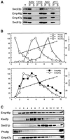
(A) Emp46p is an integral membrane protein. Spheroplasts were prepared from cells (emp46 Δ emp47 Δ/3HA-EMP46/2myc-EMP47) and treated with buffer 88 (Buffer), 1% Triton X-100 (TX100), 0.5 M NaCl (NaCl), or 0.1 M sodium carbonate (pH 11) in buffer 88. Samples were centrifuged at 100,000 × g, and totals before centrifugation (T), supernatant (S), and pellet (P) fractions were resolved by SDS-PAGE and immunoblotted for Sec23p, Sec61p, anti-HA, or anti-myc. (B) Sucrose gradient fractionation of Emp46p, Emp47p, Kex2p, Pho8p, and Sec61p. A whole-cell lysate from cells expressing both 2myc-Emp47p and 3HA-YLR080w was separated on a 20–60% sucrose density gradient, and fractions were collected, starting with fraction 1 at the top. Fractions were probed by immunoblotting. All data are from a single gradient, plotted in two parts for clarity. Relative levels of Emp46p, Kex2p, Sec61p, Pho8p, and Emp47p as determined by densitometry of the immunoblots are shown in C. Twenty percent of a total lysate is shown as T.
Figure 4
Figure 4
GFP-3HA-Emp46p is fully functional and shows wild-type subcellular distribution. (A) Cells expressing 3HA-Emp46p (EMP46) or GFP-3HA-Emp46p (GFP-EMP46) were spotted onto a YPD plate containing 0.25 M CaCl2 at 30°C as described in the legend to Figure 2. (B) Sucrose gradient fractionation of GFP-3HA-Emp46p. Whole-cell lysates from cells expressing 3HA-Emp46p or GFP-3HA-Emp46p were separated on sucrose density gradients (20–60%) and fractions were collected from the top. Relative levels of 3HA-Emp46p (□) and GFP-3HA-Emp46p (▪) in each fraction were quantified by densitometry of immunoblots.
Figure 5
Figure 5
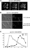
The dynamics of intracellular distribution of GFP-Emp46p in sec12-4 cells. (A) sec12-4 cells (MBY10-14C) harboring pKSY126 (GFP-Emp46p) were first observed at 20°C (a) and then the temperature of the microscope stage was raised and kept at 37°C for the indicated times (b and c). The same cells are shown. Images were visualized by confocal laser microscopy. Bar, 5 μm. (B) sec12-4 cells expressing GFP-Emp46p were incubated at 37°C for 30 min (upper panel) and and were then shifted to room temperature (RT) and observed 60 min later (lower panel). The left panels show the corresponding Nomarski images. Bar, 5 μm. (C) sec12-4 cells expressing 3HA-Emp46p were incubated at either 23°C (□) or 37°C (▪) for 90 min before spheroplasting. Lysates were loaded onto sucrose gradients as described in “Materials and Methods,” and then fractions were analyzed by SDS-PAGE and immunoblotting with anti-HA.
Figure 6
Figure 6
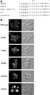
(A) The C-terminal region of Emp46p and other traffic lectins. ER-targeting dilysine signals are boldfaced, and the tyrosine-containing motifs are underlined. Numbering is from the carboxy-terminal end with the last amino acid indicated as −1. (B) Influence of the C-terminal region of Emp46p on its intracellular steady-state localization. Wild-type GFP-Emp46p (a), a series of point mutants in the tyrosine-containing motif (b-d), or a mutated dilysine motif (e and f) was expressed in KSY007 (emp46Δ) containing pRS412(ADE2). Each construct is denoted by mutated tyrosine-containing motif (YYMF at positions 13–16 from the C-terminal end) or last five amino acids (KVKLL at positions 1–5 from the C-terminal end). The left panels show the GFP patterns, and the right panels show the corresponding Nomarski images. Bar, 5 μm.
Figure 7
Figure 7
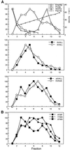
(A) Subcellular fractionation of Emp46p-KXKXX mutants in emp46 Δ cells. A whole-cell lysate from cells expressing 3HA-Emp46p with a wild-type or mutated dilysine motif was fractionated on a sucrose density gradient identical to that shown in Figure 3. Fractions were analyzed by immunoblotting. Quantitation was by densitometric scanning of immunoblots. (B) Subcellular fractionation of Emp46p-Yxxφ mutants in emp46Δ cells. A whole-cell lysate from cells expressing 3HA-Emp46p with a wild-type or mutated tyrosine-containing motif was fractionated on a sucrose density gradient and analyzed as above.
Figure 8
Figure 8
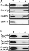
(A) In vitro budding reactions with microsomes prepared from cells expressing both 3HA-Emp46p and 2myc-Emp47p. Ten percent of the total reaction (T) and budded COPII vesicles isolated after incubation with (+) or without (−) COPII proteins was analyzed by SDS-PAGE followed by immunoblotting with an anti-HA or anti-myc. Sec61p as negative control and Sec22p as positive control were detected with polyclonal antibodies. (B) In vitro budding reactions with microsomes prepared from strains expressing C-terminal mutants of Emp46p. The same protocol as in A was used except that the reaction time was 15 min.
Figure 9
Figure 9
Binding of COPI (Ret1p and Sec21p) and COPII (Sec23p) to the C-terminal tail of Emp46p. GST fusion proteins comprising the C-terminal tail of Emp47p or Emp46p in either wild-type form (WT) or with a mutation as indicated were immobilized on glutathione-Sepharose beads and incubated with whole-cell lysate for 2 h at 4°C. The beads were washed and bound proteins were eluted from the beads and analyzed by SDS-PAGE and immunoblotting with anticoatomer, anti-Sec21p, or anti-Sec23p.
Figure 10
Figure 10
The role of the C-terminal tail of Emp46p in recruitment into the prebudding complex. Microsomes prepared from cells expressing either wild-type or mutated Emp46p were incubated with GST-Sar1p and the purified Sec23/24p complex in the presence of GMP-PNP or GDP-βS. Subsequently, digitonin-soluble prebudding complexes were recovered on glutathione beads. Emp46p or Sec22p in the prebudding complex was detected by immunoblotting. T represents 0.5% of the total solubilized membranes. Each construct is denoted as described in the legend to Figure 6B.
Figure 11
Figure 11
(A) Ca2+ sensitivity of cells expressing C-terminal tail mutants of Emp46p and Emp47p. emp46Δ emp47Δ cells were transformed with either empty plasmid (vector) or a single-copy plasmid with a C-terminal tail mutant of Emp46p or Emp47p as indicated was grown to saturation in liquid MCD-Ura-Trp, and serial dilutions were dropped on a YPD plate containing 0.25 M CaCl2 (Emp46p) or 0.5 M CaCl2 (Emp47p). (B) Expression of C-terminal tail mutants of Emp46p and Emp47p. Spheroplasts were prepared from cells used in A, resolved by SDS-PAGE, and immunoblotted with anti-HA (Emp46p) or anti-myc (Emp47p).
Figure 12
Figure 12
Secretion of glycoproteins by emp46/47 deletion strains. Cells were labeled with [35S]methionine for 10 min and were chased for 30 min. Media were collected and incubated with ConA-Sepharose to isolate extracellular glycoproteins. Proteins were separated by SDS-PAGE, and labeled proteins were detected by autoradiography. ● in A and ○ in B mark glycoproteins secreted inefficiently by mutant cells. Arrowheads designate glycoproteins secreted underglycosylated by the emp47Δ cells. (B) Low molecular weight region with increased samples.

Similar articles

Cited by

References

    1. Appenzeller C, Andersson H, Kappeler F, Hauri HP. The lectin ERGIC-53 is a cargo transport receptor for glycoproteins. Nat Cell Biol. 1999;1:330–334. - PubMed
    1. Aridor M, Fish KN, Bannykh S, Weissman J, Roberts TH, Lippincott-Schwartz J, Balch WE. The Sar1 GTPase coordinates biosynthetic cargo selection with endoplasmic reticulum export site assembly. J Cell Biol. 2001;152:213–229. - PMC - PubMed
    1. Barlowe C, Orci L, Yeung T, Hosobuchi M, Hamamoto S, Salama N, Rexach MF, Ravazzola M, Amherdt M, Schekman R. COPII: a membrane coat formed by Sec proteins that drives vesicle budding from the endoplasmic reticulum. Cell. 1994;77:895–907. - PubMed
    1. Barlowe C, Schekman R. SEC12 encodes a guanine-nucleotide-exchange factor essential for transport vesicle budding from the ER. Nature. 1993;365:347–349. - PubMed
    1. Bednarek SY, Ravazzola M, Hosobuchi M, Amherdt M, Perrelet A, Schekman R, Orci L. COPI- and COPII-coated vesicles bud directly from the endoplasmic reticulum in yeast. Cell. 1995;83:1183–1196. - PubMed

Publication types

MeSH terms