. 2002 Aug;4(8):599-604.
doi: 10.1038/ncb826.

Connective-tissue growth factor (CTGF) modulates cell signalling by BMP and TGF-beta

Affiliations

Connective-tissue growth factor (CTGF) modulates cell signalling by BMP and TGF-beta

José G Abreu et al. Nat Cell Biol. 2002 Aug.

Abstract

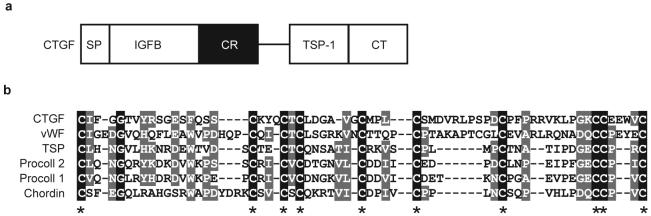
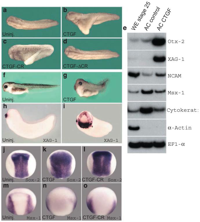
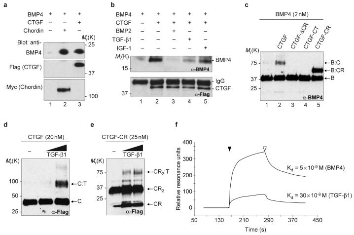
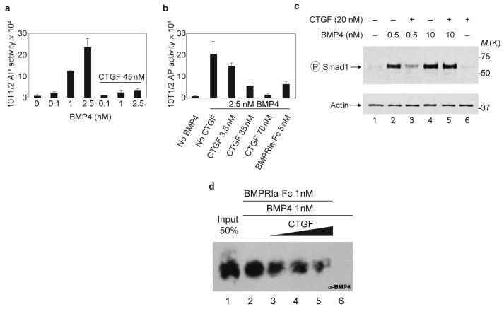
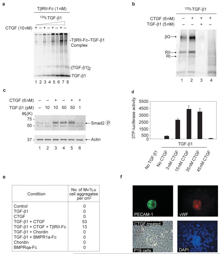
Connective-tissue growth factor (CTGF) is a secreted protein implicated in multiple cellular events including angiogenesis, skeletogenesis and wound healing. It is a member of the CCN family of secreted proteins, named after CTGF, cysteine-rich 61 (CYR61), and nephroblastoma overexpressed (NOV) proteins. The molecular mechanism by which CTGF or other CCN proteins regulate cell signalling is not known. CTGF contains a cysteine-rich domain (CR) similar to those found in chordin and other secreted proteins, which in some cases have been reported to function as bone morphogenetic protein (BMP) and TGF-beta binding domains. Here we show that CTGF directly binds BMP4 and TGF-beta 1 through its CR domain. CTGF can antagonize BMP4 activity by preventing its binding to BMP receptors and has the opposite effect, enhancement of receptor binding, on TGF-beta 1. These results show that CTGF inhibits BMP and activates TGF-beta signals by direct binding in the extracellular space.


Figures

Figure 1
Figure 1. CTGF encodes a CR-containing protein
a, The connective tissue growth factor (CTGF) protein contains four structural modules following the signal peptide (SP): insulin-like growth-factor binding (IGFB) domain, chordin-like cysteinerich (CR) domain, thrombospondin type 1 repeat (TSP-1), and a C-terminal cystineknot (CT). b, Alignment of the CR domains of CTGF, von Willebrand Factor, thrombospondin, procollagen 1 and 2 and Chordin (GenBank accession numbers XP037056, NM000552, P35448, NP001835, P02452 and AAC42222, respectively). Black boxes represent identical amino acids and gray boxes similar amino acids; dashes indicate gaps introduced to optimize the alignment. Asterisks mark the ten identical cysteine residues characteristic of CR domains.
Figure 2
Figure 2. CTGF mRNA injections induce anti-BMP phenotypes in Xenopusembryos
a-d, Dorsal views of tail-bud stage embryos. a, Uninjected; b, CTGF mRNA (150 pg); c, CTGF-CR mRNA (800 pg) and d, CTGF-ΔCR mRNA (500 pg) injected ventrally at 4-cell stage. e, RT-PCR analysis of whole embryo (WE), animal caps uninjected (AC control) or injected with 50 pg of CTGF mRNA per blastomere at the 4-cell stage (AC CTGF). Animal cap explants were dissected at stage 9 and cultured until sibling embryos reached stage 26. EF1-α was used as RNA loading control and α-Actin to monitor mesoderm in the ectodermal explants. Three-day tadpoles, f, uninjected, g, injected with 50 pg of CTGF mRNA into the animal region of each blastomere at 4-cell stage. Cement-gland marker XAG-1, h, uninjected, i, injected with 150 pg of CTGF mRNA per blastomere at 4-cell stage. j-o, in situ hybridization at neural plate stage for Sox-2 (j-l) and Msx-1 (m-o); j, m, Uninjected embryos; k, n, injected with 50 pg of CTGF; i, o, 150 pg of CTGF-CR mRNA injected per blastomere at 4-cell stage.
Figure 3
Figure 3. CTGF binds BMP4 and TGF-β1 through the CR domain
a, Westernblot analysis of BMP4 (5 nM) bound to CTGF (10 nM, lane 3) or chordin (5 nM, lane 2) after immunoprecipitation (IP) as described previously. To determine IP effectiveness, the same blots were subsequently probed with anti-Flag antibody for CTGF and anti-myc antibody for chordin. b, Competition of BMP4 (5 nM) bound to CTGF (10 nM) by IP in the absence (lane 2) or presence of 20-fold molar excess of BMP2 (lane 3), TGF-β1 (lane 4) or IGF-1 (lane 5). CTGF IP efficiency was monitored with anti-Flag antibody. c, Chemical crosslinking of CTGF and CTGF mutant constructs to BMP4. Membrane was probed with anti-BMP4 antibody. Bands corresponding to BMP4 dimer (B), BMP4-CTGF complex (B-C) and BMP4:CTGF-CR complex (B-CR) are indicated. d, e, Western blot probed with anti-Flag antibody after chemical crosslinking of CTGF and CTGF-CR to TGF-β1. Bands corresponding to CTGF monomer (C), CTGF-TGF-β1 (C-T) complex, and to CTGF-CR monomer (CR), to the dimer of CTGF-CR (CR2) and to the complex of a dimer of CTGF-CR with TGF-β1 (CR2-T) are indicated. The TGF-β1 was added at 25 nM and 50 nM. f, Sensorgrams of SPR analyses showing the binding of purified CTGF to BMP4 or to TGF-β1. CTGF protein was run (black arrowhead) over BMP4 or TGF-β1 sensor chips. After CTGF flow ended (white arrowhead), dissociation was monitored by a decrease in the resonance units. Kinetic experiments were performed using CTGF concentrations ranging between 6.5 nM and 210 nM of full-length CTGF.
Figure 4
Figure 4. CTGF antagonizes BMP4 signalling by inhibiting receptor binding
a, Dose-dependent induction of alkaline phosphatase (AP) by BMP4 in 10T1/2 cells is inhibited by CTGF. b, Adding increasing amounts of CTGF inhibits AP induced by 2.5 nM of BMP4. We estimate that CTGF can block BMP4 activity with an IC50 of about 12 nM. Error bars represent standard deviations of triplicate samples. Experiments were independently performed three times. c, Inhibition of Smad1 phosphorylation in 293T cells by CTGF at low, but not high, BMP4 concentrations. d, CTGF inhibits binding of BMP4 to its cognate receptor. BMP4 (1 nM) was preincubated 1 h at room temperature with 10, 25 and 50 nM (black triangle) of affinity-purified CTGF (lanes 3-5), and BMPRIa-Fc was added at 1 nM for 2 h. Lane 1 corresponds to the loading of 50% of the total BMP4 used in each reaction. Lane 6 shows negative control of BMP4 binding to protein A beads in the absence of the BMPRIa-Fc.
Figure 5
Figure 5. CTGF potentiates TGF-β1 receptor binding and signalling
a, Crosslinking of [125I]-TGF-β1 to 1 nM TGF-β receptor Fc fusion protein (TβRII-Fc) in the presence (+) or absence (-) of 10 nM purified CTGF. The TβRII-Fc-TGF-β1 complex is seen in the upper part of the gel. Different concentrations of [125I]-TGF-β1, ranging from 10 pM to 0.5 nM, were used. b, Labelling of cell-surface proteins by [125I]-TGF-β1 in NRK cells after chemical crosslinking with DSS. 125I-TGF-β1 (100 pM) was pre-incubated 1 h with (+) or without (-) 6 nM CTGF before adding for receptor binding. Specific binding was shown by competition with 50-fold unlabelled TGF-β1 (lane 3). Lane 4 shows negative control crosslinking of CTGF, and [I125]-TGF-β1 in the absence of cells. The three major cell surface TGF-β receptors are indicated: betaglycan (βG, 250 kDa), TGF-β-receptor II (TβRII, 80 kDa) and TGF-β-receptor I (TβRI, 68 kDa). c, Two hours after addition of proteins, Smad2 phosphorylation in Mv1Lu cells was stimulated by 6 nM CTGF in the presence of 10 pM TGF-β1. This effect was not seen when TGF-β1 was increased fivefold. Lane 6 shows that CTGF has no effect on its own. Note that Smad2 phosphorylation was induced at lower concentrations of TGF-β1 than those required in reporter-gene or cell-differentiation assays (see below), perhaps reflecting the shorter treatment times involved. d, Luciferase activity 36 h after transfection of Mv1Lu cells with the TGF-β inducible reporter p3TP-lux. Constant TGF-β1 and different concentrations of CTGF were used. Note that CTGF protein stimulates TGF-β1 signalling even when as little as 3 nM is used. e, Number of spherical cell aggregates per square centimeter observed in Mv1Lu cells after 12 h of treatment with different growth factors as described above. The concentrations of CTGF and TGF-β1 were 3 nM and 200 pM, respectively. Note that the BMP inhibitors Chordin and BMPRIa-Fc could not replace CTGF, and that soluble TGFRII-Fc inhibited spherical aggregate formation. f, Immunofluorescence showing the formation of spherical cell aggregates in P19 embryonal carcinoma cells treated with 5 nM CTGF. P19 cell aggregates were positive for PECAM-1 and vWF.

References

    1. Moussad EE, Brigstock DR. Mol. Genet. Metab. 2000;71:276–292. - PubMed
    1. Abreu J, Coffinier C, Larraín J, Oelgeschläger M, De Robertis EM. Gene. 2002;287:39–47. - PubMed
    1. Zhu Y, Oganesian A, Keene DR, Sandell LJ. J. Cell Biol. 1999;144:1069–1080. - PMC - PubMed
    1. Larraín J, et al. Development. 2000;127:821–830. - PMC - PubMed
    1. Nakayama N, et al. Dev. Biol. 2001;232:372–387. - PubMed

Publication types

MeSH terms