. 2002 Jul 15;30(14):3107-17.
doi: 10.1093/nar/gkf437.

Bone morphogenetic protein-4-induced activation of Xretpos is mediated by Smads and Olf-1/EBF associated zinc finger (OAZ)

Affiliations

Bone morphogenetic protein-4-induced activation of Xretpos is mediated by Smads and Olf-1/EBF associated zinc finger (OAZ)

Sangwoo Shim et al. Nucleic Acids Res. .

Abstract

We have previously isolated Xretpos, a novel family of long terminal repeat (LTR)-retrotransposons in Xenopus laevis, whose transcript is restricted to ventro-posterior-specific regions and induced by bone morphogenetic protein-4 (BMP-4) signaling. To explore the molecular mechanism of the transcriptional regulation, we identified and characterized Xretpos promoter regions consisting of LTRs and a 5'-untranslated region. We demonstrated that this promoter region contains all the necessary regulatory elements for the spatial and temporal expression of XRETPOS: Sequence analysis of the Xretpos promoter revealed multiple Smad-binding elements and Olf-1/EBF-associated zinc finger (OAZ) binding sites similar to BMP-4 response element, which were identified and proved to be required for BMP-4 induction in the Xvent2 promoter. We further demonstrated that Smads and OAZ proteins bind to their response elements in the promoter and these bindings are essential for the BMP-4-induced activation of the Xretpos promoter. Furthermore, we showed that the endogenous expression of Xretpos protein indeed occurred and was temporally regulated and BMP-4-inducible during the early Xenopus development. Finally, overexpression and partial loss-of-function study revealed that Xretpos has a posterio-ventralizing activity. Together, our results place Xretpos downstream of BMP-4 and provide evidence for the conserved mechanism of transcriptional regulation of the BMP-4 target genes.


Figures
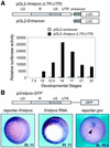
Figure 1
Figure 1
Stage- and tissue-specific expression of reporter constructs containing the LTR and 5′-UTR of Xretpos promoter. The LTR is divided into three regions, U3, R and U5. (A) Activities of luciferase reporter constructs were measured from the extracts of embryos injected with pGL2-Xretpos (LTR-UTR) or pGL2-enhancer into all four blastomeres of the four-cell stage embryos. One representative experiment is shown for this figure. (B) Xretpos reporter gene expression (left) was compared with endogenous Xretpos RNA (center). pgsc-GFP expression is shown as control (right). pXretpos-GFP was injected equatorially into all four blastomeres of four-cell stage embryos, and the expression of GFP transcripts was visualized by whole-mount in situ hybridization. The dorsal blastopore lip is indicated by arrowheads.
Figure 2
Figure 2
The Xretpos promoter is composed of positive and negative regulatory elements. Schematic representations of reporter constructs and their transcriptional activities are shown. Various deletion constructs of the Xretpos promoter were generated, injected into the two non-adjacent blastomeres of the four-cell stage (20 pg/blastomere) and allowed to develop to stage 11 for luciferase activity measurement. Fold induction was calculated as the ratio between pGL2-Xretpos (LTR-UTR) and other reporter constructs and represents the mean values from at least three independent experiments using different preparations of the plasmids. Positions of positive and negative regulatory elements in the Xretpos promoter are indicated at the top.
Figure 3
Figure 3
SBEs and the BRE-like region in the Xretpos promoter. (A) The Xretpos promoter contains three copies of consensus SBEs (SBE I–III) and Xvent BRE-like region consisting of SBE II and the 3′ box. (B) Xretpos SBE CAGAC sequences are compared with other SBEs from the responsive regions of several TGF-β, activin or BMP target genes. (C) The Xretpos BRE-like region is compared with Xvents BRE. Consensus sequences in SBE and the 3′ motif are in bold type and shaded.
Figure 4
Figure 4
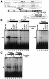
Bacterially expressed Smad proteins bind directly to SBE I and SBE III in EMSA. (A) The position and nucleotide sequences of wild-type (U3:1–207 and 31 bp SBE III) and mutants (mt SBE I and mt SBE III) used for EMSA are shown. mt SBE I was completely mutated by the linker- substitution method, while two nucleotides were mutated in mt SBE III. The SBE sequence is upper case. (B) Smad4, but not Smad3 or Smad1, binds directly to SBE I. Smad1, Smad3 and Smad4 proteins were expressed and purified as full-length His-fusion proteins in E.coli and incubated with 32P-labeled U3:1–207 containing SBE I. The concentration of His-Smad fusion proteins was 0.5 or 1 µg. Lane 1, no protein control. In competition experiments, 100-fold excess of unlabeled wild-type or mutant (mt SBE I) duplex was added in the reaction. The positions of free probe and DNA–protein complex are indicated by arrows. (C) Smad1, Smad3 and Smad4 bind directly to SBE III. EMSA was performed in the same procedures as described above except that a 32 bp double-stranded oligonucleotide encompassing SBE III was used as probe and mt SBE III as competitor.
Figure 5
Figure 5
Bacterially expressed Smad proteins bind directly to Xretpos BRE-like region and the GCAT motif in UTR. (A) The position and nucleotide sequences of wild-type and mutants used for EMSA are shown. SBE and 3′ box are upper case and underlined. In GCAT motif, GCAT sequences are upper case and AT-rich flanking sequences are underlined. (B) Smad1 bound this fragment with higher affinity than Smad4 and Smad3. EMSA was performed with 32P-labeled 32 bp Xretpos BRE-like region containing SBE II and 3′ box. Lanes 1 and 14, no protein control. In competition experiments, 100-fold excess of unlabeled wild-type or mutant duplex as indicated above the lanes was added in the reaction. (C) Smad1 binds directly to the GCAT motif (and Smad4 binds weakly). EMSA was performed with 32P-labeled UTR encompassing GCAT sequences and AT-rich flanking sequences. Lane 1, no protein control.
Figure 6
Figure 6
OAZ, Smad1 and Smad4 expressed and isolated from the embryos bind to Xretpos-BRE. (A and B) Aliquots of 10 µg of nuclear extracts were prepared from stage 13 embryos injected with RNAs encoding 6Myc-OAZ-ZF(9–19), 6Myc-Smad4 or Flag-Smad1 and incubated with the 32 bp Xretpos BRE-like probe. Addition of 2 µl of anti-Myc (0.2 µg/µl) or anti-Flag antibodies (1 µg/µl) as well as unlabeled probe (100-fold) disrupted the binding complex. Lanes 1 and 7, no protein control; lanes 2 and 3, uninjected control embryo extracts. (C) Immunoblot analysis was performed to visualize the expression and localization of epitope-tagged proteins. Aliquots of 10 µg of nuclear extracts or cytoplasmic extracts from stage 13 embryos injected with epitope-tagged RNAs were separated by 10% SDS–PAGE and blotted with an anti-epitope antibody.
Figure 7
Figure 7
Smads and OAZ binding to their response element are essential for the BMP-4-induced activation of Xretpos promoter. Xenopus embryos were co-injected into one dorsal blastomere at the four-cell stage with the indicated RNAs and reporter constructs and allowed to develop until stage 13 for luciferase activity measurement. (A) Smad1/Smad4 and OAZ functionally cooperated in BMP-4-induced activation of Xretpos promoter. RNAs for Smad1, Smad4 and OAZ were synthesized in vitro from pCS2-Flag-Smad1, pCS2-Smad4 and pCS2-6Myc-OAZ-ZF(9–19), respectively. RNA concentrations were 500 pg/blastomere. (B) OAZ is required for the BMP-4-induced activation of Xretpos. Dominant-negative OAZ mutant RNA was synthesized in vitro from pCS2-Flag-OAZ-ZF(1–13). RNA concentrations were 500 pg/blastomere for BMP-4, and 1 ng/blastomere for ZF(9–19) and ZF(1–13), respectively. (C) SBE II, 3′ box and GCAT motif contribute to the BMP-4/Smad1/4-induced transcriptional activation of Xretpos. RNA concentrations were the same as described in (A). Linker-substitution mutations were introduced into SBE II, 3′ box and GCAT motif (left); the wild-type SBE II GAGCAGACAT sequences were replaced by mutated cgGaAttCcg sequences (pGL2-Xretpos-LS-SBE II), the wild-type 3′ box GGTGGGGCAG sequences were replaced by GcTctaGagc sequences (pGL2-Xretpos-LS-3box), and the wild-type GCAT motif GCATGGCATT sequences were replaced by mutated cggaattccg sequences (pGL2-Xretpos-LS-2XGCAT), respectively.
Figure 8
Figure 8
Detection of endogenous Xretpos protein by western blot analysis. Aliquots of 50 µg of total cell extracts (A) or 20 µg of nuclear or cytoplasmic extracts (B) from the indicated stage embryos were separated by 10% SDS–PAGE, Ponceau stained, electroblotted onto nitrocellulose and probed with anti-Xretpos antibody. The position of Xretpos protein is indicated by an arrow. Note that Xretpos protein expression is temporally regulated (A) and BMP-4-incucible (B), and localized only to cytoplasmic fractions (B). Ponceau staining was performed as a loading control.
Figure 9
Figure 9
Phenotypic effects of overexpression (A) and partial loss (B and C) of Xretpos function. (A) Overexpression of Xretpos RNA resulted in reduced anterior head structures. Four-cell stage embryos were injected into two dorsal animal or marginal regions at the four-cell stage embryos with 4 ng of Xretpos RNA and allowed to develop until stage 43. Control embryos injected with 4 ng of PPL RNA produced normal tadpoles. (B) Rescue of axial structures in UV-irradiated embryos by antisense Xretpos RNA. Partial twinned axis structures were seen in UV-irradiated embryos injected into two non-adjacent blastomeres at the four-cell stage with antisense Xretpos RNA (1 ng/blastomere), but not in uninjected UV-irradiated controls. (C) Phenotypic effects of antisense Xretpos RNA injection in wild-type embryos. Antero-dorsalized structures were observed in embryos which were injected diagonally at the four-cell stage with antisense Xretpos RNA (1 ng/blastomere). Uninjected control embryos developed normally. (D) Reduction of endogenous Xretpos protein by antisense Xretpos RNA injection. The position of Xretpos protein is indicated by an arrow and Ponceau staining was performed as a loading control.

References

    1. Massagué J., Blain,S.W. and Lo,R.S. (2000) TGFβ signaling in growth control, cancer, and heritable disorders. Cell, 103, 295–309. - PubMed
    1. Hill C.S., (2001) TGF-β signalling pathways in early Xenopus development. Curr. Opin. Genet. Dev., 11, 533–540. - PubMed
    1. Whitman M., (1998) Smads and early developmental signaling by the TGFβ superfamily. Genes Dev., 12, 2445–2462. - PubMed
    1. Gawantka V., Delius,H., Hirschfeld,K., Blumenstock,C. and Niehrs,C. (1995) Antagonizing the Spemann organizer: role of the homeobox gene Xvent-1. EMBO J., 14, 6268–6279. - PMC - PubMed
    1. Ault K.T., Dirksen,M.-L. and Jamrich,M.A. (1996) A novel homeobox gene PV.1 mediates induction of ventral mesoderm in Xenopus embryos. Proc. Natl Acad. Sci. USA, 93, 6415–6420. - PMC - PubMed

Publication types

MeSH terms