. 2002 Jul 15;30(14):3202-13.
doi: 10.1093/nar/gkf435.

Identification of Sox-2 regulatory region which is under the control of Oct-3/4-Sox-2 complex

Affiliations

Identification of Sox-2 regulatory region which is under the control of Oct-3/4-Sox-2 complex

Mizuho Tomioka et al. Nucleic Acids Res. .

Abstract

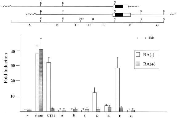
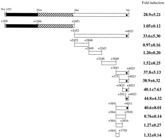
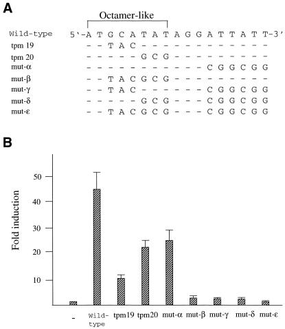
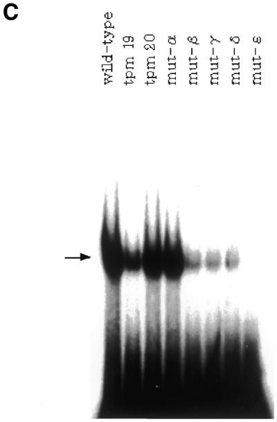
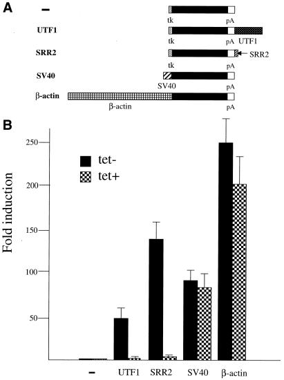
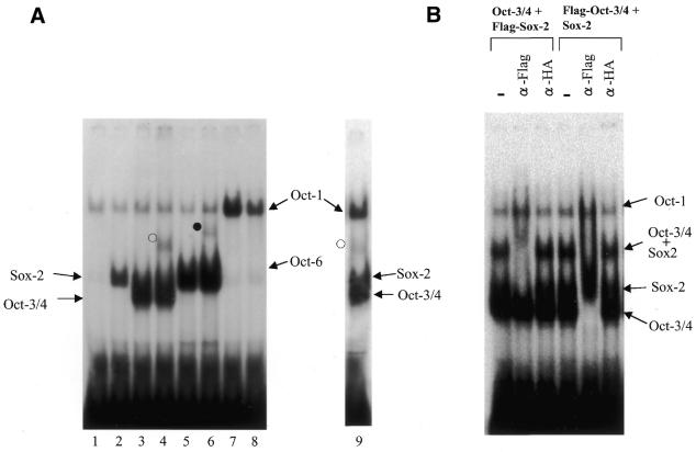
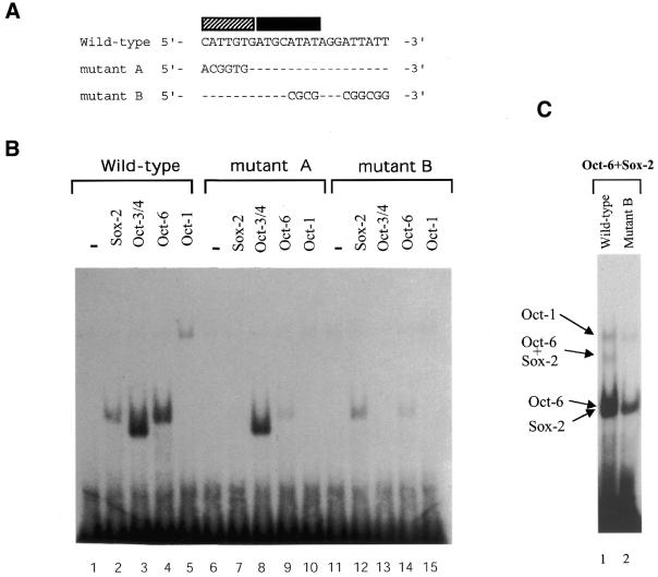
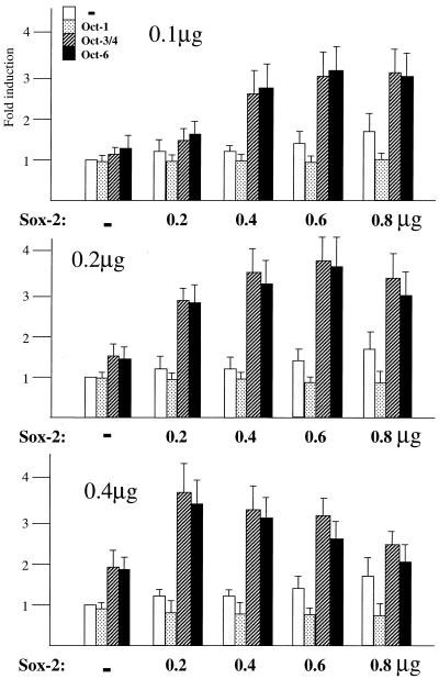
Sox-2 is a transcriptional cofactor expressed in embryonic stem (ES) cells as well as in neuronal cells. It has been demonstrated that Sox-2 plays an important role in supporting gene expression in ES cells, especially by forming a complex with embryonic Octamer factor, Oct-3/4. Here, we have analyzed the regulatory regions of the Sox-2 gene and identified two enhancers which stimulate transcription in ES cells as well as in embryonal carcinoma cells. These regulatory regions, which we termed Sox regulatory regions (SRR) 1 and 2, exert their function specifically when cells are in an undifferentiated state. Interestingly, like the regulatory elements of FGF-4 and UTF1 genes, combinatorial action of Octamer and Sox-2 binding sites support the SRR2 activity. However, biochemical analyses reveal that, due to the unique sequence and/or its organization, the SRR2 bears distinct characteristics from those of FGF-4 and UTF1 regulatory elements. That is, unlike the FGF-4 gene enhancer, the SRR2 precludes the binding of the Oct-1-Sox-2 complex. The difference between the SRR2 and UTF1 regulatory element is in the ability of SRR2 to recruit the Oct-6-Sox-2 complex as well as the Oct-3/4-Sox-2 complex. Co-transfection analyses confirm that both complexes are able to stimulate transcription through the SRR2 element.


Figures

Figure 1
Figure 1
Identification of two regulatory regions which exert their activities in F9 EC cells in an undifferentiated state-specific manner. Open and filled boxes indicate Sox-2 gene non-coding and coding regions, respectively, while wavy lines represent Lambda phage vector portions. Restriction enzymes are abbreviated as follows: B, BamHI; E, EcoRI; H, HindIII; N, NotI; Nhe, NheI; S, SalI. The Sox-2 gene and its flanking regions (A–G) were individually subcloned into the downstream of the luciferase gene of tk-Luc reporter plasmid. Each plasmid construct was introduced into F9 EC cells together with neomycin-resistant gene which is under the control of the β-actin promoter and an internal control luciferase gene of Renilla reniformis. After selection with G418, approximately 1000 of the drug-resistant colonies were pooled and expanded. Subsequently, the cells were incubated with medium containing charcoal-treated serum supplemented with or without 1 µM retinoic acid. After 48 h, whole-cell extracts were prepared from such treated cells and levels of transcription were determined by the dual-luciferase system according to the manufacturer (Promega). The intrinsic activity of the control tk-Luc plasmid in undifferentiated and differentiated cells is arbitrarily set as one and fold induction due to the presence of each regulatory element was calculated. Data were obtained from five independent experiments with comparable results.
Figure 2
Figure 2
Localization of the SRR2 core region involved in Sox-2 expression in F9 EC cells. The filled and shaded portions represent Sox-2 coding and non-coding regions, respectively. The filled circle indicates the region which is found to contain the SRR2 core region. The plasmid constructions of the reporter gene bearing the indicated portions of SRR2 were described in Materials and Methods. F9 EC cells were transfected as in Figure 1 with tk-Luc reporter gene carrying portions of SRR2 which are depicted and an internal control luciferase gene of R.reniformis. After 48 h post-transfection, the transcriptional stimulating activity of each deletion mutant of SRR2 was estimated by the dual-luciferase system according to the manufacturer (Promega). The data were obtained as in Figure 1.
Figure 3
Figure 3
The Sox-2 site is involved in potentiating SRR2-mediated transcription. (A) The sequence of the SRR2 core region. A synthetic DNA containing wild-type and triple point mutants of the SRR2 sequence was generated by assembly of overlapping oligonucleotides as described in Materials and Methods. The mutants which changed the nucleotides as consecutive triplets (tpm1–27) to non-complementary ones (A ⇔ C, G ⇔ T) are indicated below the sequence. Filled and shaded boxes indicate Octamer and Sox-2 site-like sequences, respectively. (B) Localization of sequence which is crucially involved in SRR2 activity. The tk-Luc reporter gene bearing wild-type or a series of triple point mutants of SRR2 was individually introduced into F9 EC cells together with an internal control luciferase gene of R.reniformis. Forty-eight hours after transfection, the activity of these mutants as well as wild-type SRR2 was evaluated by the dual-luciferase system. The data were obtained as in Figure 1.
Figure 4
Figure 4
Correlation between the ability of SRR2 to interact with Oct-3/4 protein and its transcriptional stimulating activity in F9 EC cells. (A) The portion of SRR2 sequence containing an Octamer-like element and its immediate downstream sequence. Mutations introduced into the wild-type sequence are indicated. For the tranfection analyses, these mutations were introduced in the context of an 81-bp SRR2 core sequence shown in Figure 3A. However, the probes for the gel-shift analyses shown in (C) lack 21 bp of the 5′ portion of SRR2 sequence (see Materials and Methods for details). (B) The AT-rich sequence located downstream of the Octamer-like sequence is involved in elevating transcriptional stimulating activity of SRR2. The tk-Luc reporter gene bearing wild-type as well as mutated SRR2 sequences are individually introduced into F9 EC cells. Transcriptional stimulating activity of each mutant was obtained as in Figure 1. (C) Effect of mutations on the binding of Oct-3/4 to the SRR2. Radiolabeled DNA probes carrying wild-type and mutated SRR2 sequences shown in (A) were prepared and individually used for gel-shift analyses with whole-cell extracts prepared from COS cells which had been transfected with Oct-3/4 expression vector.
Figure 4
Figure 4
Correlation between the ability of SRR2 to interact with Oct-3/4 protein and its transcriptional stimulating activity in F9 EC cells. (A) The portion of SRR2 sequence containing an Octamer-like element and its immediate downstream sequence. Mutations introduced into the wild-type sequence are indicated. For the tranfection analyses, these mutations were introduced in the context of an 81-bp SRR2 core sequence shown in Figure 3A. However, the probes for the gel-shift analyses shown in (C) lack 21 bp of the 5′ portion of SRR2 sequence (see Materials and Methods for details). (B) The AT-rich sequence located downstream of the Octamer-like sequence is involved in elevating transcriptional stimulating activity of SRR2. The tk-Luc reporter gene bearing wild-type as well as mutated SRR2 sequences are individually introduced into F9 EC cells. Transcriptional stimulating activity of each mutant was obtained as in Figure 1. (C) Effect of mutations on the binding of Oct-3/4 to the SRR2. Radiolabeled DNA probes carrying wild-type and mutated SRR2 sequences shown in (A) were prepared and individually used for gel-shift analyses with whole-cell extracts prepared from COS cells which had been transfected with Oct-3/4 expression vector.
Figure 5
Figure 5
The extinction of Oct-3/4 is accompanied by loss of SRR2 activity in ES cells. (A) Schematic representation of five different lucifease reporter plasmids bearing distinct regulatory regions. The filled box represents the luciferase reporter gene. tk and pA represent the thymidine kinase gene promoter (–109 to +51) and poly(A) addition signal from SV40 virus, respectively. UTF1, SV40 and β-actin indicate the UTF1 regulatory element-containing 1.2 kb BamHI/SalI genomic DNA fragment (18), the SV40 early promoter, and the human β-actin promoter, respectively. (B) SRR2 and the UTF1 regulatory element require Oct-3/4 expression to exert their activity in ES cells. The five different plasmids bearing the luciferase reporter gene were individually introduced into ZHBTc4 ES cells together with the Puromycin-resistant gene and an internal control luciferase gene of R.reniformis by a lipofection method. After selection with Puromycin, approximately 1000 of the drug-resistant colonies were pooled and expanded. Subsequently, the cells were incubated with medium with or without tetracycline (1 µg/ml). After 48 h, whole-cell extracts were prepared from such treated cells and levels of transcription were determined by the dual-luciferase system according to the manufacturer (Promega). The data were obtained as in Figure 1.
Figure 6
Figure 6
Gel-shift analyses of SRR2 with Oct and Sox proteins. (A) Both Oct-3/4 and Oct-6, but not Oct-1, are able to form complexes with Sox-2 on SRR2. The Oct-1, Oct-3/4 and Oct-6 were individually expressed in COS cells with or without Sox-2 expression, and gel-shift analyses were performed with whole-cell extracts prepared from such transfected cells by using wild-type SRR2 sequence used in Figure 4C. However, in lane 9, nuclear extract from E14 ES cells was used. The open and filled circles indicate Oct-3/4–Sox-2 and Oct-6–Sox-2 complexes bound to the probe, respectively. The experiments with the specific antibody revealed that bands observed in lanes 1–6 which co-migrate with the bands obtained with exogenously expressed Oct-1 were generated due to the binding of endogenous Oct-1 protein expressed in COS cells (data not shown). (B) Super-shift analyses with antibodies. Gel-shift analyses were done with Oct-3/4 and Sox-2 in which either one of the proteins is Flag-tagged as indicated on top of the panel. The reaction mixture contains no or a certain (anti-Flag or HA) antibody.
Figure 7
Figure 7
Each Octamer factor shows different sequence requirements of SRR2 for binding. (A) Nucleotide sequences of oligonucleotide probes used for the gel-shift analyses. A portion of the oligonucleotide sequence used for gel-shift analyses is shown. Filled and shaded boxes represent Octamer and Sox-2 site-like sequences, respectively. In the mutant probes, only mutated nucleotides are indicated. In the Oct site mutant, an AT-rich sequence downstream of the Octamer-like sequence is also mutagenized. (B) Effect of mutations of SRR2 sequence on the interaction with Octamer factors. The Oct-1, Oct-3/4 and Oct-6 were individually expressed in COS cells. Whole-cell extracts were prepared from such transfected cells and gel-shift analyses were performed as in Figure 6A using either wild-type or one of two different mutants. (C) The Oct-6 fails to bind to mutant B probe together with Sox-2. Gel-shift analyses were performed with the wild-type (lane 1) or mutant B probe (lane 2). The reaction mixture contains both Oct-6 and Sox-2. Even with long exposure, a band corresponding to the simultaneous binding of Oct-6 and Sox-2 was not detected in lane 2 (data not shown).
Figure 8
Figure 8
Both Oct-3/4 and Oct-6, but not Oct-1, are able to potentiate transcription through SRR2. COS cells were transfected with 0.1, 0.2 or 0.4 µg of Oct-1, Oct-3/4 or Oct-6 expression vectors, and the increasing amounts of Sox-2 vector as indicated. In addition, an internal control luciferase gene (2 µg) of R.reniformis and the tk-luc reporter plasmid (2 µg) bearing SRR2 were also introduced. After 48 h post-transfection, transcriptional level was estimated by the dual-luciferase system according to the manufacturer (Promega). The data were obtained as in Figure 1.

Similar articles

Cited by

References

    1. Dooley T.P., Miranda,M., Jones,N.C. and DePamphilis,M.L. (1989) Transactivation of the adenovirus EIIa promoter in the absence of adenovirus E1A protein is restricted to mouse oocytes and preimplantation embryos. Development, 107, 945–956. - PubMed
    1. Gilbert S.F., (1991) Developmental Biology, 3rd Edn. Sinauer Associates, Sunderland, MA.
    1. La Thangue N.B. and Rigby,P.W.J. (1987) An adenovirus E1A-like transcription factor is regulated during the differentiation of murine embryonal carcinoma stem cells. Cell, 49, 507–513. - PubMed
    1. Majunder S., Zhao,Z., Kaneko,K. and DePamphilis,M.L. (1997) Developmental acquisition of enhancer function requires coactivator activity. EMBO J., 16, 1721–1731. - PMC - PubMed
    1. Kaneko K.J., and DePamphilis,M.L. (1998) Regulation of gene expression at the beginning of mammalian development and the TEAD family of transcription factors. Dev. Genet., 22, 43–55. - PubMed

Publication types

MeSH terms

Associated data