. 2002 Jul 15;30(14):e66.
doi: 10.1093/nar/gnf065.

Real-time monitoring of rolling-circle amplification using a modified molecular beacon design

Affiliations

Real-time monitoring of rolling-circle amplification using a modified molecular beacon design

Mats Nilsson et al. Nucleic Acids Res. .

Abstract

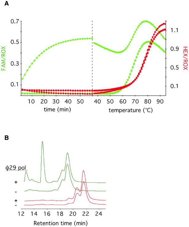
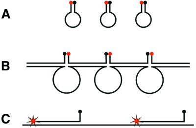
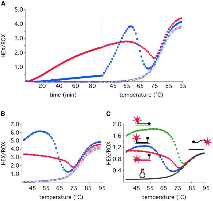
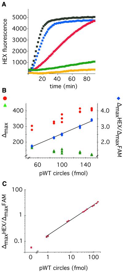
We describe a method to monitor rolling-circle replication of circular oligonucleotides in dual-color and in real-time using molecular beacons. The method can be used to study the kinetics of the polymerization reaction and to amplify and quantify circularized oligonucleotide probes in a rolling-circle amplification (RCA) reaction. Modified molecular beacons were made of 2'-O-Me-RNA to prevent 3' exonucleolytic degradation by the polymerase used. Moreover, the complement of one of the stem sequences of the molecular beacon was included in the RCA products to avoid fluorescence quenching due to inter-molecular hybridization of neighboring molecular beacons hybridizing to the concatemeric polymerization product. The method allows highly accurate quantification of circularized DNA over a broad concentration range by relating the signal from the test DNA circle to an internal reference DNA circle reporting in a distinct fluorescence color.


Figures

Figure 1
Figure 1
Non-specific accumulation of fluorescence due to exonucleolytic degradation of molecular beacons can be avoided by replacing all DNA residues of the molecular beacon with 2′-O-Me-RNA residues. (A) The DNA molecular beacon mbDNAfam labeled with a FAM fluorophore (green) and the 2′-O-Me-RNA molecular beacon mbRNAhex labeled with a HEX fluorophore (red) were added to the same test tube in the presence (circles) or absence (squares) of Φ29 DNA polymerase. The left portion of the graph shows the variation of fluorescence over time at 37°C. The right portion shows the variation of fluorescence in response to increasing temperature. This temperature profile gives an indication whether the stem parts of the molecular beacons are intact at the end of the 60 min incubation. The different molecular beacons have slightly different melting characteristics because of different hybrid stability of their respective stem sequences. (B) HPLC chromatograms showing the fluorescence from mbDNAfam (green line) and mbRNAfam (red line) molecular beacons before and after a 1 h treatment with Φ29 DNA polymerase at 37°C.
Figure 2
Figure 2
A schematic representation of the expected intra- and inter- molecular hairpin structures that quench fluorescence from molecular beacons when free in solution, or when hybridized to RCA products, and how the inter-molecular quenching structures can be avoided by modifying the molecular beacon design. (A) Molecular beacons form intra-molecular stem–loop structures when they are free in solution. (B) When the molecular beacons hybridize with a concatemeric RCA product, they can also hybridize to neighboring molecular beacons, thereby forming the stems in inter-molecular stem–loop structures, where the loops are formed by the RCA product. (C) The inter-molecular stem–loop structure can be avoided by introducing a stem-complementary sequence in the RCA product.
Figure 3
Figure 3
Comparison of real-time RCA of circularized padlock probes that generate quenching or non-quenching products, and demonstration of the intra-molecular quenching structure by varying the temperature or by restriction digestion of the RCA products. (A) A real-time fluorescence measurement (left) was followed by a temperature gradient to study the temperature dependence of the fluorescence (right). Analysis of the fluorescence temperature profile demonstrates the inter-molecular quenching structure. RCA was performed of padlock probes subjected to ligation (filled symbols) or no ligation (open symbols). The RCA reactions were templated by 10 fmol of either the pp90 (blue squares) or the pp93 (red circles) padlock probes, producing quenching and non-quenching RCA products, respectively. In the left portion of the graph the fluorescence from the mbRNAhex molecular beacon was followed in real-time at 37°C. The right portion shows the fluorescence temperature profile of the products at the end of the RCA. (B) Temperature profiles of the products generated in (A) after restriction digestion. (C) Temperature profiles of the mbRNAhex molecular beacon in presence of no hybridization target (black rectangles), or hybridization targets that in addition to the loop sequence also hybridizes to the quencher-carrying stem sequence (QUENCH, green circles), the fluorophore-carrying stem sequence (FLUO, red circles), or none of the stem sequences (NONE, blue squares).
Figure 4
Figure 4
Real-time quantification of probe circles using RCA and molecular beacons. (A) HEX generated fluorescence from mbRNAhex molecular beacons plotted against time from RCA reactions containing 0 (orange), 0.87 (green), 8 (red), 50 (blue) and 100 (black) fmol ppWT probe circles, respectively. (B) ΔmaxHEX (red circles) and ΔmaxFAM (green triangles) signal values from the mbRNAhex and mbRNAfam molecular beacons, respectively, plotted against the amount of ppWT probe circles present in triplicate RCA reactions containing 30 fmol ppMUT reference circles. The corresponding ΔmaxHEX/ΔmaxFAM signal ratios were calculated for the individual reactions and plotted as blue diamonds. (C) The ΔmaxHEX/ΔmaxFAM signal ratio is plotted against the amount of ppWT probe circles (HEX signal) present in RCA reactions containing 30 fmol ppMUT reference circles (FAM signal). The data points are from reactions containing 0.87, 1, 7, 8, 33, 38, 44, 50, 87, 100, 130 and 150 fmol of ppWT probe circles. The error bars represent the standard deviation of triplicate reactions at each concentration of ppWT probe circles.

Similar articles

Cited by

References

    1. Heid C.A., Stevens,J., Livak,K.J. and Williams,P.M. (1996) Real time quantitative PCR. Genome Res., 6, 986–994. - PubMed
    1. Tyagi S., and Kramer,F.R. (1996) Molecular beacons: probes that fluoresce upon hybridization. Nat. Biotechnol., 14, 303–308. - PubMed
    1. Li J.J., Geyer,R. and Tan,W. (2000) Using molecular beacons as a sensitive fluorescence assay for enzymatic cleavage of single-stranded DNA. Nucleic Acids Res., 28, e52. - PMC - PubMed
    1. Li J.J., Fang,X., Schuster,S.M. and Tan,W. (2000) Molecular beacons: a novel approach to detect protein–DNA interactions. Angew. Chem. Int. Ed., 39, 1049–1052. - PubMed
    1. Lizardi P.M., Huang,X., Zhu,Z., Bray-Ward,P., Thomas,D.C. and Ward,D.C. (1998) Mutation detection and single-molecule counting using isothermal rolling-circle amplification. Nature Genet., 19, 225–232. - PubMed

Publication types

MeSH terms