. 2002 Aug 6;99(16):10819-24.
doi: 10.1073/pnas.152334899. Epub 2002 Jul 22.

Brain monoglyceride lipase participating in endocannabinoid inactivation

Affiliations

Brain monoglyceride lipase participating in endocannabinoid inactivation

T P Dinh et al. Proc Natl Acad Sci U S A. .

Erratum in

  • Proc Natl Acad Sci U S A 2002 Oct 15;99(21):13961

Abstract

The endogenous cannabinoids (endocannabinoids) are lipid molecules that may mediate retrograde signaling at central synapses and other forms of short-range neuronal communication. The monoglyceride 2-arachidonoylglycerol (2-AG) meets several criteria of an endocannabinoid substance: (i) it activates cannabinoid receptors; (ii) it is produced by neurons in an activity-dependent manner; and (iii) it is rapidly eliminated. 2-AG inactivation is only partially understood, but it may occur by transport into cells and enzymatic hydrolysis. Here we tested the hypothesis that monoglyceride lipase (MGL), a serine hydrolase that converts monoglycerides to fatty acid and glycerol, participates in 2-AG inactivation. We cloned MGL by homology from a rat brain cDNA library. Its cDNA sequence encoded for a 303-aa protein with a calculated molecular weight of 33,367 daltons. Northern blot and in situ hybridization analyses revealed that MGL mRNA is heterogeneously expressed in the rat brain, with highest levels in regions where CB(1) cannabinoid receptors are also present (hippocampus, cortex, anterior thalamus, and cerebellum). Immunohistochemical studies in the hippocampus showed that MGL distribution has striking laminar specificity, suggesting a presynaptic localization of the enzyme. Adenovirus-mediated transfer of MGL cDNA into rat cortical neurons increased MGL expression and attenuated N-methyl-D-aspartate/carbachol-induced 2-AG accumulation in these cells. No such effect was observed on the accumulation of anandamide, another endocannabinoid lipid. The results suggest that hydrolysis by means of MGL is a primary mechanism for 2-AG inactivation in intact neurons.


Figures
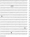
Fig 1.
Fig 1.
Nucleotide and deduced amino acid sequence of rat brain MGL cDNA. Closed circles mark amino acid residues comprising the putative catalytic triad. The HG dipeptide motif often found in lipases is boxed.
Fig 2.
Fig 2.
MGL expression in the rat brain. (a) Northern blot analysis of MGL mRNA from various regions of the rat brain; glyceraldehyde-3-phosphate dehydrogenase mRNA was also measured to control for loading conditions. (b) Western blot analysis of soluble protein from rat brain, with an immunopurified polyclonal antibody to the N-terminal region of MGL. Both immunoreactive bands were abolished after adsorption with immunizing peptide (data not shown).
Fig 3.
Fig 3.
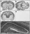
Localization of MGL mRNA in the rat brain by in situ hybridization. Coronal (a and c) and horizontal (b) sections hybridized with an MGL antisense riboprobe labeled with [35S]UTP. (d) Horizontal section hybridized with a sense probe. (e) Dark-field micrograph of MGL-positive cells in the hippocampus. AcB, nucleus accumbens; AdN, anterodorsal nucleus of the thalamus; Cb, cerebellum; CdP, caudate-putamen; Ctx, cortex; DG, dentate gyrus; Thl, thalamus. (Bar: a = 3 mm; b = 4 mm; e = 500 μm.)
Fig 4.
Fig 4.
Localization of MGL protein in the rat brain by immunohistochemistry. (a) Light micrograph of a hippocampal section immunostained for MGL revealing specific laminar distribution of the enzyme. At higher magnification (bd), cell bodies of principal cells in all subfields are MGL-negative, but are surrounded by MGL-immunoreactive axon terminals (arrowheads in c) that resemble basket cell boutons. Mossy fiber terminals in CA3 stratum lucidum (arrows in c) are strongly immunostained. (d) Light micrograph of an osmium-treated section from CA1 shows that pyramidal dendrites in stratum radiatum (arrows) can be followed as negative images in the heavily stained neuropil. DG, dentate gyrus; h., hilus; s.g., stratum granulosum; s.l., stratum lucidum; s.l.m., stratum lacunosum-moleculare; s.m., stratum moleculare; s.o., stratum oriens; s.p., stratum pyramidale; s.r., stratum radiatum. (Bar: a, 500 μm; bd, 100 μm.)
Fig 5.
Fig 5.
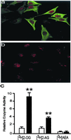
Adenovirus-mediated MGL overexpression in HeLa cells. Confocal microscopy images of cells infected with MGL-containing (a) or control (b) adenovirus. MGL immunoreactivity is shown in green; cell nuclei in red. (c) MGL activity in vector (open bars)- or MGL (closed bar)-infected cells. [3H]2-OG, 2-oleoyl-[3H]glycerol; [3H]AEA, [3H]anandamide. Results are expressed as the mean ± SEM of three experiments performed in triplicate. **, P < 0.01, Student's t test.
Fig 6.
Fig 6.
Effects of MGL overexpression on receptor-dependent 2-AG accumulation in rat cortical neurons. Expression of MGL mRNA (a) and protein (b and c) in vector- and MGL-infected neurons. The 1.2-kb mRNA in overexpressing neurons corresponds to the coding sequence of MGL. Confocal microscopy images of vector- (b) and MGL (c)-infected neurons. Note the low, but detectable levels of endogenous MGL in vector-infected cells. MGL immunoreactivity is shown in green, cell nuclei in red. (d) Time course of 2-[3H]AG accumulation after coactivation of NMDA and cholinergic receptors in vector (open bars)- and MGL (filled bars)-infected neurons. Results are from one experiment, representative of four. (e) HPLC/MS quantitation of 2-AG and anandamide accumulation in neurons. Open bars, unstimulated vector-infected neurons; filled bars, stimulated vector-infected neurons; shaded bars, stimulated MGL-infected neurons. (Left) anandamide (AEA) levels; (Right) 2-AG levels. *, P < 0.05; **, P < 0.01; n = 5; Student's t test.

References

    1. Devane W. A., Hanus, L., Breuer, A., Pertwee, R. G., Stevenson, L. A., Griffin, G., Gibson, D., Mandelbaum, A., Etinger, A. & Mechoulam, R. (1992) Science 258, 1946-1949. - PubMed
    1. Sugiura T., Kondo, S., Sukagawa, A., Nakane, S., Shinoda, A., Itoh, K., Yamashita, A. & Waku, K. (1995) Biochem. Biophys. Res. Commun. 215, 89-97. - PubMed
    1. Mechoulam R., Ben-Shabat, S., Hanus, L., Ligumsky, M., Kaminski, N. E., Schatz, A. R., Gopher, A., Almog, S., Martin, B. R., Compton, D. R., et al. (1995) Biochem. Pharmacol. 50, 83-90. - PubMed
    1. Di Marzo V., Fontana, A., Cadas, H., Schinelli, S., Cimino, G., Schwartz, J. C. & Piomelli, D. (1994) Nature (London) 372, 686-691. - PubMed
    1. Stella N., Schweitzer, P. & Piomelli, D. (1997) Nature (London) 388, 773-778. - PubMed

Publication types

MeSH terms

Associated data