. 2002 Aug 6;99(16):10405-10.
doi: 10.1073/pnas.162250899. Epub 2002 Jul 22.

The atomic structure of adeno-associated virus (AAV-2), a vector for human gene therapy

Affiliations

The atomic structure of adeno-associated virus (AAV-2), a vector for human gene therapy

Qing Xie et al. Proc Natl Acad Sci U S A. .

Abstract

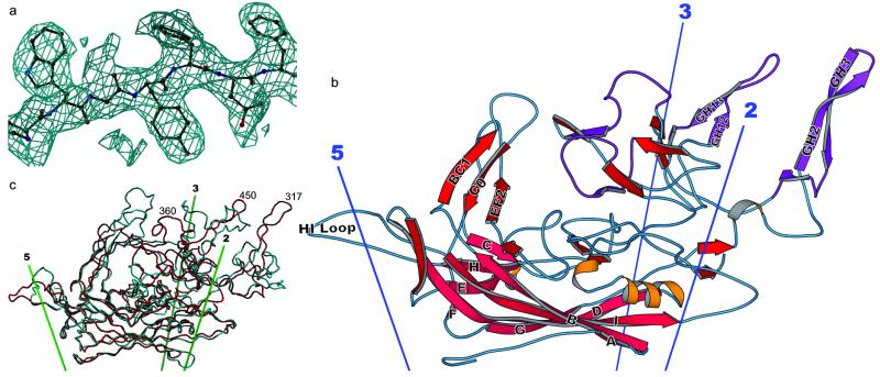
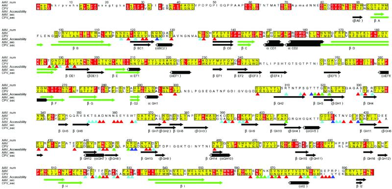
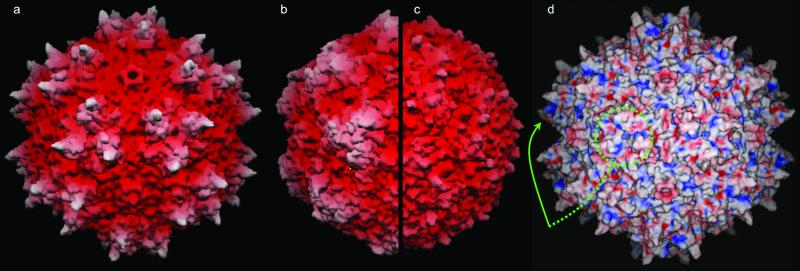
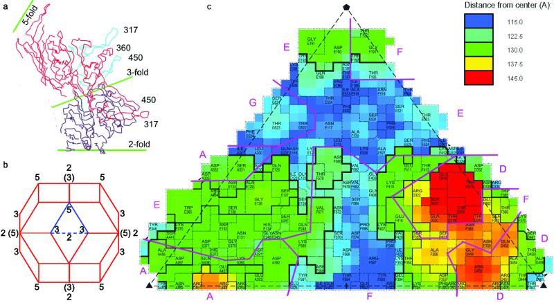
The structure of the adeno-associated virus (AAV-2) has been determined to 3-A resolution by x-ray crystallography. AAV is being developed as a vector for gene therapy to treat diseases including hemophilia, cancer, and cystic fibrosis. As in the distantly related autonomous parvoviruses, the capsid protein has a beta-barrel fold, but long loops between the beta-strands share little structural homology with other parvoviruses, leading to unique surface features. Most prominent are groups of threefold-related peaks, each an intimate association of loops from two neighboring subunits. Mutations affecting cell entry and receptor binding are clustered near the positively charged side of each peak, implicating the region in attachment to the cellular receptor, heparan sulfate proteoglycan. Amino acids involved in antibody binding are in the same general vicinity. The structure will guide rational engineering of vector capsids to tailor cellular targeting and to avoid immediate neutralization by an immune system sensitized by prior exposure to AAV.


Figures

Fig 1.
Fig 1.
Structure of the AAV-2 subunit and comparison with related structures. (a) Experimental electron density for AAV-2. Phases for this 3-Å resolution electron density map are independent of the AAV-2 model, having been obtained by symmetry averaging and extension from a CPV model at 15-Å resolution. Density is clear and allows an unambiguous fitting of the chemical sequence throughout VP3. (b) Ribbon drawing of the AAV-2 subunit. The locations of the neighboring symmetry axes are shown. The β-barrel is on the inner surface of the capsid (pink) with strands of the two sheets labeled conventionally as A, B, I, D, and G, and C, H, E, and F. Loops are labeled according to the flanking strands—e.g., GH loop. Regions where the sequence differs greatest between the AAV serotypes are colored purple (61). (c) Comparison of the backbones of AAV-2 (red) and canine parvovirus (cyan). The loop structure, which is responsible for many of the viral-host interactions differs substantially between AAV-2 and canine parvovirus, is largely absent from insect densoviruses (not shown).
Fig 2.
Fig 2.
Conservation of sequence and secondary structure. Sequences of AAV-2 and CPV were aligned according to structure, then improved in loop regions according to sequence [Programs STAMP, PILEUP, and ALSCRIPT (62–66)]. Residues that are identical in AAV-2 and CPV are red. Those with an ALSCRIPT similarity score >5 (measured from 0 to 10) are yellow. Secondary structures of AAV-2 and CPV are compared, the core β-barrel highlighted in green, and labeled according to AAV-2 (CPV in parentheses where different). Side chain (outer) surface accessibility was calculated with a 1.5 Å radius probe, and is shown with triangles shaded according to each residue's accessibility: 20 Å2 < red <50 Å2 < cyan <80 Å2 < green <100 Å2 < blue.
Fig 3.
Fig 3.
Surface topology and electrostatics. (ac) show grasp (67) surface renderings of AAV-2, canine parvovirus, and the insect densovirus, respectively, drawn to scale, and colored according to distance from the viral center. The view is down a twofold axis (center of the virus) with threefolds left and right of center, fivefolds above and below (see Fig. 4b). (d) shows the electrostatic surface potential of AAV-2 calculated with spock (68) running from −10 (red) to +10 (blue). The putative receptor-binding sites are positively charged patches on the side of each threefold-proximal peak, viewed edge-on (arrowed) or head-on where 3 equivalent patches join at each threefold (circled).
Fig 4.
Fig 4.
Interdigitation of subunits: (a) shows a thin equatorial cross-section of AAV-2 (Cα traces) orthogonal to a twofold near the threefold axes. It emphasizes the elevation of the peaks, each of which comprises loops from two subunits that are colored differently. (b) a rhombic triacontahedron showing the viral orientation for Figs. 3 and 4. The three- and fivefold axes are at vertices joining three and five faces, respectively, and twofolds bisect neighboring threefolds, some of which are labeled. (Axes in parentheses superimpose, at an angle, upon twofolds behind.) The highlighted triangle (used in c) may be repeated sixtyfold with icosahedral symmetry operators to generate the entire capsid. (c) A schematic projection of one of the 60 triangular facets shows the surface amino acids with different subunits separated by purple lines. roadmap (69) was used to color as in a topographical map, with blue closest to the virus center, red farthest from the virus center. VP2 numbering is used with a letter prefix denoting the symmetry equivalent subunit.

References

    1. Orkin S. H. & Motulsky, A. G., (1995) Report and Recommendations of the Panel to Access the NIH Investment in Research on Gene Therapy (National Institutes of Health, Bethesda).
    1. Pfeifer A. & Verma, I. (2001) in Virology, eds. Fields, B. N., Knipe, D. M. & Howley, P. M. (Lippincott, Philadelphia), pp. 469–492.
    1. Carter B. J. (2000) in Parvoviruses: From Molecular Biology to Pathology and Therapeutic Uses, eds. Faisst, S. & Rommelaere, J. (Karger, Basel), pp. 85–106.
    1. Flotte T. R. & Carter, B. J. (1998) Methods Enzymol. 292, 717-732. - PubMed
    1. Kay M. A., Manno, C. S., Ragni, M. V., Larson, P. J., Couto, L. B., McClelland, A., Glader, B., Chew, A. J., Tai, S. J., Herzog, R. W., et al. (2000) Nat. Genet. 24, 257-261. - PubMed

Publication types

Associated data